ADVERTISEMENTS:
In this article we will discuss about the dissection of pigeon. Also learn about:- 1. Dissection of Muscles of Flight 2. Dissection of Air Sacs 3. Dissection of Alimentary System 4. Dissection of Circulatory System 5. The Venous System 6. The Arterial System 7. Dissection of Cranial Nerves 8. Dissection of Urinogenital System 9. The Urinary (Excretory) System 10. The Genital System.
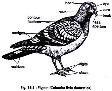
The pigeon (Fig. 18.1) is a typical flying bird. It is a preferred laboratory animal for low cost and easy availability.
Killing:
ADVERTISEMENTS:
Pigeons are killed with chloroform.
Dissection:
Pluck the feathers and put those in a basket in a corner of the laboratory. Lay the bird on a dissecting tray with the ventral surface up. Fix the pigeon in that position by pushing pins through wings and hind limbs.
ADVERTISEMENTS:
Give a longitudinal incision on the skin of the breast along the mid-ventral line. Continue the incision both anteriorly and posteriorly up to the extremities of the trunk. Give lateral incisions on the skin along the limbs. Separate the skin from the underlying muscles and remove it.
Dissection of Muscles of Flight:
All the flight muscles narrow down anteriorly. While dissecting a muscle, separate it from its attachment but never cut the insertion. The breast of the pigeon is boat- shaped. The muscles exposed after the removal of the skin are the two pectoralis major, which nearly fill the wedge-shaped space between the body and the keel of the sternum, and form the breast.
With a sharp scalpel cut the attachment of one pectoralis major to the keel, either on the right or left side of it. Posteriorly and laterally the muscle is attached to the body wall by membranous tissue. Cut along the line of attachment and turn it upwards.
The pectoralis minor is shorter and narrower and dorsal to the pectoralis major. Cut its attachment to the anterior part of the sternum to separate it from the latter. The coracobrachialis or the third pectoralis is the smallest breast muscle for flight. It is attached to the coracoid and the sternum. Separate it from the coracoid and sternum (Fig. 18.2).
Pectoralis major (depressor muscle):
It is the largest and most ventral flight muscle. The width is maximum at about the middle, where it is thickest also. The fibres of the muscle converge anteriorly to be inserted into the ventral aspect of the humerus, which it depresses.
Pectoralis minor (Supracoracoideus or elevator muscle):
It is wide and thick at the middle. The much narrowed anterior end sends its tendon through the foramen triosseum to be inserted into the dorsal aspect of the humerus, which it elevates.
ADVERTISEMENTS:
Coracobrachialis (Scapulo-humeral):
A small, narrow muscle, inserted into the ventral surface of the humerus through its tendon. It helps in depression of the wing.
Dissection of Air Sacs:
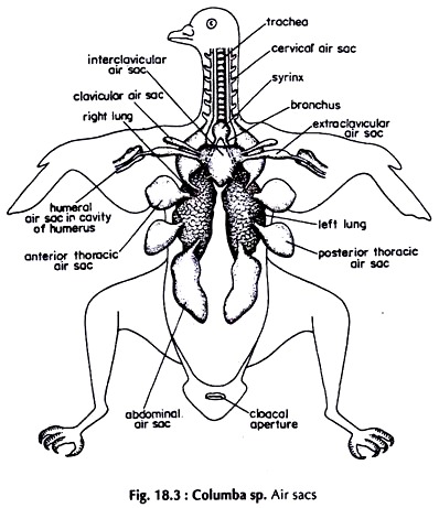
Remove the skin covering the ventral and lateral sides of the breast and the limbs, following the procedure described previously. Continue the mid-ventral incision in the skin of the neck up to the anterior end. Separate the skin of the neck on each side of the incision and pin down the flaps. Locate the trachea, which is made up of close set cartilaginous rings.
Posteriorly, the trachea divides into two bronchi. Each bronchus is continuous with the air sacs of the side through the lung. Cut the trachea and plug the cut end with water soaked cotton wool. If necessary, the air sacs can be inflated by blowing air through the trachea after removing the plug.
ADVERTISEMENTS:
Cut the ventral abdominal wall transversely posterior to the sternum. Proceed laterally and anteriorly cutting the ribs up to the anterior border of the coelom. Cut the pectoral girdle and carefully remove the sternum.
The lung and air sacs are dorsal to the visceral organs. Remove the visceral organs, taking great care that the air sacs, which are very thin, are not punctured. To make out the outlines of air sacs, if necessary, inflate them occasionally by blowing air through the trachea. Never attempt to dissect air sacs in fully inflated state. There are nine major air sacs (Fig. 18.3).
Abdominal:
ADVERTISEMENTS:
Paired, large, elongated sacs lying in the abdominal cavity. They are most posterior in location. The air sacs are connected with the ventral surface of the lungs, close to the posterior border.
Posterior thoracic:
Paired, medium-sized sacs, anterior to the abdominals and closely applied to the side walls of the body.
Anterior thoracic:
ADVERTISEMENTS:
Paired, medium-sized sacs, slightly smaller than the posterior thoracics. They are anterior to the posterior thoracics and closely applied to the body walls.
Inter-clavicular:
A median sac, dorsal to the bronchi, and anteromedian to the anterior thoracics. It is connected to the ventral surface of both the lungs. On each side it sends two branches, the clavicular, in the armpit, and the axillary (extra-clavicular) continuing in the humerus.
Dissection of Alimentary System:
With the removal of the skin on the ventral surface of the neck, the anterior part of the alimentary canal is exposed. The rest of the structures of the alimentary system lies in the coelom (Fig. 18.4). Remove the heart with the pericardium.
Mouth:
ADVERTISEMENTS:
A fairly large opening bounded by two horny beaks. Teeth absent. The tongue on the floor of the mouth cavity is large and pointed.
Pharynx:
A small chamber, posterior to the mouth cavity.
Oesophagus:
A fairly long, thick walled tube extending from pharynx to the proventriculus. The major part of the oesophagus lies in the neck running by the side of the trachea.
Crop:
ADVERTISEMENTS:
The posterior part of the oesophagus is dilated into a large, thin walled sac at the base of the neck, between the skin and the muscles, and immediately in front of the sternum. The crop is a reservoir for food, consists of grains. In pigeon the stomach is represented by the proventriculus and gizzard.
Proventriculus:
It is a slightly dilated, small tube between the crop and the gizzard.
Gizzard:
A highly muscular, biconvex body, round in outline (The cavity always contains small stones, which are swallowed by the bird for grinding food in gizzard.).
Duodenum:
The first part of the intestine. It leaves the gizzard close to the entrance of the proventriculus and form a U-shaped loop.
Ileum:
The rest of the intestine. It forms a loop at the beginning. The major part of the rest is spirally coiled. In the posterior region it again forms a loop to end in the rectum.
Rectum:
It is short and slightly wider than the diameter of the intestine. It opens into the coprodoeum of the cloaca. A pair of small outgrowths, the rectal caeca are present at the junction of the rectum and the ileum.
Liver:
A large, reddish brown, bilobed gland. Gall bladder is absent. Ducts from the liver open directly into the duodenum.
Pancreas:
A compact reddish gland lying in the loop of the duodenum. Its secretion is discharged into the duodenum by the three ducts.
Dissection of Circulatory System:
The components of the circulatory system are the heart, veins, arteries and capillaries. The heart is a part of both the venous and arterial systems.
Remove the skin covering the breast and ventral surface of the wings and legs. Give a transverse incision on the abdominal wall posterior to the sternum. Proceed with the incision anterolaterally, in both the sides up to about the middle of the sternum by cutting the ribs. Give a transverse incision on the breast at about its middle but behind the heart. Remove the posterior part of the sternum with the muscles of flight.
Carefully remove the tissues at the base of the neck, give an incision in the crop, squeeze out the grains, if any, in it and clear the space between the oesophagus and the sternum. Cut the attachment of the remaining portions of flight muscles to the keel by giving a longitudinal incision by the side of the keel going deep up to the ventral surface of the sternal bone, but do not remove them.
By this, damage to the pectoral artery, supplying breast muscles can be avoided. Push the blunt arm of a pair of stout scissors through the space between the oesophagus and the sternum, at the anterior border of the latter and cut one of the clavicles near the base of the furcula.
Proceed backwards and cut the sternum lengthwise, close to the keel, the incision running between the articulation of the two coracoids with the sternum. The body cavity is fully exposed.
Slowly push each half of the sternum outwards with the pectoral girdle of the side by cutting tissues and muscles wherever necessary, taking care not to damage blood vessels. Damage of some minor vessels cannot be avoided but this does not interfere with the dissection of circulatory system.
The Heart:
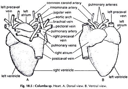
It is located at the anterior end of the coelom, oriented obliquely dorsoventrally, the anterior end being directed dorsally. Remove the pericardium to expose the heart which is reddish brown, massive and four chambered (Fig. 18.5).
Atria (auricles):
Two thin walled, muscular chambers in front of the ventricle. The right atrium receives the right and left precavals and postcaval vein bringing deoxygenated blood and the left receives oxygenated blood through a common vessel formed by the joining of four large pulmonary veins.
Ventricle:
A strongly muscular, thick walled structure, posterior to the atria. The apex of the ventricle is directed posteroventrally.
The Venous System:
Remove the pericardium. Lift the tip of the heart and push it a little forward. The space between the heart and the liver comes in view. Remove the tissues present in this area and the postcaval vein can be identified. By removing tissues just posterior to the aortic arches, the precaval veins are identified.
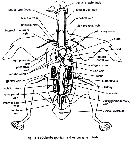
Turn the heart a little to the left and the joining of both the precavals and post caval vein to the right atrium can be seen. The vessels forming the precaval veins are similar. Expose the vessels forming the postcaval vein and hepatic portal system. The renal portal system is absent in pigeon (Fig. 18.6).
A. Precaval vein:
Each precaval receives the following veins from one side of the anterior part of the body.
Jugular:
Arises from the head and runs backward along the neck close to the vertebral column and dorsal to the oesophagus. Anteriorly the two jugulars right and left, are joined by a transverse vessel.
Posteriorly it receives a vertebral vein and joins the precaval vein, which receives:
Brachial from the wing,
Pectoral from the pectoral muscles,
Internal mammary from the inner surface of the thoracic wall.
The right precaval vein opens into the right anterior angle and the left precaval into the left border of the right auricle.
B. Postcaval vein:
A large vein, situated a little to the right of the heart. It is formed by the union of veins coming from the middle and posterior half of the body.
Caudal:
Brings back blood from the tail region. In the posterior part of the abdomen it bifurcates to two renal portal veins.
Coccygeomesenteric:
A large vein returning blood from the rectal region. It joins the caudal vein, close to the point of its bifurcation.
Renal portal:
Each receives a few small renal veins from the kidney and the main vein runs forward through the substance of the kidney, sending a few small branches to it. (To trace its course, the kidney has to be partially cut open).
Sciatic:
Brings back blood from the thigh and joins the renal portal vein of the side.
Femoral:
Brings blood from the leg of the side and joins renal portal vein to form the iliac vein.
Gonadial:
Short, runs from gonad to end in the iliac vein. Two in male; only left one present in female.
Postcaval:
Two iliacs unite to form the postcaval vein.
Epigastric:
Returns blood from the great omentum and ends in one of the hepatic veins of the left side.
Hepatic:
Three pairs of veins from the lobes of the liver. The postcaval vein runs forward through the right lobe of the liver for some distance (the liver is to be cut open to expose it) and opens on the posterior surface of the right atrium.
Hepatic portal system:
It consists of veins opening in the liver.
The hepatic portal vein is formed by the joining:
i. Inferior mesenteric from the rectal region
ii. Gastro-duodenal from the intestine and right side of the gizzard,
iii. Anterior mesenteric from the greater part of the intestine.
The hepatic portal vein opens in the lobes of the liver through branches.
C. Pulmonary veins:
Small but wide veins not connected with the major veins. Two veins from each lung, dorsal to the heart and open into the left atrium on its dorsal surface.
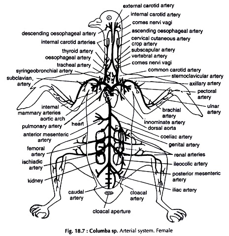
The Arterial System:
Remove the pericardium and identify the pulmonary arch arising from the right ventricle, and the aortic arch leaving the left ventricle. The left aortic arch is absent. The arteries appear as white vessels. Trace the arteries along their courses (Fig.18.7).
A. Pulmonary arch:
Soon after its origin from the right ventricle it divides into two, the right and left pulmonary arteries. Each artery loops over the heart anterodorsally, runs backward to end in the lung of the side.
B. Aortic arch:
It curves over the right bronchus, reaches the dorsal body wall and runs backward as the dorsal aorta along the mid-dorsal line.
Branches of the aortic arch:
Innominate:
The right and left innominate arteries arise from the aortic arch, close to each other, before it turns backward. Each innominate divides into two, the subclavian running laterally and the common carotid running anteriorly.
Subclavian:
Immediately after its origin it divides into two, the axillary and pectoral.
Axillary curves forward and runs dorsolaterally to enter the fore limb. It runs outward as the brachial.
Pectoral sends two slender internal mammary arteries and itself divides into three branches in the pectoral muscle.
Common carotid:
After giving out a short mesial branch supplying oesophagus, trachea and bronchus it divides into three, a slender comes nervivagi and two stout vessels, the vertebral and internal carotid. The ascending oesophageal arises mesialy from the comes nervivagi.
Internal carotid:
The two internal carotids converge anteromesially, overlap for some distance anteriorly and then run forward side by side. Further anteriorly, they move away, each gives off an external carotid and proceeds forward.
External carotid:
Close to its origin it receives the comes nervivagi and immediately sends out the descending oesophageal branch. The external carotid proceeds forward as facial artery.
Dorsal aorta:
Runs posteromedially sending following arteries to different organs.
Coeliac:
Unpaired, runs to the stomach and liver.
Anterior mesenteric:
Unpaired, ends in the intestine.
In correspondence with the position of the legs, the femoral and sciatic arteries arise far forward.
Femoral:
Paired, end in the anterior part of the thigh.
Sciatic (ischiadic):
Paired, running to the posterior portion of the thigh.
Renal:
Three pairs, one pair arise from the dorsal aorta and the other two pairs from the sciatics. The renals of each side end in the three lobes of the kidney of the side.
Gonadial (genital):
Paired in males. Arise from the anterior pair of renal arteries and end in the testes and their ducts. In female, only the left one is present.
Iliac:
Paired, end in pelvis.
Posterior mesenteric:
Unpaired, supplies the rectum and the cloaca.
Caudal:
A small vessel supplying the caudal vertebrae.
Dissection of Cranial Nerves:
The cranium is bony. The bones, however, are not very hard as in toad and lizard. To expose the brain and roots of the cranial nerves follow the technique adopted for Channa punctatus (Fig. 18.8). The cranial nerves are twelve pairs in pigeon.
Fifth (V) cranial nerve:
The fifth or trigeminal is the first nerve of the medulla oblongata and arises from the ventrolateral side of the anterior end. Immediately after its origin it divides into three branches.
a. Ophthalmic runs forward through the nasal area and innervates the upper beak region.
b. Maxillary proceeds forward along the floor of the orbit and supplies maxillary region and palate.
c. Mandibular turns downward, runs forward and supplies the lower beak.
Seventh (VII) cranial nerve:
The seventh or facial is the third nerve of the medulla oblongata. It arises from the side of the medulla behind the fifth nerve.
The nerve divides into two branches:
a. Palatine a slender branch, proceeds forward along the roof of the orbit and innervates the roof of the mouth cavity.
b. Hyomandibular a stout branch, runs backward and downward and then forward round the tympanic region, bifurcates at the angle of the jaws and supply the post-tympanic area and lower beak.
Ninth (IX) cranial nerve:
The ninth or glossopharyngeal is the fifth nerve of the medulla oblongata. It arises from the posterolateral side of the medulla behind the auditory nerve. The nerve comes out of the cranium through a small foramen present between the ear and the hypoglossal foramen.
Outside the cranium the nerve divides into two branches. The anterior branch is small and joins the hyomandibular branch of the VII or facial nerve. The posterior branch is large and innervates the floor of the buccal cavity and pharynx.
Tenth (X) cranial nerve:
The tenth or vagus nerve arises posteroventrally from the medulla oblongata.
It sends four major branches:
a. Laryngeal innervates the larynx, runs posteriorly in the neck to supply the syrinx. In the coelom it divides into three branches.
b. Pulmonary innervates the lung.
c. Cardiac supplies the heart.
d. Gastric innervates the crop, proventriculus and gizzard.
Dissection of Urinogenital System:
The urinary and genital systems are collectively described as urinogenital system. Unlike those in Amphibia and Reptilia the two systems are however, independent in Columba.
The system is located in the posterior region of the abdomen. Remove the alimentary system, heart and major portion of the circulatory system except the posterior part of the dorsal aorta and the postcaval vein.
The Urinary (Excretory) System:
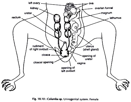
(Figs. 18.9 & 18.10)
Kidneys:
Two dark red, elongated, flat, three lobed bodies, dorsal in position and fit closely into the hollow of the pelvis.
Ureters:
The urinary ducts are narrow tubes, arise from the ventral surface of the kidneys at some distance from the anterior end, and run backward along their mid-ventral surface. Posteriorly, they open separately on the dorsal surface of the urodaeum or middle chamber of the cloaca.
The Genital System:
Male Genital System:
(Fig. 18.9)
Testes:
White, ovoid bodies, attached to the ventral surface of the anterior end of the kidneys by peritoneum.
Vasa deferentia:
Paired, narrow, convoluted tubes, arise from the inner border of the testes. The vas deferens runs posteriorly, parallel and close to the ureter.
Vesiculae seminalis:
The vas deferens enlarges slightly at its posterior end before opening on the dorsal surface of the urodaeum, lateral to urinary opening.
Female Genital System:
(Fig. 18.10)
The right ovary and oviduct are atrophied. The oviduct is represented by a fair- sized vestige, opening in the right side of the cloaca. Vestige of right ovary is often present.
Ovary:
The ovary (left) is irregularly oval with the surface raised up into rounded elevations. It is attached to the dorsal body wall of the abdomen by mesovarium.
Oviduct:
A long, stout, convoluted tube, lateral to the ovary. It bears a large funnel, the infundibulum at the anterior end.
Uterus:
The glandular posterior portion of the oviduct. It opens on the dosal surface of the urodaeum, lateral to the ureter opening.

