ADVERTISEMENTS:
In this article we will discuss about the modifications of aortic arches in vertebrates.
The basic fundamental plan of the aortic arches is similar in different vertebrates during embryonic stages. But in adult the condition of the arrangement is changed either being lost or modified considerably. The number of aortic arches is gradually reduced as the scale of evolution of vertebrates is ascended.
Cyclostomes:
ADVERTISEMENTS:
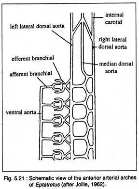
In lampreys (Petromyzon) there are eight pairs of aortic arches and in hag fishes (Bdellostoma) there are fifteen pairs. The aortic arch is divided into afferent branchial artery and efferent branchial artery.
In lampreys each aortic arch divides and sends branches to the posterior hemi-branch and anterior hemi- branch of the adjacent gill pouch (Fig. 5.13). In hagfishes each arch supplies to the hemi-branch of a single gill-pouch (Fig. 5.21).
Elasmobranchs:
ADVERTISEMENTS:
Generally there are five pairs of aortic arches in elasmobranchs but in some cases there is a variation. In Hexanchus there are six pairs of aortic arches. In Heptranchias there are seven pairs. In elasmobranchs (Fig. 6.10) the first pair of aortic arches (mandibular) disappear. Second to sixth pair of aortic arches (II-VI) persist as branchial arteries. Each aortic arch is divided into afferent and efferent branchial arteries.
Five pairs afferent arteries arise from ventral aorta and supply deoxygenated blood to the respective gills. The ventral aorta divides into two branches, called innominate arteries which again bifurcate into the first and second afferent branchial arteries. From the gills the oxygenated blood is collected by efferent branchial arteries.
In elasmobranchs there are nine pair’s efferent branchial arteries of which the first eight arteries form a series of four complete loops but ninth efferent branchial artery collects blood from the demi- branch of the fifth gill pouch.
Teleosts:
In teleosts there are four pairs of aortic arches. First pair (mandibular) and second pair (hyoidean) are lost, only four pairs (third to sixth) persist as branchial arteries (Fig. 10.145). Four pairs afferent branchial arteries arise from the ventral aorta.
They supply deoxygenated blood to the gills for aeration. The ventral aorta bifurcates anteriorly to form the first pair of afferent branchial arteries. In sturgeon (Acipenser) and Amia each afferent branchial arch bifurcates as in elasmobranchs.
Dipnoans:
ADVERTISEMENTS:
Among dipnoans there are four or five aortic arches that develop from the ventral aorta which supply blood to the gills. In the Protopterus the aortic arches retain second, third, fourth, fifth and sixth. The arches like the fish have afferent and efferent divisions. They have two pulmonary arteries which develop from the efferent division of the sixth arch. There is no second efferent branch in Neoceratodus.
Amphibians:
In amphibians the aortic arches retain the bilateral symmetry. Of the six pairs of embryonic aortic arches, the fifth one is observed in adult Cryptobranchus (Urodele). The sixth arch becomes small. It gives rise to the pulmonary artery and continues as the ductus arteriosus to the dorsal stem. The pulmonary artery may give rise to musculocutaneous artery.
In Necturus (Fig. 10.146C) the first external gill is supplied by the third afferent arch. The base of the sixth arch is absent in Necturus. The ductus arteriosus connects pulmonary artery with the dorsal stem.
ADVERTISEMENTS:
In anurans (adult), only three pairs of aortic arches (III, IV and VI) are present (Fig. 10.146D). But in the tadpole the development of aortic arches correspond to the emergence of external and internal gills (Fig. 10.146A). Fig. 10.146 shows the fate of aortic arches in some amphibians.
Reptiles:
Only three pairs of aortic arches persist such as third, fourth and sixth (Fig. 10.147A). The first, second and fifth pairs of aortic arches disappear. The fifth arch is present in reduced form in some reptiles. The remnant of the radix of aorta between third and fourth arches is present on each side in some snakes. The ill-defined conus arteriosus is splitted into three vessels.
The fourth arch on the left side which becomes the left systemic arises from the right side of the partially divided ventricle. The fourth arch on the right side arises from the left side of the ventricle. It establishes a connection with a portion of the radix aorta of the right side and becomes the right aortic arch.
The common carotid arch arises from the right aortic arch and becomes divided into external and internal carotid arteries. The sixth aortic arch loses all its connection with radices of aorta and becomes the pulmonary arteries.
The radix of aorta between carotid and systemic arches is present as ductus caroticus in many lizards (Fig. 10.147A). Similarly a part of the radix may remain connected with the sixth aortic arch.
This connecting part is known as ductus arteriosus. Ductus caroticus and ductus arteriosus are both present’ in Sphenodon. In crocodiles the right systemic arch develops from the left ventricle which gives rise to subclavian and innominate arteries.
ADVERTISEMENTS:
Birds:
The birds retain three pairs of aortic arches. These are lllrd, IVth and VIth (Fig. 10.147B). Rest three pairs such as Ist, llnd, and Vth are lost. The third becomes carotid artery, the fourth becomes systemic aorta but retains only the systemic arch on the right side.
The systemic arch on the left side disappears. The right systemic arch originates from the systemic aorta that develops from left ventricle. The sixth arch becomes the pulmonary aorta that divides into two pulmonary arteries and each artery goes to each lung.
Mammals:
The Ist, llnd and Vth aortic arches disappear. The lllrd, IVth and Vlth aortic arches persist of which the right aortic arch of IVth disappears. Only aortic arch on the left side persists (Fig. 10.147C).
The IVth becomes the systemic aorta and Vlth arch becomes pulmonary aorta. The Ist and llnd aortic arches disappear but the basal stem becomes the external carotid and lllrd aortic arches become the internal carotid. The dorsal connection between the lllrd and IVth aortic arches disappears.
ADVERTISEMENTS:
The right side of IVth aortic arch becomes right subclavian artery. The left subclavian artery arises from the upper part of the left systemic arch. Right and left common carotid arteries with the right subclavian develop from a common aortic arch, called brachiocephalic artery.
In the embryonic stages ductus arteriosus is seen on sides but ductus arteriosus in the left side remains after birth as fibrous band, called ligamentum arteriosum or Botalli.


