ADVERTISEMENTS:
In this article we will discuss about the dissection of rat. Also learn about:- 1. Dissection of Alimentary System 2. Dissection of Circulatory System 3. Dissection of Venous System 4. The Arterial System 5. Dissection of Cranial Nerves 6. Dissection of Brain 7. Dissection of Neck Region 8. Dissection of Urinogenital System 9. The Urinary (Excretory) System 10. Dissection of Genital System.
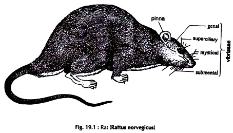
The rat is a typical mammal. Formerly guinea pig (Cavia sp.) (Fig. 19.1) were used for dissection in most of the undergraduate and postgraduate colleges in Indian Universities. Of late, due to prevailing high cost, guinea pig is being replaced by rat.
Four species of rats are common in India, of which three are wild. Rattus rattus are the large-sized ones. R. rattus and R. bengalensis are commonly found in godowns domestic houses in towns and cities. Rattus norvegicus (Fig. 19.1) are medium-sized ones common in fields, domestic houses and godowns of rural areas.
ADVERTISEMENTS:
Rattus norvegicus Albinus or white rats are still not a wild form. They are reared and bred chiefly for use in the laboratories or as pets. The organ-systems of guinea pig are almost similar to those of rats, so far requirements of routine dissection are concerned. The differences in structures, wherever present, are noted in appropriate places.
Killing:
Rats are killed with chloroform. House rats often carry internal and external parasites. The narcotized animals should be disinfected with phenol, Lysol or some other disinfectants, and thoroughly washed with water before use.
ADVERTISEMENTS:
Dissection:
Put the specimen on its back on a dissecting tray. Fix it with pins passing through the limbs. Lift the skin of the abdomen with a pair of forceps and make a small cut at about the middle of the abdomen. Starting from the cut give an incision extending up to the snout anteriorly and the genital opening posteriorly. Give transverse incisions along the length of the limbs. Separate the skin from the underlying muscles and pin it down.
Give a transverse incision on the abdominal wall in the posterior region. Proceed laterally and anteriorly on both sides cutting the ribs up to the anterior border of the thoracic cavity. Cut the clavicles and the muscles there. Lift the sternum and cut the attachment of the diaphragm to the abdominal wall.
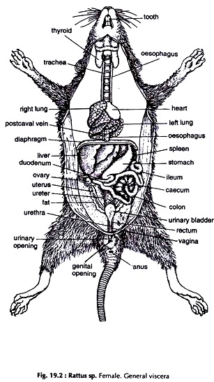
The ventral wall of the thorax and abdomen with the sternum is separated and the organs in the thoracic and abdominal cavities are exposed (Fig. 19.2). The structures in the neck region have already been exposed with the removal of the skin.
General Viscera:
Coelom:
A large cavity in the thoracic and abdominal region bounded by the ventral abdominal wall, lateral body walls and dorsal body wall.
Diaphragm:
ADVERTISEMENTS:
A muscular partition divides the coelom into a small thoracic cavity in front and a large abdominal cavity behind. The organs lodged in the coelome are collectively termed viscera.
The thoracic cavity is divided into three chambers by membranous tissue and accommodate the following organs:
Pleural cavities:
Two, each lodges a lung.
ADVERTISEMENTS:
Mediastinum:
The space between the two pleural cavities in which the pericardial cavity, the oesophagus and blood vessels are lodged.
Pericardial cavity:
A small chamber in which the heart enclosed by a double layered pericardium is lodged.
ADVERTISEMENTS:
Lungs:
Paired, spongy, bright pink in colour, located on either side of the heart. The right lung is four-lobed while the left one is un-lobed.
Oesophagus:
A narrow, straight tube, dorsal to the heart and median to the lungs, runs from the pharynx, passes through the diaphragm to join the stomach.
ADVERTISEMENTS:
The following organs are lodged in the abdominal cavity:
Liver:
A large, reddish brown gland with four lobes, the right, the left, a median cystic and a posterior caudate. It is attached to the posterior face of the diaphragm by falciform ligament.
Stomach:
A large, somewhat elongated, bend sac lying on the left side, posterolateral to the liver, being partly covered by it.
Small intestine:
ADVERTISEMENTS:
A long narrow tube, the first part forming the loop is the duodenum and the rest coiled portion is ileum.
Pancreas:
A diffused gland with small lobes, scattered on the mesentery of the duodenal loop.
Caecum:
A fairly large, blind outgrowth at the junction of the small and large intestine.
Large intestine:
ADVERTISEMENTS:
A fairly long tube. The first part is colon and the rest rectum.
Spleen:
A dark red body lying in the greater curvature of the stomach.
Kidneys:
Paired, bean-shaped, dark red bodies. The right kidney is slightly anterior to the left. The ureter is a narrow tube emerging from the hilus on the inner side.
Adrenal glands:
Paired, round bodies, a little anterior to the kidneys.
Urinary bladder:
A pear-shaped, muscular sac with a narrow neck.
Ovaries:
Paired, compact, yellow bodies, ventrolateral to the kidneys in female.
Dissection of Alimentary System:
With the removal of the skin on the ventral surface of the neck, the anterior part of the alimentary canal is partially exposed. The rest of the canal and its associated glands are lodged in the thoracic and abdominal cavities (Fig. 19.3). Remove the heart with the pericardium and the lungs.
Mouth:
It is a fairly large opening at the anterior end of the snout and bounded by two lips with corresponding jaws. The teeth are heterodont and thecodont. The muscular tongue on the floor of the mouth cavity fits into the groove of the palate.
Pharynx:
A small chamber between the mouth cavity and the oesophagus. (It is common to both the digestive and respiratory system).
Oesophagus:
A narrow, straight tube, running from the pharynx, perforates the diaphragm and joins the stomach in the abdominal cavity. Anteriorly it is dorsal to the larynx. In the neck region it runs parallel to the trachea. In the thoracic cavity it is dorsal to the heart and lungs.
Stomach:
It lies on the left side of the abdominal cavity, close to the posterior face of the diaphragm. The cardiac stomach is thin walled, transparent and on the left side. The pyloric stomach is muscular, opaque and on the right side.
Small intestine:
A very long tube of uniform diameter. It is divisible into two zones, the duodenum and ileum.
Duodenum:
The first part of the small intestine and forms a U-shaped loop.
Ileum:
It is a much coiled tube ending in the large intestine.
Caecum:
A fairly large, blind outgrowth at the junction of the small and large intestine.
Large intestine:
It has two zones, colon and rectum.
Colon:
The first part of the large intestine. It is short in length.
Rectum:
The last part of the large intestine. It is longer and opens through the anus.
Liver (see general viscera):
Hepatic ducts from the lobes join to form a common bile duct which opens in the duodenum.
Pancreas:
A diffuse type gland made up of numerous scattered lobules on the mesentery in the duodenal loop. Ducts from the lobules unite into a common duct which joins the common bile duct to open in the duodenum.
Dissection of Circulatory System:
The components of the circulatory system are the heart, veins, arteries and capillaries. The heart is a part of both the venous and arterial systems.
Expose the viscera and the neck region following the procedure described previously.
The Heart:
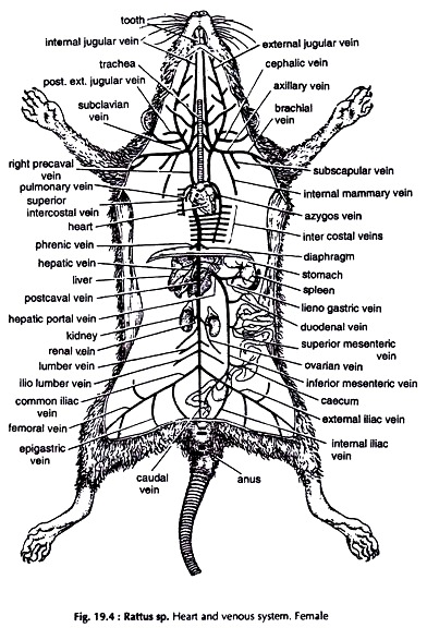
It is lodged in the pericardial cavity on the left side, at the anterior end of the thoracic cavity and oriented slightly obliquely. Remove the fat and the double layered pericardium around the heart. It is an ovoid, reddish brown muscular, four chambered structure (Figs. 19.4 & 19.6).
Atria (auricles):
Two muscular chambers, smaller in size and located in front of the ventricles. A large pulmonary artery lies between the two atria on the ventral side. The three systemic veins, two precavals and a postcaval open directly into the right atrium. The two pulmonary veins open in the left atrium through a common opening.
Ventricles:
The two ventricles together form a thick walled muscular structure constituting the major portion of the heart. An oblique groove separates the two ventricles. The left ventricle is larger than the right one. The pulmonary artery arises from the right and the aortic arch from the left ventricle. The right aortic arch is absent.
Dissection of Venous System:
Turn the ventricle upwards and locate the opening of the two systemic veins, viz. right and left precavals (cranial vena cava) and the post caval vein (caudal vena cava) the third systemic vein into the right atrium, and the opening of the common pulmonary vein into the left atrium.
Trace the two precavals. The veins they are receiving are not exactly similar. Expose the vessels forming the postcaval vein and hepatic portal system (Fig. 19.4). The renal portal system is absent.
A. Precaval veins:
Precaval veins bring back blood from the anterior region of the body. The right precaval is short and the left one is quite long. A precaval vein is formed by the union of a number of veins. The veins forming right and left precavals are similar except superior intercostal in the right and azygos in the left.
a. External jugular is formed by the union of anterior and posterior facial veins from the muscles of the head, jaws, tongue and salivary glands.
In its way back it receives:
i. Posterior external jugular from the muscles of the back and neck.
ii. Cephalic from the shoulder.
b. Internal jugular drains blood from the sternal region.
c. Subclavian is a short vein.
It receives:
i. Internal mammary from the sternal
ii. Axillary is formed by the union of brachial and subscapular, region and axillary vein.
iii. Brachial from the forelimb.
iv. Subscapular from the shoulder region.
Precaval vein is formed by the union of external and internal jugulars and subclavian vein.
Right precaval:
In addition to the above veins it receives superior intercostal from the upper part of the thoracic wall.
Left precaval:
In addition to the above veins it receives azygos formed by the union of intercostal and subcostals from the lower pari of the thoracic wall.
B. Postcaval vein:
It is a large, median vessel formed by the joining of a number of veins bringing blood from the organs in the posterior and middle region of the body.
Caudal drains blood from the tail and joins the postcaval at the point of its formation.
Internal iliac from the bladder and pelvic region.
External iliac formed by the union of:
i. Femoral from the hind limb.
ii. Epigastric from abdominal muscles and urinogenital system.
The two iliacs join to form a common iliac. The two common iliacs unite and form the postcaval vein. The caudal joins the postcaval at their junction.
Lumbar and iliolumbar:
A few pairs from the lumbar region.
Genital:
Paired, arise from the testes or ovaries and their associated structures. The right one joins precaval and the left one joins the renal vein. In male, these veins are long, since the testes are lodged in the scrotum (Fig. 19.6). In female, they are short.
Renal:
Paired, arise from the kidneys. The right renal is a little anterior in position.
Hepatic:
Paired, arise from the lobes of the liver.
The postcaval vein pierces the diaphragm and on reaching the thoracic cavity receives
Phrenic:
Paired, from the diaphragm.
Hepatic Portal System:
It consists of veins opening in the liver. It is formed near the pylorus and ends in the liver.
Inferior (posterior) mesenteric from rectum.
Superior (anterior) mesenteric from ileum, caecum and colon.
Duodenal from duodenum.
Lienogastric from stomach, spleen and pancreas.
The veins join to form the hepatic portal vein, which ends in the liver through branches.
C. Pulmonary veins:
Small but wide veins not connected with major veins. Blood is brought back by a single vein from the right lung and two veins, which join to form one, from the left lung. The two join to form a common pulmonary vein and opens in the left atrium.
The Arterial System:
A. The aortic arch (Systemic aorta):
The aortic arch arises from the base of the left ventricle. It turus left, forms an arch round the left bronchus and runs posterodorsally and medially as dorsal aorta along the mid-dorsal line of the thorax and abdomen. Trace the courses of the branches given out by the aorta (Fig. 19.7).
Innominate:
Arises from the right side of the arch and divides into two:
a. Right common carotid runs to the head lying parallel to the trachea.
It again divides into two:
i. Internal carotid lateral in position and enters the skull.
ii. External carotid medial, supplies head, jaw and tongue.
b. Right subclavian runs outward between the first rib and clavicle.
It sends following branches:
i. Vertebral courses forward and enters cranial cavity.
ii. Cervical parallel to external jugular vein and supplies muscles of the neck region.
iii. Internal mammary supplies sternal region.
iv. Costocervical supplies neck and upper part of the thoracic wall.
v. Subscapular supplies upper arm, scapula and thorax.
vi. Brachial continues in the forelimb.
The innominate artery is absent on the left side.
c. Left common carotid arises directly from the aortic arch. Branches are similar to those of the right common carotid.
d. Left subclavian emerges directly from the aortic arch. Branches are similar to those of the right subclavian.
Dorsal aorta:
Runs posteromedially sending branches to the following organs.
Intercostals:
A few pairs of small arteries supplying the thoracic walls.
Phrenic:
A pair of small arteries supplying the diaphragm.
The dorsal aorta pierces the diaphragm and enters the abdominal cavity. It sends off
Coeliac:
Unpaired, sends branches to stomach, liver and spleen.
Anterior mesenteric:
Unpaired, arises above the level of the kidneys and supplies pancreas, ileum, caecum and colon.
Renals:
Paired, supply the kidneys. Origin of right renal is anterior to that of the left one.
Genitals:
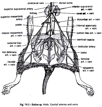
The right one arises from the dorsal aorta and the left one is a branch of the left renal artery. Supply gonads, testes or ovaries and genital ducts. In male, these arteries are long and run posteroventrally to supply testes and associated structures lodged in the scrotum (Fig. 19.5).
Lumbar:
A few pairs posterior to the renals, supplies dorsal muscles of the lumbar region.
Iliolumbar:
Unpaired, posterior to the lumbar, sends branches to iliac region and dorsal abdominal wall.
Posterior mesenteric:
Unpaired, origin close to the bifurcation of the dorsal aorta; supplies descending colon and rectum.
Common iliacs:
Paired, right and left, arise from one point.
Each divides into two major branches:
a. External iliac supplies abdominal wall and continues as femoral in the hind limb.
b. Internal iliac runs backward and supplies the pelvic region.
Caudal:
A narrow artery, continuation of the dorsal aorta and extends the whole length of the tail.
B. Pulmonary trunk:
Soon after its origin from the right ventricle it courses towards the dorsal side and bifurcates into two supplying the two lungs.
Dissection of Cranial Nerves:
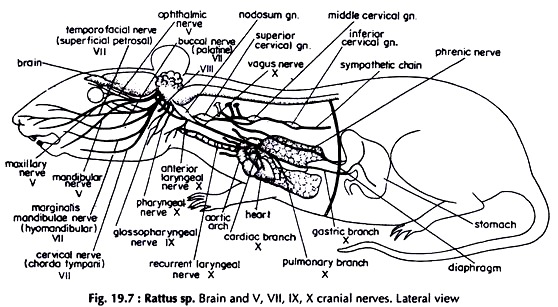
The cranium is bony and the bones are quite hard. The sutures of cranial bones are prominent. To expose the brain and roots of the cranial nerves make a hole at the junction of frontals and parietals with a pointed arm bone cutter or preferably a small hand drill. Remove the bones taking care that the roots of cranial nerves are not damaged. The cranial nerves are twelve pairs in rat (Figs. 19.7 & 19.8).
Fifth (V) cranial nerve:
The fifth or trigeminal is the first nerve of the medulla oblongata. It emerges from the lateral region of the pons.
Immediately after its origin it divides into three branches:
A. Ophthalmic:
It divides into three branches, all of which run forward:
a. Lachrymal supplies the inter-orbital lachrymal gland and the conjunctiva.
b. Frontal traverses the orbit, emerges in the region of the upper eye lid to which it sends branches and then proceeds forward to innervate the skin of the forehead.
c. Naso-ciliary enters the orbit, crosses to the medial wall and passes through the anterior ethmoidal foramen. It is now called anterior ethmoidal nerve, which enters the cranial cavity, runs forward to the nasal cavity after piercing the cribriform plate and sends branches, internal nasals to the mucous membrane. Leaving the nasal cavity as external nasal it innervates the wings and tip of the nose.
B. Maxillary proceeds forward, enters the orbit, traverses the infraorbital groove and canal and passes out through the infraorbital fissure. The nerve divides into nine branches and spread fanwise to the sides of the nose, the upper and lower lips.
C. Mandibular turns downward, runs forward and divides into two branches:
a. Nervus spinosus reenters the skull and innervates the dura mater.
b. Internal pterygoid divides into two.
i. Anterior trunk supplies the mandible.
ii. Posterior trunk innervates pinna of the ear, temporomandibular joint, skin of the temporal region, mucous membrane of the mouth, tongue and teeth and skin of the lower jaw.
Seventh (VII) cranial nerve:
The seventh or facial is the third nerve of the medulla oblongata. Arising from the anterolateral border of the medulla it enters the facial canal, from which it comes out through the stylomastoid foramen.
It divides into four branches:
a. Superficial petrosal runs along the lateral border of the carotid artery and through the middle ear. It innervates soft palate and lachrymal gland.
b. Palatine passes backward and upward between the external ear and mastoid process to the auricle and supplies palate and nasal region.
c. Hyomandibular:
The superficial branches pass anteriorly and dorsally over the zygomatic arch and branches in the neck. A deep branch supplies the muscles of the lower jaw and lip.
d. Chorda tympani enters the tympanic cavity, crosses the tympanic membrane and leaving the cavity innervates the snout, tongue and salivary glands.
Ninth (IX) cranial nerve:
The ninth or glossopharyngeal is the fifth nerve of the medulla. Arising from the posterolateral side of the medulla behind the auditory nerve it runs anteromedially between the external and internal carotid arteries.
It divides into two:
a. A small branch, pharyngeal joins the pharyngeal branch of the vagus and take part in the formation of pharyngeal plexus.
b. The main nerve, glossal runs forward and innervates the tongue, pharynx and buccal floor.
Tenth (X) cranial nerve:
The tenth or vagus nerve arises from the posteroventral part of the medulla. It comes out through the posterior lacerated foramen and bears nodosum ganglion, from which arise two nerves.
a. Pharyngeal from the anterior end of the ganglion joins the pharyngeal branch of the glossopharyngeal nerve.
b. Anterior laryngeal from the posterior end innervates larynx and thymus gland.
The vagus nerve runs backward through neck and thorax and enters abdomen. In the neck region it sends two branches.
c. Recurrent laryngeal:
In the left side it runs through the aortic arch and in the right side it makes a loop around the beginning of the subclavian artery. The nerves turn forward along the boundary between the oesophagus and trachea, to both of which they send branches and innervate the larynx and the diaphragm.
d. Cardiac innervates the heart and great vessels.
e. Pulmonary supplies the lung of the side.
f. Gastric:
The main trunk continues backward, sends minute branches to the oesophagus and along with it pierces the diaphragm. In the abdomen it sends branches to the liver. The right vagus ends in the posterior surface and the left in the lesser curvature and ventral surface of the stomach.
Dissection of Brain:
Take out the brain following the technique adopted for Channa punctatus. Put it in a high rim petri dish containing water (Fig. 19.9).
Due to the large size of the cerebrum and the cerebellum, and enormous structural changes of the latter, vision of some of the components on the dorsal surface of the brain is obliterated.
Dorsal View of the Brain:
Olfactory bulbs:
Small, paired, club- shaped, anteriormost outgrowths of the brain.
They are joined with the cerebral hemispheres. An olfactory nerve arises from each olfactory bulb.
Cerebral hemispheres:
Two large bodies, relatively long and narrow with broad bases and united mesially. They together form the cerebrum. By shallow depressions the cerebral hemisphere is divided into a lateral temporal lobe, anterior frontal lobe, median parietal lobe and posterior occipital lobe. Posteriorly the cerebral hemispheres cover the thalamencephalon and the optic lobes.
Corpus callosum:
A transverse commissural structure connecting the two cerebral hemispheres.
Pineal body:
A small, round mass at the posterior junction of the two cerebral hemispheres.
Mesencephalon (mid brain):
Only a small part of it is visible.
Cerebellum:
It is very large and consists of a central and two lateral lobes, divided by numerous fissures. Each lateral lobe bears an irregularly shaped prominence, the flocculus.
Medulla oblongata:
It is swollen in front and narrows down posteriorly to continue as spinal cord.
Ventral View of the Brain:
Hippocampal lobe:
A ridge-like structure, narrow anteriorly, extends from the base of the cerebral hemispheres.
Corpora striata:
A transverse band of white fibres situated in the posterior region of the cerebral hemispheres.
Optic chiasma:
It is not a part of the brain. The optic nerves arising from the optic lobes cross each other in front of the infundibulum forming optic chiasma.
Infundibulum:
A mesial downward process from the floor of the diencephalon.
Pituitary body (Hypophysis):
An ovoid body attached to the infundibulum.
Crura cerebri:
These are prominent elevations on the floor of the mid brain.
Pons varoli:
A flat band of transverse fibres connecting the lateral parts of the cerebellum.
Dissection of Neck Region:
Expose the arteries, veins and nerves in the neck region (Fig. 19.9) following the procedure described previously.
Arteries:
Common carotid:
The right and left common carotids arise from the innominate artery and the aortic arch respectively and run forward through the neck, along the outer sides of the trachea. Near the level of the larynx each carotid divides into two branches, external and internal carotids.
Veins:
1. External jugular:
It is the outermost vein in the neck region.
2. Internal jugular:
It is medial to the external jugular.
Both the jugulars run along the sides of the trachea.
Nerves:
1. Glossopharyngeal (IX cranial nerve):
After its emergence from the cranium it runs inward and forward to the larynx and divides into pharyngeal and glossal nerves.
2. Vagus (X cranial nerve):
a. Vagus ganglion:
Immediately after coming out of the cranium the vagus forms a large ganglion, the nodosum ganglion.
b. Anterior laryngeal:
It is the first branch of vagus, close to the posterior end of the vagus ganglion. The laryngeal runs medially and forward to end in the larynx.
c. Recurrent laryngeal:
Immediately after origin from the vagus the left one runs through the aortic arch and the right one round the subclavian artery. The recurrent laryngeals run forward on either sides of the trachea close to it and enter the larynx.
d. Cardiac depressor:
This branch of vagus runs backward to the heart through the neck region. In the neck it is placed between the recurrent laryngeal and the cervical sympathetic nerve.
e. Vagus:
The main trunk of the vagus, which is lateral in position, runs ventral to the aortic arch on the left side and the subclavian artery on the right side.
The main trunk of the vagus enters thorax posteriorly.
3. Cervical sympathetic:
It is the outermost nerve of the neck region. Anteriorly it lies between the cardiac depressor and the main trunk of the vagus nerve, but just anterior to the aortic arch it is lateral to the vagus nerve. The nerve bears three ganglia, the superior cervical ganglion at about the level of the bifurcation of the common carotid, the middle cervical ganglion at the level of the first rib and the inferior cervical ganglion between the second and third ribs.
Dissection of Urinogenital System:
The urinary and genital systems in rat are not completely separate. They are collectively described as urinogenital system. In this species interdependence of the two systems is restricted to the urethra in male only, which acts as a common passage for both urine and sperms.
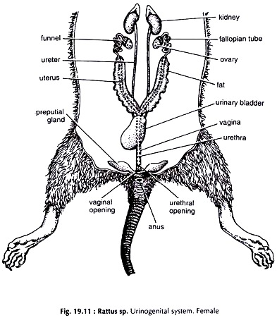
The system is located in the posterior region of the abdomen. Follow the procedure adopted for Calotes sp. to expose the organs of the urinogenital system (Figs. 19.10 & 19.11). Remove the fat covering the testes, which are lodged in the scrotum.
The Urinary (Excretory) System:
Kidneys:
Two dark red, bean-shaped bodies with a notch, the hilus on the inner side. The right kidney is slightly anterior to the left one.
Ureters:
The urinary ducts arise from the hilus. They are long, thin, tubular and lie against the dorsal abdominal wall. They run backward and open separately on the dorsal surface of the narrow neck of the urinary bladder.
Urinary bladder:
A small, pear-shaped, muscular sac with a narrow neck opening in the urethra.
Urethra:
A tubular continuation of the neck of the bladder. In male, the urethra is long and muscular and passes through the penis to open at its tip. In female, it opens to the exterior through an aperture, immediately posterior to the clitoris.
Dissection of Genital System:
Male Genital System:
The scrotum is present in front of the anus, between the hind legs. Cut open the scrotum giving a longitudinal incision along the mid-ventral line to expose the scrotal sacs. The testes are lodged in the cushion-like muscular portion of the scrotal sacs (Fig. 19.10).
Testes:
White, ovoid, paired bodies attached to the scrotal sacs at the posterior end by a band of tissue, the gubernaculum.
Epididymis:
Attached to the inner border of the testes. An epididymis is formed by the convolutions of numerous narrow tubules, the vasa efferent.
Vasa deferentia:
Paired tubes. The vas deferens arises from the posterior end of the epididymis, passes forward through the inguinal canal and reaches the abdominal cavity. It loops round the ureter and opens in the urinary bladder near the base of the urethra.
Seminal vesicles:
A pair of large, curved and sacculated organs associated with vasa deferentia.
Urethra:
A common passage for urine and sperms. It runs through the penis.
Coagulating glands:
A pair of fairly large, lobed glands attached to the inner concave surface of the seminal vesicles.
Prostate glands:
Two small, lobed bodies. The glands are attached to the junction of the bladder and urethra by stalk.
Penis:
A long, median, muscular organ, en-sheathed distally by a fold of skin, the prepuce. The terminal region of the penis is called glans.
Cowper’s glands:
Two small, flask-shaped glands at the base of the penis.
Preputial glands:
Flat bodies lying between the prepuce and the skin on either side of the penis.
Female Genital System:
(Fig. 19.11)
Ovaries:
Paired, compact, oval, yellow bodies in the abdominal cavity, ventrolateral to the kidneys. They are suspended with mesovarium.
Oviducts:
Two tubes corresponding to the ovaries.
The oviduct is differentiated into two distinct zones:
a. Fallopian tube:
The anterior, narrow, looped region opening in the body cavity through a ciliated funnel, facing the ovary.
b. Uterus:
The thick walled, dilated portion following the fallopian tube:
Vagina:
The two uteri join to form a median vagina. It is a muscular passage, ventral to the rectum but dorsal to the urethra. It opens to the exterior by vulva, immediately posterior to the urinary opening.
Preputial glands:
As in male.
Clitoris:
A small, rod-like body anterior to the vulva.
The Endocrine Glands:
(Fig. 19.12)
Hormones:
Complex organic chemicals are secreted by certain glands. Since the glands have no ducts like other glands (exocrine glands), they are named ductless glands or endocrine glands, and their secretion as hormones.
Dissection:
Expose the thoracic and abdominal cavity following the procedure adopted for rat.
ADVERTISEMENTS:
1. Thyroid gland:
Situated at the anterior end of the trachea, behind the thyroid cartilage and larynx. Remove fat, lymph nodes and salivary glands. Cut the sternohyoid muscles along the mid-ventral line and the thyroid gland is exposed.
Thyroid is the largest endocrine gland. It is enclosed in a two layered capsule. Septa of the fibro-elastic capsule extend inwards from the inner capsule and divide the gland into tubules.
2. Parathyroid gland:
Two pairs. Small, yellowish glands closely associated with thyroid. A pair lies on either side of thyroid.
3. Thymus gland:
A fairly large pinkish gland in the thorax enclosing the base of the aorta. It is relatively large in young but gradually atrophies with age.
4. Adrenal (Suprarenal) gland:
The glands are a pair, each embedded in the fatty tissue attached to the anterior end of a kidney. The gland is enclosed in a thick capsule of fibrous connective tissue.
5. Islet of Langerhans:
The islets are seathered in the pancreatic tissue and can be identified only in histological sections of pancreas.
6. Pituitary gland:
It is an oval body attached to the ventral surface of the brain, just posterior to the crossing of optic nerves. It is connected to the brain by infundibulum and covered with duramater. The gland has three lobes-anterior, intermediate and posterior.
7. Gonads:
Testes and ovaries are paired and secrete hormones. In male the testis are lodged in scrotum. In female the ovaries are ventrolateral to the kidneys.

