ADVERTISEMENTS:
In this article we will discuss about the dissection of giant African land snail. Also learn about:- 1. Dissection of Alimentary System 2. Dissection of Nervous System 3. Dissection of Reproductive System.
Killing:
Achatina is a land snail (Fig. 11.1). The only successful method of killing it in stretched condition is by prolonged drowning. About 24 hours time is required to kill a healthy snail by this method.
Wash the snails with water. Put them in a large glass jar. Fill it with water and place a lid on the jar taking care that no air is left between the lid and the water level. Put some heavy weight on the lid as the snails are capable of exerting considerable pressure on the lid to throw it off.
Snails are killed by asphyxia. They drop down at the bottom of the jar sometimes before death. The dead snails should be thoroughly washed to get rid of the mucus on their body. At the time of dissection the students must be provided with a good amount of non-absorbent cotton wool to wipe out the thick mucus from fingers and instruments to facilitate dissection.
Dissection:
Remove the shell using the method adopted for Pila. The pulmonary aperture (pneumostome) is a large orifice near the right corner of the mantle collar. Insert one arm of a pair of scissors through the pulmonary aperture and cut the mantle collar along its line of fusion with the visceral stalk.
ADVERTISEMENTS:
Starting from the pulmonary aperture give another incision along the junction of the mantle and the perivisceral mass in the body whorl. Turn the mantle left. The pallial complex is exposed.
Dissection of Alimentary System:
Fix the specimen with pins passing through the foot and the flap of the mantle. Give an incision on the middle of the dorsal wall of the visceral stalk. Proceed anteriorly up to the head and posteriorly to the junction of the body whorl and the visceral mass cutting the diaphragm, constituting the floor of the mantle cavity.
The organs in the visceral stalk and in the perivisceral region are exposed. Partially uncoil the spirally twisted digestive gland to expose the stomach and the intestine (Fig. 11.2).
Mouth:
A semicircular ventral opening at the anterior end of the snout.
Buccal mass:
A large, highly muscular body, narrower anteriorly and broader posteriorly. An odontophore is lodged in the posterior region of the buccal cavity. A semilunar cartilagenous jaw is present at the anterior end of the dorsal border.
Salivary glands:
ADVERTISEMENTS:
Paired, elongate oval, cream white glands, united posteriorly but free anteriorly, on the dorsal surface of the crop. The ducts open in the buccal cavity near the base of the oesophagus.
Oesophagus:
A narrow tube running backwards to the crop from the posterior end of the buccal mass.
Crop:
ADVERTISEMENTS:
A thin walled elongated sac divided into a large anterior and a small posterior crop. A digestive duct from the digestive gland opens in the posterior crop at its junction with the stomach.
Stomach:
A thick walled, heart-shaped sac, partially embedded in the digestive gland. The crop and the intestine are placed side by side and form the limbs of a U, while the stomach forms the base. A digestive duct from the digestive gland opens in the stomach.
Intestine:
ADVERTISEMENTS:
A fairly long, thin walled, round tube. It forms a loop in the first part. The posterior half is coiled and embedded in the digestive gland.
Rectum:
An almost straight tube, adherent to the posterior wall of the mantle cavity. It opens to the exterior by anus in the pulmonary aperture or pneumostome.
Digestive gland:
ADVERTISEMENTS:
A large, spirally twisted, blackish brown structure occupying major portion of the visceral mass. It has two lobes. The digestive ducts are two—one opening in the crop and the other in the stomach.
Dissection of Nervous System:
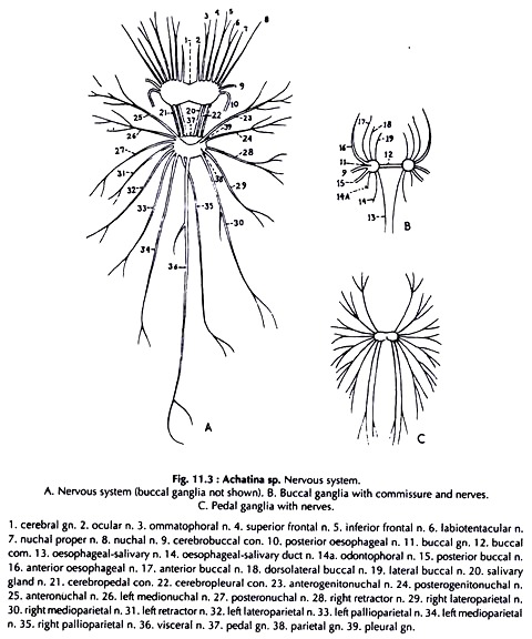
Expose the organs in the visceral stalk and in the perivisceral region following the procedure described previously. The nervous system is greatly condensed. The ganglia are closely aggregated, the commissures and connectives are shortened or even absent (Fig. 11.3).
Remove the thin membrane covering the cerebral ganglia, which are dorsal to the oesophagus and posterior to the buccal mass. Cut the oesophagus near its origin and remove a portion of its anterior part to expose the ventrally situated visceral and pedal ganglionic ring. Expose the buccal ganglia on the posterior face of the buccal mass between the oesophagus and the radular sac.
Trace the commissure and connectives. While tracing the nerves extra care should be taken to expose the statocyst nerves which are extremely fine. The statocyst nerve and the cerebropedal connective of a side are enclosed in a common sheath. Of all the nerves, the visceral nerve is the longest.
After its origin from the ganglionic mass it runs posteriorly sending out branches all along the way to different organs. Piercing the transverse septum dorsal to the spermathecal sac it enters the posterior haemocoelomic chamber. Leaving the chamber at the apical end, it runs up to the tip of the digestive gland along its inner concave border.
ADVERTISEMENTS:
Cerebral ganglia:
Two. almost round, fused at the narrower medial side. Each ganglion gives out ocular, ommatophoral, superior frontal, inferior frontal, posterior oesophageal, nuchal proper, nuchal and labiotentacular nerves.
Buccal ganglia:
Two, small and round; on the posterior face of the buccal mass. They send nerves to buccal mass, oesophagus and salivary glands.
Cerebrobuccal connectives:
Two, Each runs from the posterior border of a cerebral ganglion to join the buccal ganglion of the side.
ADVERTISEMENTS:
Visceral and pedal ganglionic mass:
Paired parietal, pleural, pedal and unpaired abdominal ganglia fuse to form a stout ring through which the cephalic aorta runs.
Cerebropedal connectives:
Two. Each arises from the posteroventral surface of the cerebral ganglion, the origin being lateral to that of the cerebropleural connective and runs around the oesophagus to join the anterior border of the pedal ganglion of the side in the visceral and pedal ganglionic mass.
Cerebropleural connectives:
Two, Each runs from the posteroventral surface of the cerebral ganglion around the oesophagus to join the anterior border of the pleural ganglion of the side in the visceral and pedal ganglionic mass.
Buccal commissure:
A slender, transverse nerve connecting the two buccal ganglia on the posterior face of the buccal mass.
Statocyst nerves:
Two slender nerves. Origin close to that of the cerebropedal connective. Joins the statocyst of the side.
Pleural ganglia:
They send anterogenito nuchal, posterogenito nuchal, anteronuchal, medionuchal and posteronuchal nerves.
Visceral ganglionic mass:
It is formed by the fusion of paired parietal and unpaired abdominal ganglion and sends nerves to retractors, lateroparietals, medioparietals, pallioparietals and one visceral nerve.
Visceral nerve:
One. Arises from a slightly dorsal position of the posterior border of the visceral and pedal ganglionic mass and runs backward. It sends branches to the majority of the organs of the visceral mass including ovotestis.
Pedal ganglia:
Nine pairs of nerves arise from the ganglia. They form regular plexuses in the sole of the foot along the borders.
Dissection of Reproductive System:
Achatina is hermaphrodite (Fig. 11.4).
It is better to dissect reproductive system in a freshly killed specimen. Carefully uncoil the spirally coiled digestive gland, which constitutes major portion of the visceral mass. The ovotestis is embedded in the posterior lobe of the digestive gland. It is visible through the transparent mantle.
Follow the ovotestis duct leaving the ovotestis at its posterior end, and ending in a Carrefour attached to the albumen gland. Trace the large sperm oviduct from the albumen gland proceeding anteriorly. In the visceral stalk the sperm oviduct divides into a vas deferens and an oviduct.
Further anteriorly a spermatheca is connected with the oviduct. The vas deferens joins the penis and the oviduct joins the vagina. The gonopore is located on the right side at the anterior end of the visceral stalk, posterior to the ocular tentacle.
Ovotestis:
Consists of 4 to 5 light cream, finger-like lobes covered by transparent, membraneous mantle.
Ovotestis duct:
A duct from each lobe joins to form the duct.
It has three zones:
a. Apical ovotestis duct:
A narrow and short portion, the first part.
b. Ovisperm vesicle:
Enlarged and much folded middle region.
c. Basal ovotestis duct:
The last part. A short tube joining the Carrefour.
Carrefour:
A minute chamber connected with the ovotestis duct, albumen duct, sperm oviduct, (sulcus (male portion of the genital duct) and uterus (female portion of the genital duct)) and talon. It is lodged in a depression on the concave face of the albumen gland.
Talon:
A small, curved hollow body having the shape of a claw of hawk.
Albumen gland:
A cream white, slightly curved, fairly large gland connected with the Carrefour by a short duct.
Sperm oviduct:
ADVERTISEMENTS:
A fairly large, long sac formed by the sulcus and the uterus. The uterus has two zones. The cream white apical part— apical uterus, and the light yellow distal part— the basal uterus.
Prostatic acini:
More than 100 yellowish, small, maize grain-like bodies attached to the sulcus part of the sperm oviduct.
Oviduct:
A muscular tube, joins the basal uterus posteriorly and the vagina anteriorly. It receives the duct of the spermatheca.
Spermatheca:
A fairly large, thin walled oval sac, attached to the anterior end of the sperm oviduct. The duct is quite long and joins oviduct.
Vagina:
A muscular chamber, the wide end connected with the oviduct and the other end is narrow.
Vas deferens:
A narrow muscular tube, runs from sulcus and joins the penis.
Penis:
A slightly curved, muscular structure.
Genital atrium:
A small chamber connected with the penis and the vagina. It opens through the gonopore.


