ADVERTISEMENTS:
The following points highlight the top nine types of system in pigeons. The types are: 1. Muscular System 2. Digestive System 3. Respiratory System 4. Circulatory System 5. Lymphatic System 6. Nervous System 7. Endocrine System 8. Excretory System 9. Reproductive System.
Type # 1. Muscular System:
The muscular system of pigeon is extremely modified to meet the requirements of its peculiar way of life. The muscles of the active regions, i.e., breast and wings, legs, neck, tail are highly developed, whereas in the comparatively immobile regions, partly the back muscles become atrophied.
Some muscles in pigeon’s body have changed their function due to the modifications for flight. The myofibrils composing the muscles are extremely elongated and they can withstand fatigueless during prolonged activity.
ADVERTISEMENTS:
There are many muscles in pigeon, of which the following muscles, connected with the activities of the wings and legs, are described below:
Breast and Wings Muscles:
The muscles which are concerned with the activity of the wings are called flight muscles (Fig. 9.10).
Flight in pigeon is caused by the action of the following muscles:
ADVERTISEMENTS:
(1) Pectoralis major:
These are paired muscles of immense size. These are highly vascularized and constitute about one-fifth of the total weight of the body. Each muscle arises from the whole of keel of sternum and from the clavicle.
It is present over the entire area of the breast bone and its fibres are inserted into the great trochanter of humerus. They constitute the main depressor muscles of the wings and the powerful down- stroke is caused by these muscles.
(2) Pectoralis minor (or Subclavius or Supracoracoideus):
These paired muscles are situated on the dorsal side of the pectoralis major and on the anterior side of the sternum proper rather than the keel. Each muscle sends its tendon through the foramen triosseum (aperture between furcula, scapula and coracoid) and inserts itself into the anterodorsal surface of humerus. The foramen acts as a pulley. These are the main elevator muscles of the wings.
(3) Scapulo-humeralis (Posterior and anterior):
These small muscles are stretched from the pectoral girdle to humerus and are responsible for the rotatory motion of the wing.
ADVERTISEMENTS:
(4) Tensor patagialis longus:
It originates from pectoralis major and remains inserted into the patagial membrane.
It acts as a tensor of patagial membrane.
(5) Rhomboideus externus:
ADVERTISEMENTS:
It originates from the spines of all thoracic vertebrae and last few cervical vertebrae. It is attached with the ventral border of scapula and draws the scapula medially.
(6) Rhomboideus pro-fundus:
ADVERTISEMENTS:
It originates from all thoracic vertebrae and is inserted into the vertebral border of scapula beneath the rhomboideus externus. It also draws the scapula medially.
(7) Scapulo-humeralis anterior:
This muscle arises from the anterior axillary border and head of scapula. It is attached with the humerus just proximal to pneumatic foramen and draws the humerus medially.
(8) Scapulo-humeralis posterior:
ADVERTISEMENTS:
It originates from the entire lateral surface of the scapula and remains inserted into the humerus distal to pneumatic foramen.
It also draws the humerus medially.
(9) Latissimus dorsi:
It arises from the spines of thoracic vertebrae and remains attached on the humerus between the attachment of triceps and deltoideus major. It draws the humerus medially.
ADVERTISEMENTS:
(10) Serratus anterior:
This muscle originates from the fascia covering third rib ventral to uncinate process. It is inserted into the ventral border of scapula anterior to the origin of scapulo-humeralis anterior muscle. It depresses the scapula.
(11) Serratus posterior:
It arises from the fascia covering fourth, fifth and sixth ribs near the level of uncinate process. It is connected with the medial border of the posterior one- third of the scapula. It also depresses the scapula.
(12) Tensor patagialis brevis:
Arising from the dorsal surface of scapula superficial to deltoideus major, this muscle is inserted into the tendinous to the lateral surface of the elbow to elevate the humerus.
ADVERTISEMENTS:
(13) Scapulo-humeralis pro-fundus:
It takes the origin from the neck of scapula and is attached to the anterior distal one-half of humerus. It draws the humerus medially and posteriorly.
(14) Tensor accessories patagialis:
It originates from the fascia covering the biceps muscle and remains inserted into the patagial membrane. It keeps the patagial membrane tensely stretched when the wing is extended.
(15) Expansor secundariorum:
This is a smooth muscle that originates from the long tendon from scapula and fascia of scapulo-humeralis posterior muscle and distal end of the humerus. It is connected with the bases of the proximal five secondary remiges and ventral coverts. It helps the expansion of the remiges.
(16) Deltoideus major:
It elevates the humerus and arises from the dorsoanterior border of scapula to insert into the great trochanter of humerus.
(17) Carocobrachialis anterior:
It originates from the distal end of the pro-coracoid and attaches itself with the great trochanter of humerus to assist the action of deltoideus muscles.
(18) Coracobrachialis longus:
This muscle draws the humerus medially and also depresses the wings. It arises from the coracoid and remains attached with the humerus, proximal to pneumatic foramen.
(19) Coracobrachialis brevis:
It also arises from the coracoid and inserted into the humerus to assist the action of coracobrachialis longus.
(20) Triceps:
It is a large, fleshy muscle and originates by two heads from the humerus. It is connected with the tendon of anconeus muscle to the olecranon process of ulna. It acts to extend the anti-brachium.
(21) Biceps:
It acts as the flexor of the anti-brachium. Its long head arises from scapuloclavicular joint, while the short head originates from the head of the humerus. It is inserted into the anteroproximal surface of radius.
(22) Brachialis inferior:
This muscle arises from the distal end of humerus and attaches itself with the proximal ventral surface of ulna. It assists the activity of biceps muscle.
(23) Pronator sublimus:
It helps the pronation of wing. It arises from the distal end of humerus and is attached to the anterodistal surface of radius.
(24) Pronator pro-fundus:
It also assists the pronation of wing. It arises from the proximal one-third of ulna and attaches itself with the distal end of radius.
(25) Ectepicondyloradialis:
It helps the activity of biceps. Originating from the lateral epicondyle of humerus, it is inserted to the shaft of radius.
(26) Anconeus:
It assists the activity of triceps. It also arises from the lateral epicondyle of humerus and is attached with the lateral surface of ulna.
(27) Extensor digitorum communis:
Arising from the distal end of humerus, it is attached with the posterodistal surface of carpometacarpus and pollex. It flexes the pollex, extends the index digit, elevates and flexes the hand.
(28) Flexor metacarpi radialis:
It flexes the hand. This muscle originates from the fascia of ancones muscle and is inserted into the outer surface of carpometacarpals.
(29) Flexor digitorum sublimus:
It also flexes the hand. It originates from the distal end of hurries and remains attached with the proximal and distal anterior surface of carpometacarpus.
(30) Flexor digitorum pro-fundus:
It arises from the proximal one-third of ulna and attaches itself to the anterodistal border of carpometacarpus by an elongated tendon. It flexes the hand.
(31) Flexor carpi ulnaris:
Originating from the distal end of humerus, it attaches to the posterior proximal surface of carpometacarpus to flex the hand.
(32) Abductor pollicis:
It abducts the pollex. This small muscle arises from the anterior surface of carpometacarpus distal to pollex and inserted to the ventral surface of pollex.
(33) Adductor pollicis:
This muscle adducts the pollex. It originates from the anterior surface of carpometacarpus proximal to pollex and attaches to the posterior surface of pollex.
(34) Extensor metacarpi radialis:
This muscle extends the wing. It originates from the lower end of humerus and remains inserted into the carpometacarpus, proximal to first digit.
(35) Ulnimetacarpalis ventralis:
It arises from the middle third of ulna and attaches to the carpometacarpus proximal to pollex by a strong tendon. It acts as pronator.
(36) Ulnimetacarpalis dorsalis:
It elevates and flexes the hand. It arises from the distal end of ulna and inserted to the proximal carpometacarpus.
(37) Interosseus palmaris:
It elevates the index digit. After originating from the ventral surface of carpometacarpus it is connected with second and third digits.
(38) Interosseus dorsalis:
It arises from the dorsal surface of carpometacarpus and is inserted to the second and third digits. It also elevates the index digit.
(39) Extensor pollicis longus:
This muscle extends the hand and arises from the distal half of radius to insert into the carpometacarpus proximal to the first digit.
(40) Deltoideus minor:
It originates from the head of scapula and attaches to the great trochanter of humerus. It elevates the humerus.
Leg muscles:
The leg musculature of pigeon is highly specialised and assists to perch on the branches of the tree, besides walking on land.
The muscles are:
(1) Peroneus longus:
It originates from tibia and remains inserted to the tendon of flexor perforans et perforatus digiti (III). It flexes the digits.
(2) Peroneus brevis:
This muscle assists in extension and abduction of tarsometatarsus. It arises from the posterior proximal portion of tibia and unites with the lateral surface of ankle.
(3) Gastrocnemius:
It arises from three heads:
(i) Lateral condyle of femur,
(ii) Medial surface of head and neck of tibia and
(iii) Ilium near the femurocaudalis muscle. It joins with the tarsometatarsus and phalanges and assists in the extension of tarsometatarsus.
(4) Plantaris:
After originating from the medioproximal shaft of tibia, it is attached with the tibial cartilage (heel) to help extension of tarsometatarsus.
(5) Tibialis anticus:
It has its start from the external condyle of femur and anterior surface of the head of tibia and joins with the metatarsus. It flexes the tarsometatarsus against shank.
(6) External digitorum longus:
This muscle acts as a common extensor of the digits. It starts from the anterior surface of tibia beneath the peroneus brevis and ends in the distal phalanges of all digits.
(7) Flexor perforans et perforatus digiti (III):
It acts as a flexor of the third digit. It arises from the medial condyle of tibia and remains inserted into the second phalanx of the third digit.
(8) Flexor hallicus longus and Flexor hallicus brevis:
The former arises from the lateral condyle of femur and the latter originates from the tibia. Both the muscles join with the ventral surface of the first digit and act as flexors of the first digit.
(9) Flexor perforatus digiti (II):
It originates from the tibia and joins to the median surface of the first phalanx of the second digit. It acts as adductor of the second digit.
(10) Flexor perforatus digiti (IV):
It acts as adductor of the fourth digit. After arising from the tibia, it is inserted to the median surface of the first phalanx of the fourth digit.
(11) Extensor hallicus:
This muscle arises from the metatarsus and attaches itself to the dorsal surface of the first digit. It extends the first digit.
(12) Lumbricalis:
This muscle acts as a flexor of the third digit. It arises from the tendons of flexor muscles and is inserted into the third digit.
Type # 2. Digestive System:
The digestive system of pigeon includes alimentary canal and digestive glands. This system helps in nutrition. In comparison to the size of the bird, the length of the alimentary canal is short and its different parts are modified in such a way that assimilation of simplified food occurs in a short time. The digestive system is highly modified in pigeon due to loss of teeth and also due to an adaptation for aerial life.
I. Alimentary Canal:
The alimentary canal (Fig. 9.16A) consists of the following parts:
(i) Mouth:
This inlet aperture is present at the tip of the head and is bounded by horny upper and lower beak. Teeth are totally absent.
(ii) Mouth cavity and pharynx:
Mouth leads into mouth cavity and pharynx.
These regions contain:
(a) Internal nostril:
It is a single opening on the roof of the pharynx through which both the nares open internally.
(b) Tongue:
It is a prominent structure with free pointed anterior end. Some taste buds and numerous mucous glands are present on the surface of the tongue.
(c) Glottis:
It is an aperture which leads into trachea and is placed near the base of the tongue,
(d) Gullet:
It is the last part of the pharynx which continues as an opening into the next part of the alimentary canal.
(iii) Oesophagus:
It begins from the gullet and runs through the ventral part of the neck as a straight tube. Near the junction of neck and trunk it comes between skin and muscle and enlarges to form a sac, called crop. The crop plays dual role in the life of pigeon and other birds.
It acts as a large temporary reservoir of crude food grains. The presence of crops enables the bird to ingest a considerable quantity of food grains very quickly. Food may be regurgitated from the crops to feed the young’s. Besides its normal function of storage, the crop glands present in the epithelial lining of the crop produce a proteinaceous, white slimy secretion, called pigeon’s milk.
This is produced in both the sexes during breeding season. The young squabs are fed by the parents by regurgitation of the fluid. The secretory activity of the crop-glands is controlled by the hormone, called prolactin of anterior pituitary.
The composition of pigeon’s milk is given below:
Water … 65-81%
Protein … 13-3-18-8%
Fat … 6-9-12-7%
Ash … 1-5%
It has a great nutritive value, even more efficient than mammalian milk. From the crop, the oesophagus enters the cavity of trunk and passes dorsal to the heart to open into the next part of the alimentary canal, called stomach. Thin muscle slips from the pectoralis muscle regulate the activity of the crop.
(iv) Stomach:
It is divisible into two distinct parts:
(a) Proventriculus:
It is the first part of the stomach to receive the oesophagus (Fig. 9.16A). It is tubular and internally lined by numerous gastric glands. A red coloured small spleen remains morphologically attached to the outer side of proventriculus.
(b) Gizzard:
It is the second part of stomach and is semicircular in outline. The walls of the gizzard are highly muscular. It contains very little inner space. Internally the wall contains numerous ridges and its epithelial lining becomes very thick and tough.
It has innumerable small tubular glands. A few stones, which are swallowed by the bird, remain within the lumen and are responsible for crushing the food (Fig. 9.16B). The function of gizzard is purely mechanical and acts mainly as the grinding machine to crush the food.
The gizzard compensates the role of teeth in pigeon. In grain-eating birds, the gizzard is highly muscular, while in piscivorous birds, it is simple and thin-walled.
(v) Intestine:
Intestine begins from the gizzard near the opening of proventriculus and is divisible into following parts:
(a) Duodenum,
(b) Ileum and
(c) Rectum.
(a) Duodenum:
This is the first part of the intestine and is distinctly ‘U’-shaped in appearance. Internally, the duodenum contains villi, crypts of Lieberkuhn and goblet cells,
(b) Ileum:
It is the second part of the intestine having numerous villi inside it. The length of the ileum is shorter than that of other vertebrates. The first and the last parts of the ileum are loop-like and the middle part is spirally coiled,
(c) Rectum:
It is the last part of the intestine and is of same thickness as that of ileum. Near the junction of ileum and rectum, a pair of small lateral blind leaf like caeca originates.
(vi) Cloaca:
Rectum opens into a chamber, called cloaca. The cloaca is spacious, muscular and is elaborated into three chambers: coprodaeum, urodaeum and proctodaeum (Fig. 9.16C). The coprodaeum receives the intestine. The urinogenital ducts open within the urodaeum and the proctodaeum opens externally by the vent.
In the nestlings, a special thick- walled glandular chamber, bursa fabricii, remains in close association with cloaca and communicates with the proctodaeum. It degenerates in the adult bird. The bursa fabricii may have some local protective function in young ones.
(vii) Cloacal aperture:
The proctodaeum of cloaca opens to the exterior through the cloacal aperture or vent. It is present on the ventral side and near the base of the tail.
II. Membranes and Mesenteries:
There are membranes and mesenteries which divide the body cavity into different parts and also support the viscera. The following membranes and mesenteries are prominent.
They are:
(i) Parietal peritoneum: membrane lining the body wall,
(ii) Ventral ligament: arises from the ventral body wall to the gizzard,
(iii) Falciform ligament : arises from the ventral body wall to the ventral side of the liver. It is continuous with the ventral ligament,
(iv) Oblique septum : a partition arising from the lateral body wall to the centre which separates the body cavity into an anterior and posterior compartments. This septum is continuous with the pericardial sac at the centre and encloses the posterior air-sacs,
(v) Pericardiac sac : encloses the heart. It is composed of parietal pericardium and visceral pericardium,
(vi) Coronary ligament : attaches the anterior side of the liver to the combined pericardial sac and oblique septum,
(vii) Castro-hepatic ligament: attaches the gizzard to the liver,
(viii) Mesogastric mesentery : connects the gizzard to the dorsal body wall,
(ix) Meso-duodenal ligament: connects the two halves of the duodenal loop. The pancreas is also located on this ligament,
(x) Mesentery proper: attaches the small intestine to the dorsal body wall,
(xi) Hepato-duodenal ligament: connects the duodenum to the liver.
III. Digestive Glands:
The following digestive glands are associated with the alimentary system:
i. Mucous glands:
These are present on the surface of the tongue and crop and are responsible for the moistening of the food.
ii. Salivary glands:
Paired angular and unpaired sublingual salivary glands are located in the pharyngeal region. The secretion, called saliva moistens the food and also contains diastatic enzyme.
iii. Crop glands:
The secretion, called “crop milk or pigeon’s milk” is secreted by these glands. The secretion is used for the nourishment of the young’s.
iv. Gastric glands:
Present on the internal lining of proventriculus and secretes gastric juice.
v. Tubular glands:
These glands are present on the internal lining of gizzard. The fluid secreted by these glands is thick, horny, yellowish green in colour.
vi. Liver:
Paired deep brown coloured glands of immense size are present ventral to the gizzard. It produces the bile which passes through two bile ducts, one coming from each lobe of liver. The bile ducts open separately within the two limbs of the duodenum. The gall bladder is absent in pigeon. Loss of gall bladder is an adaptation to aerial life for reducing the weight of the body.
vii. Pancreas:
It is a pinkish white gland located in between the two limbs of the duodenum. It is a combination of both exocrine and endocrine glands. The exocrine part producing pancreatic juice opens into the duodenum by two or three pancreatic ducts. The endocrine part produces insulin, which is directly poured in the blood vessel.
viii. Crypts of Lieberkuhn and Goblet cells:
These are present in the lining of the duodenum and secrete to influence the pancreas to produce pancreatic juice.
ix. Glands in the ileum:
These glands produce digestive juices which contain various enzymes.
x. Caecal glands:
Produce certain digestive juices for the digestion of vegetable fibres. They are also concerned with the absorption of water.
Physiology of Digestion:
The physiology of digestion in pigeon involves the three following steps:
Ingestion:
Pigeon is a grain or seed eater. It picks up grains very rapidly which are quickly swallowed. The shape of the beak is modified in such a way that it helps in its ingestion. The swallowed food, being moistened by mucus, is stored in the crop.
Digestion:
Within the crop the food is moistened and macerated. The passage of food from crop to proventriculus is regulated. Inside the proventriculus, the gastric juices containing HCI and pepsin act over the food. Within the gizzard the food is completely churned by the action of gizzard wall and stones. The secretion of gizzard glands helps in the process.
The churned food, while travelling through the duodenum, comes in contact with the bile and pancreatic juice. The bile neutralizes the acidity. Different enzymes are present in the various digestive juices. The completely broken-down food is absorbed through the lining of small intestine. Residual part passes into the rectum where water is absorbed and the fibres are broken down by the caecal juice.
Egestion:
Residual part, called faeces, is temporarily stored in the cloaca where it mixes with the urine and is periodically dropped through cloacal aperture.
Type # 3. Respiratory System:
The aerial mode of life requires extra energy. In order to get energy, pigeon eats a large quantity of food and to break down the assimilated food at a faster rate, the respiratory system is extensively modified. The lungs are proportionately smaller in size, but the functional efficiency is greatly increased by the development of air-sacs.
The respiratory system of pigeon has two unique features:
(i) Presence of non-elastic, compact lungs and
(ii) Possession of several air-sacs.
Following structures are present in the respiratory system of pigeon (Fig. 9.17):
a. External nares:
These are paired openings, present near the base of the upper beak and within the cere.
b. Internal nares:
Single opening which opens at the roof of pharyngeal region and both the external nares communicate through this common aperture.
c. Glottis:
This is a slit-like aperture, which is present on the floor of the mouth cavity and near the base of the tongue. It leads into the next part, called trachea.
d. Trachea:
This elongated tube begins from glottis and runs along the neck region along the ventral side of the oesophagus. The trachea is composed of complete bony tracheal rings (Fig. 9.17 A). Near its commencement, the trachea is enlarged into a chamber, called the larynx. This chamber is supported by a cricoid cartilage (which is composed of four pieces) and one pair of arytenoid cartilages. In birds, the larynx does not function as voice box.
e. Syrinx:
Near the junction of neck and trunk, the trachea is swollen into a chamber, called syrinx (Fig. 9.17 B). It is formed by the dilatation of the last three or four tracheal rings and first bony ring of each bronchus. The mucous membrane of the syrinx constitutes a pad-like thickening and is provided with several muscles and membranes.
The syrinx is actually the voice box. The syrinx is the characteristic organ of pigeon and many other flying birds. It has a complicated structural construction. A bar of cartilage, called pessulus, is present at the junction of two bronchi. It extends dorsoventrally inside the tympanum and holds a small fold of mucous membrane, called semilunar membrane or membrana semilunaris (Fig. 8.17B).
There is no semilunar membrane in many passerine and non-passerine birds (King, 1989). The inner membranous lining of the bronchi produces inconspicuous internal tympani-form membranes. The sound is produced by the vibration of the membrana semilunaris while the pitch of the sound is controlled by the action of the syringeal musculature.
The syringeal musculature includes a pair of intrinsic syringeal muscles (which arise from the lateral sides of the trachea and are attached with the syrinx) and a pair of sternotracheal muscles (originates from the sternum and is inserted into the trachea). The position of syrinx can be changed by the action of these muscles.
f. Bronchus:
Within the trunk, the trachea bifurcates into right and left bronchi (Fig. 9.17A). The first ring of each bronchus is complete and bony, while the rest are incomplete mesially. The left and right bronchi are called primary bronchi or mesobronchi.
Each meso- bronchus, in the beginning, is composed of rings of cartilages, but inside the lung such rings are absent. Each primary bronchus enters the lung through a small space, called vestibulum and extends up to the posterior extremity of lung.
Within the lung, the mesobronchus sends a pair of branches, called secondary bronchi and each secondary bronchus breaks up into a network of tertiary or Para bronchi and sends branches to the air-sacs.
Each tertiary bronchus again subdivides into numerous finer network of tubules (air capillaries), which remain in close contact with the blood capillaries. While running posteriorly, the diameter of meso- bronchus gradually decreases and it finally opens into the abdominal air-sac by an ostium.
g. Lungs:
The lungs are small in size in comparison with that of the body. These are paired pink coloured organs. The lungs are spongy organs with little elasticity (Fig. 9.17A). The dorsal surface of the lungs is fitted closely with the interspaces of ribs and lacks the peritoneal covering, i.e., pleura is absent on the dorsal side.
The ventral wall has a compact fibrous tissue sheet, called pleura or pulmonary Apo neurosis. The wall of the pleura has special fan-shaped costopulmonary muscles which originate from the joints of vertebral and sternal ribs. The alveolar lining is formed by the ramification of tertiary tubules with the distribution of blood vessels.
h. Air-sacs:
The air-sacs are bladder-like structures. These are formed by the dilation of the mucous membrane of the bronchus. The air-sacs are thin-walled membranous sacs and are devoid of blood vessels. Following air-sacs are present in the body of pigeon and all of them remain in communication with the pneumatic cavities of bones. There are nine major and four accessory air-sacs in pigeon (9.18).
Major air-sacs:
The major air-sacs (Fig. 9.18) originate directly from the lungs. Of the nine air-sacs, four are paired and one is unpaired.
(a) Paired air-sacs:
(i) Posterior or abdominal air-sacs,
(ii) Posterior thoracic air-sacs,
(iii) Anterior thoracic air-sacs and
(iv) Cervical air-sacs.
(b) Unpaired air-sac:
(i) Inter-clavicular or median air-sac.
Accessory air-sacs:
These air-sacs originate as paired diverticula from the inter-clavicular air-sac.
These paired sacs are:
(i) Clavicular air-sacs and
(ii) Humeral air-sacs.
Inspiratory Air Sacs:
a. Abdominal air-sacs:
These are also called the posterior air-sacs and lie among the coils of intestine. These are the posterior-most and largest air-sacs in birds. The right air-sac is larger than its left counterpart. These air-sacs send diverticula into the pelvic girdle, synsacrum, hind limbs and between thigh muscles.
b. Posterior thoracic air-sacs:
These paired air-sacs are placed on the posterior side of the thoracic cavity. The left sac is slightly larger than the right. Both the sacs are closely apposed with the lateral wall of the body cavity. Each bronchus, near its entrance into the lung, gives three short branches : One enters into the anterior thoracic air-sac, the second is connected with the cervical air-sac and the third enters the inter-clavicular air-sac.
c. Anterior thoracic air-sacs:
These paired air-sacs are located one on each side of the thoracic cavity towards the anterior part between the lungs and the ribs.
Expiratory Air Sacs:
a. Cervical air-sacs:
These paired air-sacs are placed near the base of the neck and lie in front of the lungs. Each sac sends diverticula into the cervical vertebrae and the skull.
b. Inter-clavicular air-sac:
This is an unpaired and median air-sac of large size. It has two ducts, one opening into each lung. Although this sac is unpaired in adult, it is formed by the fusion of two sacs which are evident by the presence of two ducts.
Each side of this air-sac gives off two extensions:
(i) Clavicular air-sac and
(ii) Humeral air-sac.
These sacs are communicated with the cavities of the bones. A layer of fibrous tissue, called oblique septum, encloses the ventral walls of both the thoracic air-sacs. This septum extends up to the pericardium and unites with the similar septum of the other side.
Such union along the middle line separates the body cavity into two chambers. One chamber houses the lungs, thoracic and inter-clavicular sacs and the other chamber contains the heart, liver, stomach, intestine and abdominal air-sacs.
Role of air-sacs:
The air-sacs play an important role in the life of flying birds. The air-sacs are not provided with capillary network, so they are not directly respiratory in function. Besides, these sacs are essential components for aerial life.
The different uses of air-sacs are:
As the lungs are anchored firmly to the dorsal wall of the thoracic cavity, the elasticity of the lungs is greatly hampered. To carry on an efficient circulation of air through lungs during respiration, some mechanical aid becomes necessary, specially during flight. The air-sacs give mechanical aid by acting as the bellows.
Act as balloons:
When the air-sacs are inflated due to intake of warm air, the specific gravity of the body is lowered to a considerable extent. As the warm air is lighter than ordinary air, the retention of such air inside the air-sacs makes the body considerably lighter. This also lessens muscular efforts to sustain the body heavier than air.
Function as ballast:
The air-sacs are so nicely arranged on the two sides of the body that proper centre of gravity is established for balanced flight. If the equilibrium is lost by chance during flight, restoration of the equilibrium is easily maintained by shifting of the contained air from one side of the body to the other.
Lessen mechanical friction:
The air-sacs send branches which are inserted between the muscles (specially the flight muscles) like pads. Such a placement of air-sacs reduces mechanical friction to a large extent and increases the flexibility of the wings during flight.
Regulate and maintain body temperature:
The skin of bird lacks integumentary glands. So the skin has no utility in the regulation and maintenance of body temperature. Retention of warm moist air inside the air-sacs helps to regulate and maintain the body temperature.
Serve as the containers of reserve air:
During rest, respiration involves the alternate depression and elevation of the breastbone caused by the activity of the intercostal muscles. But during flight the breastbone as well as the thoracic basket is kept in a rigid state and the intercostal muscles remain in tension.
The respiratory process is slightly hampered for the time. Therefore, some internal source of reservoir of air becomes indispensable. The air-sacs sub-serve this function and ventilate the lungs during flight.
Act as resonator:
The pitch of the sound is controlled to some extent by the forceful expulsion of the air from the air-sacs which act as resonator.
Regulate the moisture content of air:
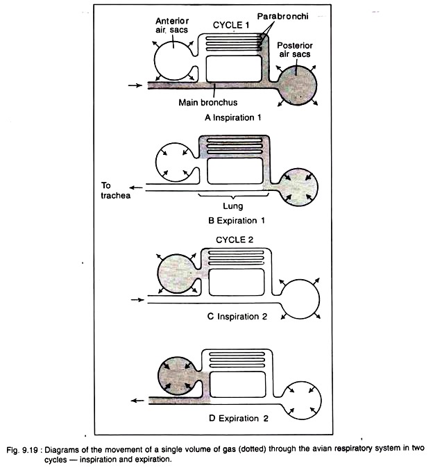
Water is evaporated from the walls of the air- sacs in birds. So the air-sacs regulate the water content of the body (Fig. 9.19).
Mechanism of Respiration:
The unique feature of avian respiration is the double supply of oxygenated air to the surface of lungs for improved aeration. For this reason, the respiration is called double respiration.
Bretz and Schmidt-Nielson (1972) measured the course of air flow through the avian lungs, using oxygen electrodes and flow meters. Two cycles of inspiration and expiration are required for a single volume of gas to move through the respiratory system (Fig. 9.19). During the first inspiration, the sternum is lowered, and the lungs and the air- sacs are expanded.
The fresh air rushes through trachea and bronchi into the posterior air sacs. During the first expiration, the sternum is raised and the air from the posterior sacs goes to the para-bronchi and air capillaries. At second inspiration, air from the Para bronchi goes to the anterior air sacs and, on the second expiration, the air is expelled to the outside (Schmidt-Nielson, 1979).
In other air-breathing animals, in between inspiration and expiration, some amount of residual air always remains within the cavity of lungs. But in birds, the residual air remains inside the air sacs and to the smaller branches of the bronchi. Because of the fact aeration of blood is complete in pigeon and thus increases the respiratory efficiency to yield extra energy.
The respiratory movements are caused by two sets of muscles one set operates during flight and the other set works at the time of rest. During flight, both inspiration and expiration are caused by the movement of pectoral muscles. At the time of rest, inspiration is caused mostly by the activities of intercostal muscles and expiration by the movement of abdominal muscles.
Type # 4. Circulatory System:
Two different fluids circulate through the body of pigeon. The blood, heart and the blood vessels constitute the blood vascular system. Another fluid, the lymph, and the lymph channels are included under the lymphatic system.
Blood-Vascular System:
Blood:
The blood contains plasma and corpuscles. The red blood corpuscles are oval in shape and nucleated. The white blood corpuscles are present in much lesser number but are of different types.
The different types of white blood corpuscles are:
(i) Lymphocytes,
(ii) Heterophils,
(iii) Polymorphonuclear- pseudo-eosinophilic granulocytes,
(iv) Basophils,
(v) Eosinophil’s and
(vi) Monocytes.
The blood platelets are absent in pigeon, but the blood clots quickly. New blood cells are formed in the bone marrow and the blood corpuscles are destroyed within spleen. The spleen is a red body of oval shape situated on the right side of the proventriculus and attached with it by peritoneum.
Heart:
The heart is an oval organ placed in the anterior part of the thoracic cavity but ventral to the oesophagus. The heart is quite large in size in proportion to body size. It is enclosed by a thin white membranous pericardium and the pericardial cavity contains a serous fluid. The auricles and ventricles are distinctly separated by a groove, called the coronary sulcus.
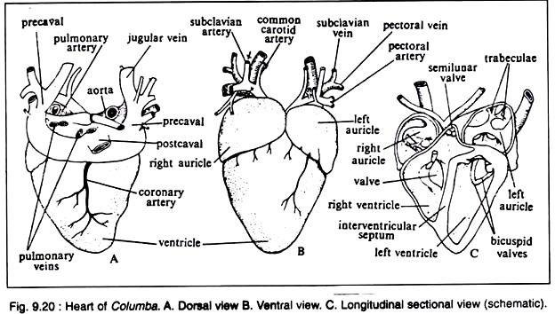
The sinus venosus is absent and is absorbed in the wall of the right auricle. Both auricle and ventricle are completely divided into right and left chambers (Fig. 9.20). Thus heart is completely four-chambered and all the chambers are lined by endocardium.
The right auricle is slightly larger than the left auricle. The ventricles are very powerful. The left ventricle contains a round cavity while the right one has a crescentic cavity partly surrounding the left. The auricles and ventricles are divided internally by inter-auricular and inter-ventricular septa, respectively. The right auriculoventricular valve is flap-like and muscular in nature.
The presence of single right auriculoventricular valve is a diagnostic feature of pigeon. The left auriculoventricular valve is membranous and provided with two cusps (bicuspids) which are attached with the ridges of the ventricular wall. Cordlike fibres (chordae tendineae) are attached to the margins of the auriculoventricular valves and to the walls of ventricles by papillary muscles.
These muscles control the activity of the auriculoventricular valves via the chordae tendineae. The right auricle receives deoxygenated blood from three caval veins and the left auricle receives oxygenated blood through four pulmonary veins.
From the left ventricle the single right aortic arch originates and conveys oxygenated blood to the different parts of the body. The right ventricle gives rise to pulmonary arch which carries deoxygenated blood to the lungs. The left ventricle is usually called systemic ventricle and the right is called pulmonary ventricle. The openings of the arches are guarded by three cup-like thick semilunar valves.
 The working of heart is controlled by elaborate intrinsic nervous system of heart. The wall of the right auricle bears sinuauricular node (or pacemaker) and the atrial septum bears auriculoventricular node. A special ring of Purkinje fibres is also present around right auriculoventricular wall. The rate of systole and diastole is much faster than that in other vertebrates.
Mechanism of Circulation through Heart:
During the diastolic phase, the heart relaxes and the auricles receive blood from the veins. The right auricle gets the deoxygenated blood and the left auricle is filled up with oxygenated blood from the lungs via the pulmonary veins. The systolic action starts from the right auricle.
It actually begins from the sinuauricular node and passes to the auriculoventricular node. This wave then spreads to the remaining parts of the heart. At the time of auricular systole, the blood comes to ventricles through the auriculoventricular aperture.
When the ventricles start contraction, the deoxygenated blood from the right ventricle is pushed to the lungs by the pulmonary arches. Single right aortic arch from the left ventricle conveys the oxygenated blood to the different parts of the body.
The heart of pigeon is a double circuit heart and there is no chance of mixing up of oxygenated and deoxygenated blood except in the capillaries. This is a significant evolutionary advancement in birds over reptiles.
Blood Vessels:
The blood vessels include the arteries, veins and capillaries. The arteries supply blood to the different parts of the body and break up into arterioles and finally to finer anastomosing branches—the capillaries. The capillaries reunite to form the venules which ultimately form the veins.
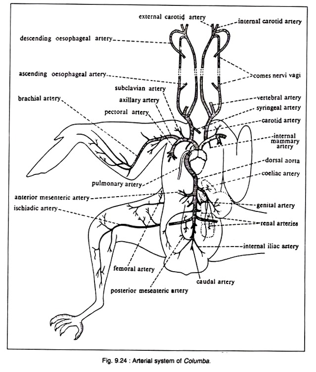
Arterial System (Fig. 9.24):
In pigeon, only the right aortic arch is present. It arises from the left ventricle and passes backward between the auricles arching over the bronchus of the corresponding side. It then reaches the mid-dorsal line of dorsal body wall and runs backward as the dorsal aorta.
The innominate or brachiocephalic arteries are unequal in length, the right one is smaller than its left counterpart. The innominate arteries originate from the same regoing of the emergence of right aortic arch.
The left systemic arch is absent in all adult birds. But vestige of the left systemic arch is present in the form of a solid ligamentous tissue extending obliquely forward (Fig. 9.20). Existence of the vestige has been reported by Glenny (1941), Bhaduri et. al., (1957).
The arterial system of pigeon comprises of the following aortae and their branches:
Aortic Arch:
An aortic arch originates from the left ventricle and then curves over the right bronchus. It reaches the dorsal body wall and then proceeds backwards as the dorsal aorta. This right aortic arch, immediately gives rise to two stout innominate or brachiocephalic arteries. Each innominate artery gives rise to common carotid and subclavian arteries.
The common carotid arteries run paralleled with each other along the neck region. Each common carotid artery at the region of the thyroid gland divides into (a) a stout vertebral artery, (b) a slender comes nervi vagi and (c) an internal carotid artery.
The internal carotid arteries — after their emergence — converge anteromedially and run forward side by side through hypapophysial canal of the cervical vertebrae. In the anterior region of the neck the paired internal carotid arteries come out of the hypapophysial canal and depart laterally to give off external carotid arteries.
The other important arteries are:
A slender syringobronchial artery—supplies oesophagus, trachea, syrinx and bronchus. Comes nervi vagi artery—gives many branches to thyroid, crop, oesophagus, skin of neck, etc. The comes nervi vagi artery passes alongside the vagus nerve and opens into the external carotid very near to its origin from the internal carotid artery.
A small anterolateral branch of comes nervi vagi gives off many smaller arteries. The external carotid artery gives origin to (i) hyomandibular artery and (ii) facial artery. Both these arteries give off many tributaries (Fig. 9.21).
Subclavian artery:
The subclavian artery is a very stout vessel and gives rise to many arteries. After its origin it divides into (i) an axillary artery and (ii) a pectoral artery (Fig. 9.22).
Pectoral artery:
This artery branches profusely and supplies the breast muscles. Axillary artery. This artery is the continuation of the subclavian artery in the armpit or axilla. The axillary artery makes a slight curve and penetrates the brachial plexus and finally runs outward as the brachial artery to the arm.
The pectoral artery ramifies into the pectoral muscles. The pectoral artery gives off a slender internal mammary artery (outer) which gives blood to the outer wall of the thoracic cavity.
Some of the branches of the subclavian artery are (Fig. 9.22):
(i) Sterno-clavicular artery gives branches to sternum, coracoid and clavicle,
(ii) Accessory sterno-clavicular artery supplies blood to the adjacent muscles
(iii) Internal mammary artery (outer) supplies the inner wall of the chest cavity,
(iv) Axillary artery proceeds to the arm as the brachial artery and gives off
(a) A coracoscapular branch
(b) A pro-funda brachii,
(c) A circum flexahumeri and
(d) A superficial brachial.
Anteriorly, the axillary artery near the elbow joint region divides into two unequal branches.
The branches are:
(i) Ulnar artery:
This is a larger branch and gives cubital artery to the elbow joint and runs between the extensor and flexor muscles of the ulna,
(ii) Inter-osseous artery:
It gives off a superficial ante-brachial artery into the prepatigial muscle and proceeds anteriorly through the pronator muscles.
Dorsal aorta and its branches (Fig. 9.23):
The dorsal aorta runs along the mid-dorsal wall of the body cavity and sends the following branches:
(i) Dorsal intercostal artery:
It supplies the intercostal muscles,
(ii) Coeliac artery:
It arises from the dorsal aorta as a single artery to supply the abdominal viscera. It gives a short splenic artery to the spleen,
(iii) Anterior mesenteric artery:
It supplies the small intestine.
(iv) Genital artery:
This artery supplies the gonad. In male, the testis gets the spermatic artery, while the female gets the ovarian artery to the ovary,
(v) Renal arteries:
The renal arteries comprise of three pairs of arteries supplying the three lobes of the kidney, (a) Anterior renal arteries. These paired arteries supply blood to the anterior lobe of the kidney, (b) Median and posterior renal arteries. Both these arteries are paired and supply the median and posterior lobes of the kidney,
(vi) Femoral artery:
These paired elongated branches pass through the kidney to supply blood to the proximal region of the hind limbs.
(vii) Ischiadic artery:
These paired arteries supply blood to the posterior part of the hind limbs.
(viii) Internal iliac artery:
The dorsal aorta divides posteriorly to form two internal iliac arteries, a posterior mesenteric artery and a single caudal artery,
(ix) Posterior mesenteric artery:
This single artery supplies the mesenteries of the posterior side,
(x) Caudal artery:
Single slender vessel originates as continuation of the dorsal aorta to supply the tail region.
Pulmonary arch:
The pulmonary arch arises from the right ventricle and immediately after coming out of the heart, it bifurcates to send pulmonary arteries to the lungs (Fig. 9.24). The pulmonary arch conveys deoxygenated blood from the heart to the lungs for oxygenation.
Venous System:
The venous system of pigeon is peculiar and shows the following characteristics:
(i) Each lung gives out two pulmonary veins opening into the left auricle,
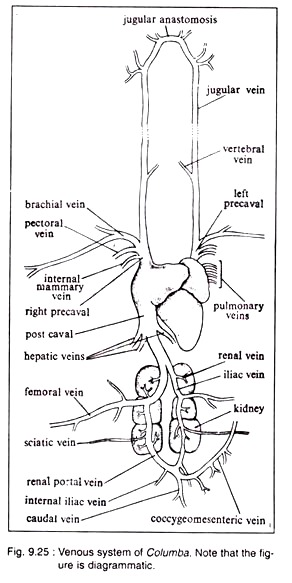
(ii) Two Percival’s and one postcaval open directly into the right auricle (Fig. 9.25). There is no trace of sinus venosus.
(iii) Considerable reduction of renal portal vein.
The veins in pigeon may be divided into three categories:
i. Pulmonary,
ii. Systemic and
iii. Portal veins (Fig. 9.25).
i. Pulmonary veins:
The pulmonary veins constitute a very short circulatory circuit and carry oxygenated or pure blood from the lungs. These veins enter the left auricle.
ii. Systemic veins:
Three principal systemic veins—two precavals and one postcaval — drain deoxygenated blood from the capillaries of the body and open separately into the right auricle.
Veins anterior to the heart:
The paired precavals with all the veins opening into them are included under this category. Each precaval receives (i) Jugular vein, (ii) Brachial vein, (iii) Pectoral vein and (iv) Internal mammary vein.
Jugular vein:
This vein receives several small veins from the crop and the shoulder, the vertebral vein and other veins from the head and neck. The vertebral vein brings blood from the vertebral column and spinal cord to the jugular vein. The veins from the crop and shoulder are small and numerous.
Their number and disposition are variable—so they are not given specific names. The left and right jugular veins are connected anteriorly by a small transverse connecting vein, called jugular anastomosis. The anastomosis gets veins from the venous sinuses of the brain. This cross-connection in the jugular veins is a special adaptation for the flexibility of neck.
The connection below the head prevents stoppage of blood circulation if one jugular vein is compressed during universal movement of the neck or head.
The jugular vein receives facial vein (carrying blood from the skin and muscles of the head), tracheal vein (brings blood from the trachea), cervical cutaneous vein (originates from a plexus in the skin of neck) and oesophageal vein (gets blood from the oesophagus). These small veins are not shown in Fig. 9.25.
The precaval vein is formed by the union of the following three veins: Brachial vein. The brachial vein receives blood from the corresponding wing. Some small branches from the shoulder also open into it. Pectoral vein. This vein is formed by the union of profusely branched veins from the pectoral region. Internal mammary vein. This vein brings blood from the sternum, coracoid region and the ribs.
Veins posterior to the heart:
The veins which are posterior to the heart include the following: Postcaval vein. This vein is formed by the fusion of two iliac veins. Each iliac vein is the continuation of the femoral vein bringing blood from the leg region. The femoral vein passes through the kidney tissue.
The postcaval receives few hepatic veins from the liver and a small vein from the ligament of the gizzard. Genital veins (spermatic vein in case of male and ovarian vein in female) are short veins which emptily in the iliac veins. Renal veins.
These veins bring blood from the kidneys and open into the iliacs as well as into the renal portal vein. Sciatic vein. This vein from the thigh opens into the renal portal vein. Internal iliac veins. These paired veins bring blood from the dorsal pelvic region.
Caudal vein:
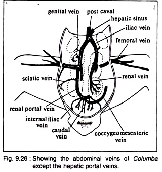
This small vein comes from the uropodium. Coccygeomesenteric or inferior mesenteric vein. This vessel runs anteriorly in the mesentery to participate in the hepatic portal system. It also gets branches from the rectum. The blood from this vein also flows to the renal portal vein (Fig. 9.26).
iii. Portal veins:
The hepatic and renal portal veins are also considered under the posterior veins. The renal portal vein originates at the junction of the coccygeomesenteric, internal iliac and caudal veins. Each renal portal vein passes through the kidney tissue of that side and opens into the femoral vein and also receives sciatic vein.
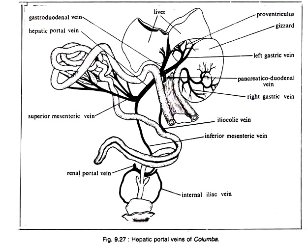
The renal portal vein is peculiar, because it never breaks up into capillaries in the kidney, but sends off a few small branches. Small renal veins open to this vessel. The hepatic portal vein forms an elaborate system. This system drains blood into the liver from the abdominal viscera (Fig. 9.27).
The hepatic portal system includes: Castro-duodenal vein which is formed by the pancreaticoduodenal vein and left gastric vein. The pancreaticoduodenal vein also gets a vein from the last part of the small intestine and the right gastric vein. The mesenteric veins are included under this system.
Type # 5. Lymphatic System:
The lymphatic system is well-developed and elaborate. Numerous lacteal vessels emerge from the small intestine. These vessels unite to form paired thoracic ducts. These ducts eventually open into the precaval veins.
Type # 6. Nervous System:
The nervous system of pigeon is divisible into:
(i) Central Nervous System,
(ii) Peripheral Nervous System and
(iii) Autonomous Nervous System.
i. Central Nervous System:
The Central Nervous System includes the brain and the spinal cord.
Brain:
The brain of pigeon is peculiar for its short and rounded form. It is built upon the same structural plan as that of other vertebrates.
Coverings:
The brain is covered by two meninges, an inner piamater and an outer duramater.
Parts of the brain:
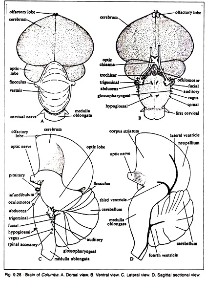
The olfactory lobes are poorly developed and form the most anterior part of the brain (Fig. 9.28A). The cerebral hemispheres are large, situated just posterior and dorsal to the olfactory lobes. The cerebral hemispheres consist largely of corpora striata (Fig. 9.28D).
The corpora striata are composed of solid mass of tissue and control the reflex behaviours of the animal. Each corpus striatum is differentiated into three regions— the upper portion is called the hyper striatum, the lateral region as the mesostriatum and the lower part as the paleostriatum.
The roof of each cerebral hemisphere is called the neopallium. The neopallium is un-convoluted. The cerebral hemisphere becomes expanded posteriorly to meet the cerebellum.
The diencephalon is inconspicuous and remains completely covered by the cerebral hemispheres and cerebellum. From the hypothalamus, a hypophysis arises. On the roof of the diencephalon, a small pineal body projects between the cerebral hemispheres and cerebellum.
The optic lobes are large in size and spherical in shape. They are pushed to ‘the lateral side due to the backward growth of the cerebral hemispheres. The optic nerves are prominent and form optic chiasma (Fig. 9.28B) situated ventral to the mid-brain.
A major portion of the optic nerve passes into the thalamus and the rest into the mid-brain. The mid-brain as well as the thalami have reciprocal connections with the corpus striata of the cerebral hemispheres.
The cerebellum is highly developed and consists of a large central vermis and two small lateral lobes — the flocculi. Transverse grooves are present on the surface of the vermis. The over-development of cerebellum is possibly connected with the control of movement and the precise timing during flight.
Like that of other vertebrates, pigeon possesses both spinocerebellar and vestibulocerebellar nerve tracts. In addition to these two tracts, tecto-cerebellar and strio-cerebellar tracts are present. The cerebellum is solid, because the fourth ventricle does not extend into it. The medulla oblongata has a prominent ventral fissure.
Ventricles:
The lateral ventricles are greatly reduced and represented by small crescent-shaped cavities located at the posterior end of cerebral hemispheres. The third ventricle (Fig. 9.28D) is a small slit-like cavity situated posterior to the corpus striatum.
The lateral ventricles and the third ventricle are communicated by foramen of Monro. Each optic lobe contains an extension from the aqueduct of Sylvius, a narrow passage contained in the mid-brain connecting the third ventricle with the fourth ventricle. The fourth ventricle is a small cavity between the cerebellum and medulla oblongata.
Spinal cord:
The spinal cord is covered by Pia mater and Dura mater like that of brain. Along its length, the spinal cord is enlarged in the cervical and lumbosacral regions. Each such enlargement gives origin to a pair of plexi which, in turn, send nerves to the limbs.
In the region of the lumbosacral enlargement, the spinal cord is open and the cavity becomes expanded to form a diamond-shaped sinus rhomboidal is. This cavity is filled with a fatty substance.
ii. Peripheral Nervous System:
The cranial and spinal nerves constitute the peripheral nervous system. This system is essentially same as in Calotes already described. 12 pairs of cranial nerves emerge out of the brain. The cranial nerve ‘O’ (Terminal nerve) is absent in Columba.
Fig. 9.29 shows the origin and distribution of the trigeminal (V) and facial (VII) cranial nerves. The trigeminal originates by many roots from the lateral part of medulla oblongata ventral to the optic lobes. It has three branches: ophthalmic, maxillary and mandibular.
The trigeminal nerve passes through the orbital fissure and innervates the skin, Herbst and Crandy corpuscles and muscles of tongue and larynx. The facial nerve — after arising from the lateral surface of medulla but posterior to trigeminal — sends nerve to the pharynx and muscles for mastication. Fig. 9.30 represents the origin and distribution of glossopharyngeal (IX) and vagus (X) nerves.
The glossopharyngeal originates superficially from the lateral surface of the medulla posterior to the auditory nerve and comes out of the cranium through a small foramen between ear and hypoglossal foramen. It sends branches to palate, pharynx, larynx and taste-buds on the tongue.
The vagus nerve originates from the lateral side of the medulla, posterior to glossopharyngeal. The main nerve trunk is found in the neck sheathed in the connective tissue. It runs posteriorly along the neck region adjacent to jugular vein. Near the brachial plexus it sends branches to the oesophagus and crop.
The main trunk sends branches to heart, lungs and abdominal viscera. The spinal accessory (XI) is a motor nerve. It arises from the lateral surface of medulla oblongata posterior to vagus. It supplies nerves to the neck muscles.
The hypogossal (XII) nerve arises from the ventral surface of the medulla posterior to abducens nerve and passes through the hypoglossal foramen just lateral to the occipital condyle. It sends branches to the neck and tongue muscles.
Spinal nerves:
There are thirty eight pairs of spinal nerves in pigeon. Paired spinal nerves pass through the intervertebral foramina to the body and limb musculature. Each spinal nerve has a sensory dorsal root and a motor ventral root.
These nerves are named according to the zones of the vertebral column from which they arise. They are: cervical, thoracic, lumbosacral and caudal nerves. The number of the spinal nerves in each region is: cervical = twelve pairs, thoracic = eight pairs, lumbosacral = twelve pairs and caudal = six pairs.
On each side, first nine cervical nerves serve the neck musculature, while the last three cervical together with first two thoracic nerves form the brachial plexus. This plexus gives rise to (1) Brachialis superior to the wing and (2) Brachialis inferior to the pectoral muscles, membrane between radius and ulna and posterior border of ulna.
The brachialis superior is formed of three main trunks:
(a) The eleventh cervical nerve,
(b) The twelfth cervical and first thoracic nerves and
(c) The second thoracic nerve with some nerve fibres from the third thoracic nerve.
These three nerve trunks, before joining to form the brachialis superior, give two nerve trunks to form the brachial inferior. Besides these nerves, the eleventh cervical nerve gives nerves to innervate the anterior shoulder muscles.
The brachial superior nerve continues into the forearm as the radialis nerve. The brachialis inferior is formed of two branches: (a) combined twelfth cervical and first thoracic nerves and (b) from the second thoracic nerve. These two trunks, before joining, give nerves to the pectoral muscles.
The brachialis inferior bifurcates on the median region of the elbow into:
(i) Medianus nerve in the membrane between the radius and ulna and
(ii) Ulnaris nerve supplies the posterior border of ulna.
The third to seventh thoracic nerves innervate the body and spinal muscles. The eighth thoracic and the first lumbosacral nerves unite to give branches to the muscles of the hip and thigh. The second lumbosacral nerve sends a small branch to the first lumbosacral nerve. The second, third, fourth and fifth lumbosacral nerves unite to supply small branches to the hip and a stout sciatic nerve.
The sciatic nerve extends along the lateral face of the thigh to the knee and divides into tibial and fibular nerves. The sixth lumbosacral nerve gives a small branch to the fifth lumbosacral and unites with the seventh, eighth and ninth lumbosacral nerves to form the pudendal plexus (Fig. 9.31). The rest of the three lumbosacral nerves and the six caudal nerves serve the tail muscles.
iii. Autonomous Nervous System:
It includes the sympathetic cords, one passes over the ventral surface of the ribs and the other passes dorsal to each rib. In between the ribs these two cords become fused. The nerves from the sympathetic cords between the third and fourth, fourth and fifth, fifth and sixth thoracic nerves together with a branch from the sympathetic ganglion join to from the coeliac plexus.
Two small autonomic nerve trunks run posteriorly to the cloaca and adjoining viscera. The vagus nerve belongs functionally to the sympathetic nervous system.
Type # 7. Endocrine System:
The endocrine system is well-developed in pigeon.
The following endocrine organs constitute the endocrine system:
Thyroid gland:
These paired glands are present one on each side of the trachea near the junction of neck and trunk. The thyroid hormone, thyroxin, regulates the general metabolism and the periodic moulting of pigeon. The functioning of thyroid glands is controlled by the thyroid stimulating hormone of the anterior pituitary.
Parathyroid glands:
These paired glands are present in the vicinity of thyroids. These are small in size and control the calcium and phosphate metabolism of the body.
Thymus glands:
These paired glands are present on the sides of the throat. They are well-developed and elongated in young stage. They become greatly reduced in matured adult. The physiological function of these glands is not properly ascertained. It is believed to be associated with the growth of the animal.
Adrenal glands:
These paired glands are prominent yellowish bodies, situated anterior to the kidneys. Each gland is irregular in shape and is composed of chromaffin tissue intermingled with fatty tissue. The secretion of the adrenal gland — adrenalin — controls some vital functions of the animal.
Pituitary gland:
This highly specialised gland remains within a skeletal cage formed by the sphenoid bone. This inner skeletal cage is called the sella turcica. The pituitary gland possesses two major divisions—(i) adenohypophysis (originates from the roof of the embryonic mouth cavity) and (ii) neurohypophysis (arises from the floor of diencephalon).
The adenohypophysis is subdivided into three parts—pars distalis, pars tuberalis and pars intermedia. The neurohypophysis has two subdivisions— lobus nervosus and infundibulum. The pars distalis represents the anterior pituitary, and the pars intermedia and lobus nervosus constitute the posterior pituitary.
The pars tuberalis together with infundibulum constitute the stalk of pituitary gland. The bipartite lobes of the anterior pituitary secrete different hormones like gonadotrophins, adrenocorticotrophin, thyrotrophin, prolactin, etc. Various external stimuli influence the secretory activities of the anterior pituitary.
Islets of langerhans:
The pancreas contains endocrine islets of Langerhans. These islets discharge insulin into the blood stream.
Gonads:
The gonads, besides their normal function of producing gametes, secrete sex hormones. The testes secrete testosterone and the ovaries produce the oestrogen. The testosterone is secreted by the Interstitial of Leydig cells of testis and controls the growth of sex organ and secondary sexual characters. The oestrogen regulates the secondary sexual characters and the behaviour of the bird.
Type # 8. Excretory System:
The excretory system comprises of a pair of kidneys and a pair of ureters opening into the cloaca. Absence of urinary bladder is a notable feature in the anatomy of pigeon.
Kidneys:
Each kidney is a flattened body which is divided into three lobes. These kidneys are of metanephric type and remain closely fitted into the dorsal wall of the pelvis. The nephrons are highly specialised. The glomeruli are supplied by renal artery and the loop of Henle is quite extensive.
This loop helps to reabsorb water from the glomerular filtrate. The urine contains a little quantity of water with high concentration of uric acid precipitate.
Ureters:
Each ureter originates from the first and second lobes of the kidney and passes down to open into the middle chamber (urodaeum) of the cloaca. The urine is voided with the faeces.
Type # 9. Reproductive System:
The sexes are separate. Sexual dimorphism is absent in pigeon.
Female Reproductive System:
The female reproductive system is peculiar by having only left ovary and left oviduct (Fig. 9.33 A).
Ovary:
The right ovary and oviduct are atrophied in adult. The left ovary is large and contains eggs of various sizes. The ovary is suspended to the dorsal body wall by a short mesentery, called mesovarium.
Oviduct:
The left oviduct is long and is attached with the dorsal body wall by broad ligament or mesotubarium. The anterior end of the oviduct opens to the coelom by an expanded funnel-like opening, called the oviducal funnel or ostium.
The remaining part of the oviduct is thick, muscular and coiled. Various glands are present in the inner lining of the oviduct. When an ovum is matured, the ovarian follicle bursts to liberate the ovum. The ovum enters into the cavity of the oviduct through the oviducal funnel.
While passing down the oviduct, the ovum (fertilized or unfertilized) becomes invested by the secretion of the various glands (Albumen and shell glands). The left oviduct opens into the urodaeum. A small vestigial right oviduct is found on the right side of the urodaeum.
Male Reproductive System:
The male reproductive system includes two testes and two vasa deferentia (Fig. 9.33B).
Testes:
Each testis is an ovoid body which is attached to the anteroventral end of the kidney by a fold of peritoneum, called mesorchium. The size of the testes varies greatly according to season. The testes are composed of numerous coiled seminiferous tubules. Between the tubules, groups of Leydig cells are present.
Vasa Deferentia:
From the inner side of each testis originates a much coiled duct, called vas deferens. Each vas deferens runs posteriorly, parallel with the ureter and opens into the urodaeum at the tip of a small papilla. These papillae are slightly erectile and constitute the miniature copulatory organs of many birds.
The last part of the vas deferens becomes slightly swollen to form seminal vesicle. Typical copulatory organs, observed in other vertebrates, are absent in pigeon.
Insemination and egg laying:
Fertilization is internal. Insemination is done when the proctodaea of both the sexes are averted and brought close together in a state of ‘cloacal kiss’. During this act, the sperms are ejected into the female tract which travels up to fertilize the egg.
Two eggs are generally laid at a time in pigeon. These eggs are incubated by the parents for a fortnight at a temperature of 38° to 40°C. When development becomes complete, young bird breaks the shell and comes out of the egg. The young is nourished by the parents with the pigeon’s milk.
Structure of egg:
The egg is large in size due to accumulation of great quantity of yolk material. The protoplasm forms a small round area, called germinal disc (blastodisc), containing the ovum. As it passes down the oviduct, a coat of thick albumen is accumulated and the disc becomes pushed to the upper side.
As the egg rolls during its transit, the albumen becomes coiled at the two sides to form twisted cord—the chalaza. In addition, more fluid albumen is deposited. Then a tough shell membrane and a calcareous shell are added by the secretory activities of the glands present in the oviduct.
The shell membrane is parchment-like and is composed of two layers which enclose an air-space at the broad end of the egg (Fig. 9.34). The shell is usually white in colour which may be coloured due to the deposition of special pigments. The shell consists of three layers and is provided with vertical pore canals.


