ADVERTISEMENTS:
In this article we have compiled various notes on viruses. After reading this article you will have a basic idea about:- 1. Origin of Viruses 2. Characteristics of Viruses 3. Historical Review 4. Nature 5. Virus Induced Symptoms 6. Nomenclature, Classification and Identification 7. Plant Cell Changes 8. Methods of Transmission 9. Properties 10. Control 11. Methods 12. Virus Strains and Virus Mutation and Others.
Contents:
- Notes on the Origin of Viruses
- Notes on the Characteristics of Viruses
- Notes on the Historical Review of Viruses
- Notes on the Nature of Viruses
- Notes on Virus Induced Symptoms
- Notes on the Nomenclature, Classification and Identification of Viruses
- Notes on the Plant Cell Changes Caused By Viruses
- Notes on the Methods of Transmission of Viruses
- Notes on the Properties of Viruses
- Notes on the Control of Virus Diseases
- Notes on the Methods of Study of Viruses
- Notes on Virus Strains and Virus Mutation
- Notes on Bacteriophage
- Notes on the Distinction between Virus and Cellular Organism
Note # 1. Origin of Viruses:
ADVERTISEMENTS:
Broadly speaking, only three general hypotheses of the origin of viruses are taken into consideration.
(i) The ancestors of viruses were at one time cellular organisms. As a result of parasitic existence in other cells, they gradually lost more and more of their own cellular machinery until they eventually became reduced to their present form.
(ii) The ancestors of viruses were once free-living pre-cellular forms of life, which managed to survive after the evolutionary emergence of cellular organisms only by becoming parasitic on them.
(iii) The viruses have not evolved from organisms, either pre-cellular or cellular, but have arisen from detached fragments of the genetic material of cellular organisms. These genetic fragments, as a result of detachment from the rest of the genetic system, acquired the ability to multiply more rapidly than the other constituents of the cell, and their unregulated growth caused disease and death of the cell.
ADVERTISEMENTS:
Liberated after cell death, the genetic fragments were able to ensure their own perpetuation by entering adjacent healthy cells and again multiply there.
Originally passed from cell to cell in the form of nucleic acid, they eventually acquired the capacity to direct the simultaneous synthesis of the infected cell of a special protein, which served to enclose the nucleic acid fragments, and thus made their transfer from cell to cell a much less hazardous operation.
The above hypotheses have not yet been supported by factual information.
Note # 2. Characteristics of Viruses:
The word virus is from Latin virus, a poison. As a preliminary working definition, viruses may be characterized as ultra-microscopic disease-producing entities, capable of being introduced into living cells of particular kinds of organisms, and capable of reproducing or being reproduced only within such cells. They cannot be made to multiply on artificial media.
The viruses can, however, be seen with an electron microscope. Because they are able to pass through bacterial filters, they have been called filterable viruses. A typical virus particle apparently consists of a core of nucleic acid, partly or wholly surrounded by a sheath of protein. Some of the viruses have been isolated in a pure form and even crystallized.
All thus far isolated have been found to be nucleoproteins of very large molecular size and weight.
Plant viruses have not been definitely observed in plants other than flowering plants and bacteria, but this may be due to lack of study rather than a real absence. Again, animal viruses inhabit vertebrates, arthropods, and many other animals.
Particles of both plant and animal viruses vary from spherical to slenderly rod- shaped, according to the kind of virus. Some animal viruses are brick-shaped. Some of the smallest viruses are only about 0.01 micron in length, while some of the largest ones approach 0.5 micron.
ADVERTISEMENTS:
The viruses are responsible for a large number of important plant and animal diseases. In many cases the virus is more or less latent (i.e., it exists and reproduces but causes no detectable harm) in a particular host and causes a recognizable disease only when introduced into some other kind of host.
In general, the plant viruses are transmissible by sap, by grafting, or by insects. The virus diseases and infections in plants are recognized and described on the basis of symptoms and transmissibility.
Viruses possess some of the qualities of living organisms they are able to reproduce, they occur in distinct strains or varieties, and they undergo changes similar to mutations. Unlike living organisms, they do not respire, nor do they possess cellular structures. Many Biologists regard viruses as intermediate between non-living matter and living organisms.
Note # 3. Historical Review of Viruses:
ADVERTISEMENTS:
The first known record of the existence and behaviour of virus is a variegation in the colour of tulips reported by Carolus Clusius in 1576. That the variegation might be due to a disease was suggested only in 1670. In 1715 an account of an infectious chlorosis of Jasminum was published. About fifty years later the so-called ‘curl’ disease of potatoes came into prominence. But there was a great controversy over its cause.
About 1886 Adolf Mayer described a disease of tobacco plant which occurred in the tobacco-growing regions of Holland, as Mosaikkrankheit which means mottling type of virus disease. He described the disease, and from the mosaic pattern common on leaves of affected plants, Mayer suggested the name ‘mosaic’.
Mayer showed that this mosaic disease of tobacco could be communicated to a healthy tobacco plant by inoculation with the sap of the infected plant. But Mayer did not suggest that the diseased condition was due to virus. Two years later, Erwin F. Smith showed that the disease ‘peach yellows’ was also communicable and could be transmitted by transplanting a bud from a diseased tree to a healthy tree.
The first scientific proof of the existence of a virus was given by the Russian Botanist DmitriIwanowski in 1892. Iwanowski working with the mosaic disease of tobacco, described by Mayer, proved that sap from such a diseased plant was capable of inducing the mosaic disease in healthy tobacco plants. He passed the sap through a bacteria-proof Chambeiland filter candle and found the filtrate to retain infectivity.
ADVERTISEMENTS:
It is the first record of the passage of either a plant or an animal virus through a bacteria-proof filter. Six years later in 1898 Loeffler and Frosch showed that the foot and mouth disease of cattle is caused by an agent which could pass through bacteriological filters. In 1892, a Dutch Bacteriologist Martinus Willem Beijerinck took up the study of tobacco mosaic.
He found the sap of infected plant, when filtered through bacteria-proof filter, to be sterile but still infectious, which he designated as contaginm vivum fluidum and subsequently, referred it as a virus. Beijerinck confirmed the findings of Mayer and Iwanowski and claimed more emphatically than either of them that the causal agent was not a bacterium or any conceivable corpuscular material.
The relationship between an insect and a plant virus has been experimentally established by a Japanese farmer, Hashimoto, who worked in 1894 with the dwarf disease of rice and the leafhopper Nephotettix apicalis var. cincticeps. In 1895 Takata in Japan transmitted virus by means of the leafhopper Deltocephalus dorsalis.
During 1906-07, Ball, Adams, and Shaw working on curly top of sugar-beet established the leafhopper transmission of virus. Further evidences of leafhopper transmission of virus were put forward by Boncquet and Hartung in 1915. That aphids are also responsible for the transmission of virus was demonstrated by Allard in 1914.
ADVERTISEMENTS:
Iwanowski continuing his study of tobacco mosaic virus described in 1903 certain intracellular bodies in the tissues or diseased plants. One type was amoeboid, the other was in the form of crystalline plates. Holmes in 1929 described the primary infection lesion of tobacco mosaic virus and thereby he indicated the usefulness of the symptomatology.
In the area of strain differences in viruses, McKinney (1926) is the pioneer worker. He suggested that strains arise by mutation. Takahashi and Rawlins in 1933 exhibited the physical phenomenon of tobacco mosaic virus. By 1935 it was evident that the virus was a particle distinct from any known living entity and comparable in many ways to larger molecules or colloidal particles.
In 1935 Stanley for the first time isolated a crystalline protein in more or less purified condition possessing the properties of the tobacco mosaic virus. In his opinion, tobacco mosaic virus is an auto- catalytic protein which may be assumed to require the presence of living cells for multiplication. In the succeeding five years protein crystals were isolated from preparations of several plant viruses.
These preparations were infectious and capable of producing the disease concerned upon inoculation into the respective host. Other pioneer workers in this line are Bawden and Pirie. They have shown that all virus proteins crystallized so far, are nucleoproteins.
Note # 4. Nature of Viruses:
There has been some argument as to whether viruses should be considered to be living or non-living. True, viruses are individual organic compounds whose chemical composition resembles protoplasmic constituents. They behave as microorganisms only when in association with the complex mechanisms of living cells.
ADVERTISEMENTS:
Viruses reproduce in a host cell and are capable of mutation. They contain the true essence of life by the possession of an extremely potent complement of genes and behave as microorganisms only when in associated with the complex mechanisms of living cells.
In their ability to reproduce themselves in living tissues they also resemble microorganisms. With the messages contained in the single strand of the nucleic acid, the virus is able to divert the enzyme systems of the host cells into new pathways and synthesize more virus particles instead of host substance.
In this respect viruses seem to resemble self-duplicating genes and chromosomes, but they differ in that they are able to penetrate some unicellular of multicellular hosts from the outside.
But on the other hand, viruses do not possess cellular structures. They by themselves do not carry on respiration, lack capacity for independent metabolism, and do not multiply by classical growth and fission methods from pre-existing virus particle. Those viruses which have been prepared in pure crystalline form and found to be nucleoproteins are not living organisms in the ordinary sense.
Again the virus crystals are chemically inert and can apparently be kept indefinitely without significant change.
Again viruses are often accepted as molecules having ability to duplicate themselves. But Stanley and others suggested the significant difference between viruses and other molecules pointing out that a virus comes to life the moment it infects a cell. Besides this, the term ‘molecule’ implies a precise knowledge of the structure of a compound.
ADVERTISEMENTS:
Viruses are thus ultra-microscopic disease-producing particles of organic matter which can multiply only in living plants and animals and are responsible for a large number of important plant and animal diseases. It is apparent that furthermore is necessary before assigning any particular status to the viruses.
Note # 5. Virus Induced Symptoms:
Effects of Viruses on Plants:
Viruses are similar to obligate parasites in that they cannot be grown on non-living media. They are intimately associated with the host cell and few kill the infected plant although some cause severe distortion and dwarfing.
The changes brought about by viruses are treated as symptoms which may be:
(i) Morphological changes or external symptoms,
(ii) Histological and cytological or internal symptoms, and
(iii) Metabolic changes; but these are all correlated.
The symptoms of the majority of plant virus diseases are most conspicuous on plants making rapid growth. Plants that are almost mature at the time of infection usually do not develop symptoms on any part except on new growths.
Viruses being infectious induce a variety of symptoms covering a wide range of host reactions.
Some of the symptoms frequently encountered are mentioned below:
I. External Symptoms:
The most common symptom in the green tissues of higher plants is the alteration in the normal development of chlorophyll— chlorosis. This may be accompanied with various other malformation. Besides this, necrosis of tissue and dwarfing, distortion of a particular organ or the entire plant are also common symptoms. The external symptoms may be primary or localized and systemic.
The primary or initial symptom is a local reaction at the actual site of inoculation consisting of spots or rings of various types.
They are usually necrotic but are occasionally chlorotic and known as local lesions. There may also occur a second type of primary symptom known as clearing of the veins, a condition where the veins of the youngest leaves become yellow. Whereas, the systemic symptoms are of widespread occurrence in the host tissue.
Some of the external symptoms are described below:
Chlorosis:
The dis-balance of normal development of chlorophyll leading to yellowing or formation of different shades of green without pattern is the chlorosis.
Mosaic:
The interspersion of various degrees of chlorosis with the normal green colour of the leaf resulting in a mosaic pattern of yellow and green, forms the mosaic symptom (Fig. 339A to C).
Mosaic mottling:
When the leaves show a mottling of light or dark-green, yellow or even white it is known as the mosaic mottling.
Necrosis:
Death of the host cells, or necrosis, is a symptom of many virus diseases, and may consist of small areas on the leaves, streaks on the stem or large areas of dead tissue which ultimately cause the death of the whole plant. The necrosis may spread causing various patterns as it develops.
A relatively rapid killing of a bud, branch, or the entire top of the plant is the top necrosis. The necrosis of phloem elements is known as phloem necrosis.
Ring spots:
These spots consist of various types of chlorosis and necrosis. In each ring spot numerous concentric rings develop on the leaves with a central spot. Spots of circular chlorotic areas are known as chlorotic ring spots. Whereas, in cases where necrosis appears in rings alternating with normal green are the necrotic ring spots.
Veinclearing and veinbanding:
In infected leaves, when a clearing or chlorosis of the tissue in or immediately adjacent to the veins takes place, is referred to as vein- clearing. Again, the symptom consisting of a broader band of chlorotic tissue along the veins or bands of green tissue in that position, set off by chlorosis or necrosis in the intervene parenchyma is called vein-banding.
Yellowing:
Leaves and other green parts instead of developing green colour turn yellow, this is known as yellowing.
Distortion and overgrowths:
Distortion of the leaves is a common symptom of virus disease and may take the form of crinkling and curling or upward rolling of margins. Some viruses induce the formation of outgrowth known as enations, masses of hypertrophied tissue developed on the surface of leaf or stem.
Virus attack may also induce proliferation of stem buds, or cause distinct tumours or galls on roots and stems. Often virus infection results in stimulation of dormant buds and hyperplastic tissue leading to unusual differentiation. Virus infection may also cause distortion and sterility of flowers (Fig. 339D & E).
Stunting:
This is shown by reduction in size of leaves or other organs or of the entire plant. Stunting is frequently accompanied with resetting. This is shown by shorter internodes, smaller leaves and fruits. Stunting with more or less rosetting is characteristic of bunchy top of bananas. Reduction is size also results in dwarfing.
Premature defoliation and death:
Premature dropping of leaves is also a symptom of virus disease. This may also lead to premature death of the affected plant.
Masked symptoms:
Under certain environmental conditions often no distinguishable symptoms appear in spite of the presence of virus in the infected plant. Such an infected plant is referred to as having masked symptoms. Again, when plants show symptoms for a short time when first infected, but eventually become and remain symptomless, the infected plants of this type are known as symptomless carriers.
II. Internal Symptoms:
This will include intracellular inclusions. Certain abnormal intracellular inclusions are characteristic of virus infections, they do not occur in diseases caused by other infectious agents. There are several different kinds of intracellular inclusions crystalline or fibrous or striate material; amorphous known as X-bodies; vacuolate material (Fig. 340B & C); intra-nuclear inclusions; and other types of inclusions.
Crystalline inclusions or Striate materials:
These occur mainly in the cells of plants infected with tobacco mosaic virus and are usually in the form of plates of varying size (Fig. 340C).
Amorphous inclusion bodies or X-bodies:
The amorphous bodies are protoplasmic (Fig. 340C), more or less about 10/am in length, may be several in one cell. They are relatively stable and are preserved by ordinary cytological fixatives. They often resemble the nucleus of the cell. In addition to the tobacco mosaic virus, these inclusions occur in all tissues of plants infected with Hyosyamus mosaic virus.
Intra-nuclear inclusions:
These inclusions may consist of thin rectangular plates and usually several in each nucleus, or of isometric crystals. They are common in the nuclei of leguminous plants affected with the viruses of pea mosaic and yellow bean mosaic.
Other types of inclusions:
A number of miscellaneous inclusions occur in the cell cytoplasm of virus-infected plants.
Some of them are:
A spherical hyaline and homogeneous body called a spherule, present in the wound tumour virus-infected root tumour cells of Rumex acetpsa; spindle-shaped bodies in the virus-infected cells of Epiphyllum; fusiform and variably shaped protein bodies in the cytoplasm of the virus infected epidermal cells of the leaves of Opuntia brasiliensis.
Note # 6. Nomenclature, Classification and Identification of Viruses:
In spite of continuous efforts made by Johnson (1927), Smith (1937), Fawcett (1940), Holmes (1939, 1948), Valleau (1940), Lwoff, Home and Tournier (1962), Pereira (1966), Tourinier (1966), Hansen (1956, 1968), Thornberry (1968), Gibbs (1969), Martyn (1968, 1970), Harrison (1971) and many others, there is no final agreement about nomenclature and classification of viruses as because viruses are genetically variable and new strains differ in host range, virulence and other characteristics arising from different arrangements of the nucleotides in the nucleic acid molecule.
A specific virus exhibits fixed characteristic properties. These include the size, structure, and chemical composition of the virus particles, the host range, the tissue specificity, and the nature of the infection caused. When the properties of a large number of different viruses are examined, it is found that they fall into groups, each characterized by the possession of a number of properties in common.
The major groups of viruses may broadly be separated on the basis of characters like:
Type of nucleic acid present in the virus particle, nature of host and disease induced, properties of virus particles (shape, size, etc.), and other related characters. Although nothing is known about the origin and relationships of the viruses, it is tempting to imagine that these groups are natural ones, each of which unites a series of virus that are genetically related to one another.
Some have gone so far to create families, genera, and species for virus, conferring on individual viruses Latin binomial designations, just as if they were cellular organisms. Since a homology between viruses and cellular organisms is still questionable, it is too early-for such an approach to the viruses.
The situation often becomes all the more difficult as viruses are genetically variable and new strains differing in host range, virulence and other characteristics arise, these perhaps representing different arrangements of the four nucleotides in the nucleic acid molecule.
Such aberrations no doubt occur during virus replication; many are probably harmful and disappear in a uniform environment but some survive and prosper under changed environmental conditions or in a different species and variety of plant, thus extending the host range of the virus.
There are probably other mechanisms, perhaps including some form of genetical recombination, which bring about variation in viruses. The problem is complicated by the difficulty of distinguishing between viruses and virus strains, and by the lack of any satisfactory system of nomenclature and classification.
Hence the classification of virus has been subject to change over the years. For one thing, as more is learned about the properties of different virus, their classifications change. For this reason only the principles of virus classification are considered here.
The most widely used taxonomic criteria for viruses depend upon the structure of a virus itself.
Four major criteria are used:
(i) The nature of a nucleic acid—DNA or RNA, single-stranded or double- stranded;
(ii) Particle structure—helical, icosahedral, or complex;
(iii) Presence or absence of viral envelope; and
(iv) Dimensions of the viral particle.
Beyond these physical characteristics, other criteria (immunologic, cytopathologic, or epidemiologic) are used to subdivide the groups. Such a classification provides great convenience and utility, although it is not necessarily based upon the evolutionary origin of individual viruses.
Following are methods that are helpful to identify plant viruses:
1. Viruses are inoculated into indicator plants which develop typical symptoms when infected by specific viruses and in virus assay.
2. Serological tests are carried out using antisera of known viruses.
3. Transmission aspects of the virus are considered: whether by sap inoculation, and the vectors, if any, involved; whether the virus is persistent or non-persistent in the vector; whether stylet borne, circulative or propagative, and other aspects of its transmission.
4. Such properties as the thermal inactivation point, the dilution end point, and survival outside the plant can be used to characterize viruses.
5. Interaction with other viruses is considered, notably cross-protection.
6. Host range and symptoms are studied.
7. Study of morphology and chemical constitution of the virus particle.
Note # 7. Plant Cell Changes Caused By Viruses:
The changes of plant cells caused by viruses may be indicated in the following manner:
i. Histological and Cytological Changes:
The chlorotic tissue of mottled leaves is generally thinner than the normal green tissue and has shorter palisade cells containing fewer and smaller chloroplasts. Some viruses, those which cause leaf curling and yellowing bring about necrosis of the phloem tissue whereas in others necrosis is preceded by tissue proliferation and is perhaps caused by crushing than directly by the virus.
Leaf enations caused by the cotton leaf curl virus originate in part from increased cambial activity producing extravascular tissue which may be abnormal in nature. Xylem is generally less affected by virus than is parenchyma or phloem, but tyloses and formation of gum have been described in the xylem of plants infected with viruses.
Nuclear division may fail or be abnormal in cells infected with certain viruses. Amitotic nuclear division takes place in Petunia stem enations caused by virus infection. The nuclei after elongating, constricting and cleaving produce daughter nuclei. Amitosis may be associated with a shortage of RNA.
During division of virus infected cells there is competition between the virus particles and the nuclear DNA for the RNA present in the nucleolus. Since the formation of the nuclear spindle in mitosis is dependent on RNA, any shortage of the latter could lead to amitotic nuclear division.
Other abnormalities shown by virus infected cells include lobbing and distortion of the nucleolus, and spindle breakdown resulting in either scattering of the chromosomes or their failure to separate, the latter leading to the formation of ‘giant’ nuclei. The sterility of plants infected by certain viruses may be associated with such nuclear abnormalities.
ii. Metabiolic Changes:
Viruses affect the nucleic acid metabolism of the infected cell, thereby bringing about changes in protein synthesis which in some way result in the formation of more virus. Reduced efficiency of chloroplasts in virus infected plants and the photochemical activity of chloroplasts lead to mosaic, mottling and yellowing of leaves.
Changes in the amounts and activity of several enzymes take place due to virus infection. Increased oxidase activity is a general feature of many virus infections in plants.
The abnormal growth pattern of plants infected by some viruses suggests an interference with the growth regulating processes of the plant. Virus infection causes increased respiration rate in plants. The combination of decreased photosynthesis and increased respiration results in a fall in the carbohydrate content of virus infected leaves.
Note # 8. Methods of Transmission of Viruses:
The various methods of transmission of plant viruses are as follows:
i. Seed Transmission:
Viruses may be externally seed borne as in tomato, cucumber, etc.; or internally seed borne in testa, endosperm and/or embryo as in barley, cowpea, bean (bean mosaic), etc. The internally seed borne viruses are more effective than the externally seed borne ones.
ii. Transmission by Grafting:
Since viruses are intimately associated with the living cells of the host, it is rather easy for their transmission through grafting between living cells of virus infected and virus-free plants. In fruit and ornamental trees where grafting is the normal method of propagation, transmission of virus by grafting becomes a means of natural transmission.
iii. Transmission by Vegetative Propagation:
Viruses are very commonly perpetuated in the vegetative organs of perennial plants (fruit trees). When such plants are virus-infected all the vegetative parts used for their propagation also become virus-infected. As such, viruses are readily transferred from locality to locality in virus-infected nursery stock, bulbs, tubers (leaf roll of potato) and roots.
Hence the infected perennials are the common reservoirs for perennating of many viruses.
iv. Transmission by Parasitic Phanerogams:
Species of Cuscuta when parasitizing virus-infected host plants sends haustoria into the host tissue and thereby receives virus infection.. The same virus-infected species, of Cuscuta when extends its stem to parasitize other plants, the virus may be transmitted to such plants through the newly formed penetrating haustoria. Cuscuta thus functions as the transmitting agent.
v. Transmission by Insects:
Most viruses are transmitted by insects. The insects responsible for the transmission of viruses either possess mouth parts adapted for biting or stylets for piercing and sucking. The sucking insects are the usual insect vectors.
But the principal insect vectors are: thrips, plant bugs, leafhoppers, white flies, aphids and coccids.
There is specificity of certain insects for particular viruses. Some viruses may be carried mechanically on the mouth parts of the insects and the latter remain viruliferous for a period of only a few minutes to a few hours. These are known as non- persistent viruses. They are rapidly lost by the vector, usually after a short period of feeding.
The non-persistent viruses are carried to the first plant and rarely to the second if the feeding periods are of some hours duration. Again some vectors may not transmit the viruses to a healthy plant until sometime has elapsed after they have fed upon the diseased plant. These are persistent viruses.
The persistent viruses retain infectivity for a long period of time and there is delay in the development of infective power. In such case it is possible that the virus is ingested by the insect and is later transmitted through the body into the saliva, by which channel it eventually reaches the next host plant.
The delay in the development of infective power—the latent period also known as the period of incubation, varies greatly with the different viruses. These viruses may also multiply within the body of the vectors. The persistent viruses are not transmitted to the first two or three plants, but to all the others for a considerable period.
The longest latent period, so far discovered, is that of a strawberry virus known as ‘Virus 3’ transmitted by the aphid Capitophorus fragariae Theob., which takes 10 to 19 days.
vi. Transmission by Mechanical Means:
Transmission by this means consists of transference of sap from a virus-infected plant to a healthy plant by artificial or natural means. Since infection through natural openings, like stomata is rather rare, mechanical transmission often involves wounding of the host tissue for the easy entrance of the virus from host to host. Viruses transmitted by mechanical means are usually in high concentration in the plant.
Some viruses can spread from a diseased plant to a healthy one by contact of the leaves brought about by the wind. Cultivation procedures and the movement of animals may play some part in the spread of viruses. The tobacco mosaic virus is transmitted very rapidly by rubbing the extracted juice of a diseased plant over the leaf of a healthy tobacco plant.
By this process hairs or epidermal cells are sufficiently wounded to bring about infection.
Some viruses may spread below ground by mechanical contact between the roots of infected and healthy plants. Usually viruses of mosaic group are most readily transmitted by mechanical means.
vii. Soil Transmission:
The soil-borne viruses infect host through root system. These viruses do not usually persist in the soil more than a few months at the most. The viability of the soil-borne viruses, however, depends largely on the soil texture. Roots of infected perennial hosts serve as permanent reservoirs of soil-borne viruses.
viii. Transmission by Mites:
Eriophyid mites transmit several viruses. The big- bud mite, Phytopus ribis transmits virus that causes disease of Ribes. Mites cannot fly and presumably spread viruses by crawling from plant to plant or more likely, by being dispersed by wind.
ix. Transmission by Nematodes:
Nematodes belonging to the genera Xiphinema, Longidorus and Trichodorus transmit a number of viruses. Spread might result from systemic root infection of plants with extensive root systems; the roots could thus be made available to nematodes feeding at some distance from the original site of infection. The nematodes feed on the epidermal cells near the root tip and acquire virus.
x. Transmission by Fungi:
Several viruses including those causing big-vein disease of lettuce and tobacco necrosis are transmitted by Olpidium and Synchytrium which infect plants. The virus is borne internally by the zoospores of the fungus when they are developed in the virus infected host.
xi. Pollen Transmission:
Gases of dissemination of viruses through pollen grains are few in comparison with other means. Common example is bean mosaic virus.
xii. Transmission through weeds:
Weeds serve as collateral hosts for transmission of sugarcane mosaic virus.
Note # 9. Properties of Viruses:
The properties of the plant viruses are conveniently divided into the following categories:
i. Host Range:
Some viruses like beet curly top, cucumber mosaic, and tobacco mosaic virus have wide host range and host plants may fall within widely different families. Others, have extremely restricted host range, for example corn mosaic virus. Host specificity is a genetic character of the virus and is determined by its nucleic acid.
In some cases virus is actively present in the host plant without causing obvious effect. The absence of distinctive symptoms is due to masking effects of unfavourable environmental conditions. Symptoms appear when conditions become favourable.
A virus showing the phenomenon of masking is a masked virus and the host plant is the masked carrier. Again the presence of a virus in the host with the total absence of visible symptoms over the entire range of environment (favourable and unfavourable) to which the host is exposed is designated as a latency of a virus and such a virus is a latent virus.
A latent virus never induces symptoms or makes its presence known in a host over the entire range of environmental conditions. Host plants which harbour the virus but remain symptomless throughout the entire range of environmental conditions are the symptomless carriers.
ii. Physical Properties of Viruses:
The viruses could be distinguished in part by the symptoms they produce on the infected hosts. But the symptoms very often overlap and also change appearance with variation in environment. Viruses were found to differ markedly in the point at which they were inactivated by various agents.
Those most extensively used are:
(i) The thermal inactivation point, it is the constant temperature at which a virus extract is completely inactivated when exposed for 10 minutes;
(ii) The longevity in vitro, is the number of hours or days at which a virus remains infectious at room temperature either as a virus extract or in the host tissue;
(iii) The dilution inactivation point, is the degree of dilution of the virus extract with water or buffer solution at which infection no longer occurs. These are commonly designated as physical properties.
iii. Structure of Viruses:
Each virus particle is called a virion. It is composed of a single type of nucleic acid (DNA or RNA), but never both, which gives the virion infective capability. The nucleic acid which may be either linear or circular is surrounded by a protein coat called a capsid to form a nucleocapsid (Fig. 341C). A capsid is again made up of protein subunits called capsomeres which are in turn composed of a number of protein molecules.
The virus particles or units of virus—the virions possess a true symmetry of structure. Virions may either be naked (naked virions) or surrounded by an envelope of carbohydrates and lipids or lipoproteins (fig. 341A & B). The enveloped virions are sensitive to lipid solvents such as ether and chloroform.
The nucleic acid of a virus may occur as either double-stranded DNA, single-stranded DNA, double-stranded RNA or single-stranded RNA.
Plant viruses have been found to contain only single- or double-stranded RNA. Bacterial viruses contain single- or double stranded DNA or single-stranded RNA. Animal viruses have all types of nucleic acids except single-stranded DNA.
The amount of nucleic acid in a virus particle varies enormously:
RNA 1 to 5 per cent and DNA 5 to 50 per cent. The viral protein usually forms the largest part —50 to 90 per cent. Viruses lack components necessary for energy generation and protein synthesis (for example, ribosomes).
Generally, the enzymatic capabilities of viruses are extremely limited and confined to enzymes involved in the viruses entry into cells and replication of their own nucleic acid. Because of their metabolic limitations viruses are unable to replicate independently, and they must invade a living cell and utilize the cellular ribosomes, energy sources and certain other components of this cell in order to produce new viruses.
Viruses differ considerably in size. The smallest viruses are similar in size to large protein molecules or ribosomes, and their nucleic acid codes for only a few genes. The more complex virions may be larger than some of the most minute bacteria. Largest virus is Smallpox virus 300 nm in diameter and the smallest ones are 20 nm in diameter. Some simple viruses can be prepared as crystals.
The virions conform to one of the following shapes:
i. ICOSAHEDRAL—a regular polyhedron with 20 triangular faces and 12 corners. This shape is determined by the capsid.
ii. HELICAL—these virions resemble rods. Their capsid is a hollow cylinder with a helical structure.
iii. ENVELOPED—the internal nucleocapsid of these viruses, which may be either icosahedral or helical, is surrounded by a membranous envelope. Enveloped virions are pleomorphic (have varying shapes) since the envelope is not rigid, although they generally appear somewhat spherical.
iv. COMPLEX—some virus particles have a very complicated structure. They have several coats around the nucleic acid.
v. BACTEROID—the virus particles are with a more or less spherical ‘head’ and a slender ‘tail’ of equal or greater length.
Some virus—like infectious agents, known as viroids which cause a variety of plant diseases, e.g., potato spindle tuber chrysanthemum stunt, etc., have no protein capsid and consist only of a small single-stranded RNA of low molecular weight—and are sub-viral in size. They were described by Diener in 1967.
iv. Virus Replication:
Viruses replicate only in living cells. The term ‘replicate’ actually means multiplication of virus particles which does not take place by division of existing virus particles but are formed directly by aggregation and organization of molecules within the protoplasm of the host cell. The basic steps in virus replication are similar for all viruses, whether they infect the plant, bacterial, or animal cells.
In case of plant viruses; after entry into the host through natural openings, wounds (mechanical or insect), or through pollen grains, the virus comes in contact with host cytoplasm by pinocytosis. The infective part (RNA) is freed from its protein coat soon after inoculation. This is the eclipse stage. This stage is followed by an extensive virus synthesis.
The site of viral RNA synthesis is nucleus, more correctly nucleolus; that of protein synthesis is cytoplasm in the vicinity of nucleus and/or nucleolus and that assembly of virus particles is the endoplasmic reticulum and/or nucleus. Complete virus particles gradually diffuse throughout cytoplasm and may ultimately lead to the formation of large virus aggregates or virus crystals. The details of plant virus synthesis are given below.
The degraded protein coat of the virus probably remains in the host cell and becomes a part of the host cell protein. The naked RNA then induces the host cell to form enzymes: RNA-polymerases, RNA-synthetases, or RNA-replicases. These enzymes in the presence of viral RNA and its nucleotides produce additional viral
RNA:
The new viral RNA induces the host cell to produce the specific protein molecules required for its coat. During this process the inhibition of cell protein and RNA synthesis makes nucleotides, amino acids, and free ribosomes available for synthesis of viral components.
The viral protein and nucleic acid are constructed from the same 20 amino acids and four nucleotides which occur in normal cells. Energy needed for energising various chemical reactions for synthesis of viruses comes from ATP of the cells. Thus viruses are completely dependent upon host cells for this replication.
In case of bacterial and animal viruses, the virion (virus particle) adsorbs specifically to receptors on the host cell and it is after this adsorption that the viral nucleic acid penetrates into the cell. Either free nucleic acid enters the cell (in the case of most bacteriophages) or whole virions (for all other viruses) enter and then release their nucleic acid.
Replication of viral nucleic acid and synthesis of other viral constituents follow. These viral nucleic acid and protein constituents are made separately within the host cell and are then assembled into complete virions (virus particles) during the stage of virus maturation.
Finally, the newly formed mature virions are released from the host cell (Fig. 336). The method of release varies depending on the virus in question.
Cells may be lysed releasing many mature virions, or virus particles may be gradually extruded from the living host cell. The details of replication in T-even phage which offer an excellent model for the general process of infection by a phage and destruction of the host cell by lysis, are incorporated under Bacteriophage.
Virus and host cells may often reach some sort of equilibrium with a minimum damage to the host allowing both to survive. The virus is then a commensal.
v. Serological Tests:
Viruses respond to serological tests. Antiserum could be secured by injecting a partially purified virus extract to a suitable animal. It has been demonstrated that when juice extracted from a tobacco plant infected with the tobacco mosaic virus is injected into the blood of the rabbit in proper doses, the serum taken later from the animal causes flocculation when added in proper proportion to freshly extracted juice.
Besides this, when strains of the virus differing in various disease symptom patterns are tested against the same serum, they react similarly. When a group of viruses give similar reactions to a given antiserum, they are regarded usually as strains of the same virus, whereas, when a virus gives no reaction, it is regarded as distinct from the virus used to produce the antiserum.
This response to the serological test by the viruses may be utilized as a useful tool in determining virus similarities and dissimilarities.
vi. Cross-protection Tests:
If plants affected with virus, such as white mosaic, were inoculated with common mosaic virus, the symptoms of the latter did not become evident.
It is generally assumed that when one virus, preceding another in the tissue, prevents or markedly impairs increases of virus subsequently introduced, it is an indication that the two viruses are closely related and are probably strains of a single virus. Such a protective phenomenon is known as the cross-protection test. The cross- protection test in conjunction with others may be used in the characterization of a virus.
vii. Synergistic Effect:
Two unrelated viruses when present separately incite relatively mild effects on the hosts. But the combined effect of both of the two viruses produces very severe effect on a particular host. The combined action of the two viruses is known as a synergistic effect.
viii. Special Features:
In tulips where a virus transmitted through the bulbs can cause a desirable colour variegation of the flowers. Bulbs are purposely maintained in a virus-infected state to obtain plants bearing flowers with variegation to fetch high market value.
Again virus-infected aphids or aphids fed on virus-infected plants (for example, Aphis fabae and Macrosiphum granarium) have longer life span, greater egg laying capacity, earlier attainment of adulthood and more rapid breeding.
Note # 10. Control of Virus Diseases:
Some of the general principles of controlling virus diseases are incorporated below:
I. Cultural Methods:
1. Use of Virus-free Planting Material:
The simplest way to ensure this is securing planting material from uninfected plants. Tuber indexing is one of the methods for selecting virus-free potato tubers. It consists of selecting tubers from vigorous plants appearing healthy. From these tubers eyes or buds are taken out and planted under suitable conditions.
If the plants grown from these buds are healthy, the tubers produced by them are suitable for cultivation as virus-free planting material. If not, the parent tuber should be rejected. Virus-free planting materials can also be obtained by culturing of excised stem tips or by heat treatment.
2. Isolation:
Cultivation of susceptible crops at a distance from each other delays or reduces the severity of virus diseases. In cases where insects spread the virus from old to new crops, appreciable control can be achieved by breaking the continuous pattern of cultivation by omitting a crop from the cycle.
3. Roguing and Field Sanitation:
Both these practices eliminate virus sources. Roguing of diseased plants from seed beds and fields cuts down the source of virus. Some viruses are carried in weeds which when detected should be immediately removed. Perennial plants should be dug out and destroyed just as rationing in sugarcane should be discouraged. Elimination of alternate host plants also produces effective results.
4. Cover crops and other barriers:
The non-susceptible plants towering over the under-sown economically important crops break the flight of insects as a result of which few vectors can reach the crop.
Barriers erected at intervals in the field may also have the same effect. The barriers may be screens of cloth or some other suitable material, but more often rows of plants including sunflowers, maize, oats, and barley which are neither susceptible to the virus nor colonized by vectors.
5. Density of Crop:
Dense growth of a crop reduces disease incidence because:
(i) Dense foliage generally provides an unfavourable microclimate for the insects to develop, and
(ii) Viruliferous insects infect a smaller percentage of plants.
6. Area of Field:
Insects infect mostly plants at the edges of the field resulting greater concentration of diseased plants in the periphery of a field than in its middle, as such many plants escape infection in large fields as against smaller fields.
7. Planting and Harvesting Time:
Alteration of the dates of sowing or harvesting of a crop enables it to escape disease. Early sowing leads to early maturity of plants at the time when a virus normally strikes. This causes reduced incidence of disease severity because host plants possess greater resistance to infection with age.
II. Heat Therapy:
Heat therapy has proved most effective for controlling virus diseases of vegetatively propagated crops. Plant material treated upto one hour with hot air at a temperature range of 35 to 40 °C produces good result. Sugarcane mosaic has been controlled by treating the cuttings at 53 to 54°C. Immersion of sets in water at 52°C for 20 minutes kills the virus of chlorotic streak of sugarcane.
III. Chemotherapy:
Chemotherapy for the control of plant virus diseases has two aspects:
(i) Protection of plants with chemicals decreases virus multiplication or insect vectors fail to acquire viruses from infected host tissue,
(ii) Use of insecticides for killing vectors.
IV. Apical or Meristem Tissue Culture:
Many viruses cannot attack apical meristems. Localized viruses cannot move far away from their points of entry into host tissue; though systemic viruses can do so but majority of them fail to reach the apical region where apical meristem is located. Apical meristems and a few inches of adjoining system portions of a rapidly growing host and/or axillary buds of systematically infected plants remain free from viral infection.
These when grown in tissue culture medium produce virus-free plantlets which when transferred to soil produce virus-free adult plants. This apical or meristem tissue culture technique has proved extremely valuable in plants what are propagated by cutting vegetative parts.
V. Immunization:
Cross-protection (vaccination) method in which infection of the plant with a mild strain of the virus protects it against subsequent attack by a virulent strain, may produce good result.
VI. Resistant and Tolerant Varieties:
Cultivation of virus disease resistant varieties obtained by breeding and/or selection, is the best way of controlling virus diseases.
VII. Indexing and Certification Programme:
Many countries of the world follow certification programme by certifying stocks and seeds of various plants to be healthy and free from virus infection. This is associated with indexing which is testing of plants or plant parts for virus presence or absence.
This is done by:
(i) The use of indicator plant,
(ii) By the application of polyvalent antiserum technique,
(ii) Observing the presence or absence of virus disease symptoms,
(iv) By the use of immuno diffusion procedures, and
(v) By thin layer chromatography.
VIII. Other Methods:
Plant Protection:
Thin film of oil spray on host surface inhibits both acquisition and transmission of stylet borne viruses by aphids.
Elimination of insect vectors:
The easiest way to control spread of the large number of insect-transmitted viruses would be through elimination of vectors by spraying both healthy and infected plants with insecticides.
Some of the effective insecticides are:
Parathion, Menazon, Malathion, Pyrethrum, DDT, Demeton.
Note # 11. Methods of Study of Viruses:
Since viruses cannot be cultivated on non-living media, they can be studied by cultivating in specific host cells, either bacterial, plant, or animal cells, depending on the nature of virus. From this, host range and symptoms produced by viruses on different hosts may be studied.
Besides these, certain physical and chemical properties and other features of viruses like thermal inactivation point, longevity in vitro, dilution of fend-point, transmission characteristics, cross protection reactions, serological reactivity may also be studied by using virus infected host tissue extracts.
Viruses can also be critically studied by isolating them from virus infected plant tissue extracts in a suitable environment of pH, temperature, and ionic strength by treatment with organic solvents, filtration and centrifugation. Various techniques including gel filtration, electrophoresis, and density gradient centrifugation can be employed to further purify a virus.
Again for critical studies of viruses, application of methods including electron microscopic counting, plaque assay, quantal assay and hem-agglutination produces good results.
Some of the tools that are used for the study of the structure and properties of viruses are:
Centrifuges (low-speed and high-speed), electron microscope, X-ray diffraction, equipment’s for serological studies and tissue culture techniques, and indicator plants.
For identification and grouping of viruses; characteristic features like:
Size and shape and relative percentage of nucleic acid and protein, host range and virus-symptoms, chemical and physical properties of viruses, and structural peculiarities of the virus particles are taken into consideration.
Note # 12. Virus Strains and Virus Mutation:
Viruses that resemble one another in host range, symptomatology, physical properties, chemical composition, serological reactions, and particle morphology, but still differ in some small way, are called strains.
They can differ from one another in the arrangement of amino acids in the subunits, amino acid composition, ability to be transmitted by different species of an insect, or in virulence or severity of symptoms produced on different host plants. Virus strains can be naturally occurring or can be produced by the use of mutagenic agents, all of which in some manner alter the RNA of the virus.
Again due to mutation, a virus responsible for a particular disease, may exist in several or many slightly different strains. In viruses, mutation is spontaneous and the frequency of mutation can be increased by exposure to X-rays or other known mutagens. It is probable that mutations in viruses are similar in nature to the gene mutations.
New virus strains can also be obtained by hybridization between two strains when inoculated into the same host plant. The new strains recovered possess properties different from either of the two strains originally Used for inoculation. These new strains are developed by recombination of the genetic material (RNA or DNA).
Note # 13. Bacteriophage:
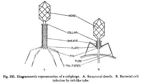
The bacteriophages or bacterial viruses or simply phases are widely distributed in nature. They are ultramicroscopic, but some (Vaccinia) are larger than small bacteria. A bacteriophage has a hexagonal to polyhedral head and a rigid tail which are almost same in length. The tail has a central core surrounded by a contractile sheath. The tail serves as an adsorption organ.
The portion of the head closer to the head has a projected structure called collar and the tail is terminated by six plates each of which again has contractile fibres (Fig. 335).
Bacteriophages occur in six morphological types (Fig. 342):
i. A head with a rigid tail having contractile sheath and tail fibres (Fig. 342A).
ii. A head with a flexible tail without contractile sheath. It may or may not have terminal appendages (Fig. 342B).
iii. A head with a short tail, the tail is without contractile apparatus and may or may not have appendages (Fig. 342G).
iv. A head with large capsomeres (individual protein subunits of capsids) at each apex of the hexagon; it has no tail (Fig. 342D).
v. A simple head without capsomeres and a tail (Fig. 342E).
vi. A long flexible tail without a head (Fig. 342F).
A bacteriophage has nucleic acid (DNA or RNA) and a protein coat. But few phages contain both DNA and RNA. Again some phages contain lipid and ribosome In most of the DNA phages the nucleic acid of the virion is double-stranded, although in a few cases it is single-stranded.
The RNA phages have single-stranded RNA in their virions. In all kinds of phages the nucleic acid is contained within the head. Bacteriophages are easily isolated and cultivated on young, actively growing cultures of bacteria in broth or on agar plates. The best and the most usual source of bacteriophage is coliphage—phage pathogenic for Escherichia coli cultures.
There are several steps of bacteriophage infection which ultimately leads to bacteriophage multiplication (Fig. 336). These are: adsorption, penetration, replication of DNA, maturation, and release. When a phage particle comes in contact with a susceptible strain of E. coli, fibres at the end of the phage tail are the adsorption sites of the phage that bind to specific receptors on the bacterial cell wall (Fig. 335B).
Following adsorption, an enzyme (phage lysozyme) located in the phage tail degrades a small portion of the bacterial cell wall. The tail sheath of the phage then contracts driving the core of the tube into the cell injecting the DNA much as a syringe injects a vaccine (Fig. 335B).
The protein coat of the phage remains outside the cell. Within minutes after penetration of phage DNA into the host cell, all transcription of RNA from the host chromosome ceases.
The host DNA is degraded. All RNA subsequently synthesized is in RNA transcribed from the phage DNA.
By this mechanism the phage subverts all metabolism of the bacterial cell to its own purpose – the synthesis of more phage. The host enzymes supply energy for phage replication through the breakdown of glucose, synthesize the subunits of protein and nucleic acid of replicating phage- and even participate in the synthesis of phage nucleic acid and phage coat protein’
For the active replication of nucleic acid and the synthesis of viral proteins viruses require cellular ATP, ribosomes, transfer RNA, enzymes, and certain biosynthetic processes. In addition, the phage DNA codes for enzymes concerned with the assembly of the phage protein capsid.
During the replication of any virus, the viral protein and nucleic acid components develop separately from each other. During maturation of phage, assembly of phage protein and phage DNA takes place independently; so also the head and tail by stepwise processes.
Once the head is formed, it is packed with phage DNA, after which the tail is attached, a new phage is assembled. The foregoing processes are repeated resulting in the formation of large number of new phages. During the latter stages of infection period, another phage-induced enzyme, coded for the phage DNA, makes its appearance.
This is the phage lysozyme that digests the host cell wall from within, resulting in the host cell lysis and release of the new phages (Fig. 336).
From the standpoint of infectivity and host relationships two types of bacteriophages are recognized: lytic or virulent and temperate (lysogenic) or avirulent. When lytic phages infect bacterial cells large number of new phages are produced. They are released bursting bacterial cells. This is called a lytic cycle. But in the temperate type, a state of lysogeny exists, and the infection may not be apparent.
The phage DNA is not reproduced by the host but is transmitted genetically from one host to others of the next generation. Hence in lysogeny, the temperate phage DNA instead of usurping the functions of the host bacterial cell’s genes, is incorporated into the host’s DNA and becomes gene in the bacterial chromosome as a pro-phage.
There are three possible ways of inheritance of bacteriophage DNA:
(i) The phage chromosome inserts itself into the host chromosome, after which it can be passively replicated and distributed at cell division as part of the host DNA;
(ii) The phage chromosome establishes itself independently, it replicates and is distributed in daughter host cells like the host chromosome keeping its separate entity; and
(iii) The phage chromosome replicates separately from the host chromosome, but the distribution of phage chromosome takes place in the daughter cells only when the number of newly replicated chromosome is large.
Actinophages:
Particular viruses attack actinomycetes and have been found in Actinomyces bovis and Nocardia farcinica. Such viruses are—actinophages—are abundant in the soil. Some possess tail and in others there is no tail at all.
Bacteriocins:
Both Gram-positive and Gram-negative bacteria produce bacteriocidal substances known as bacteriocins. The formation of bacteriocins is due to genetic determinants, the bacteriocinogens which are not integrated into the bacterial chromosome and are thus plasmids. Bacteriocins have relationship with phages and are considered as products of defective phage genomes.
The initiation of bacteriocin synthesis kills the produce cell. Bacteriocins take their specific names from the organisms producing them, e.g., colicins from Escherichia coli, pyocins from Pseudomonas aeruginosa, megacins from Bacillus megaterium.
Cyanophages:
These are viral agents that attack a wide range of blue-green algae. They were first discovered by Safferman and Morris in the year 1963. They are very similar to bacteriophages both in structure and infection cycle. Gyanophages are named according to their known hosts.
For example LPP-.1 is specific on hosts: Lyngbya, Phormidium, Plectonema. The nucleic acid of cyanophages are double-stranded DNAs.
Mycophages (Mycoviruses):
These are viruses attacking fungi. Mycophages were first discovered in Agaricus bisporus in 1957 by Sinden. Subsequendy in 1960 mycophages were discovered in Penicillium chrysogenum, P. cyaneofulvum, P. funiculosum, and P. stoloniferum. The viral particles of the mycophages are polyhedral or spherical with diameters of 33 to 41 nm.
They all contain double-stranded RNA. Young apical regions of hyphae are generally free of virus particles; older regions contain many particles enclosed in vesicles.
Viruses enter plants either through damaged cells including leaf hairs, or are introduced into the plant by vectors. Slight damage is necessary for successful establishment of the virus within the cell. This involves removal of the proteinaceous coat of the particle and liberation of nucleic acid within host cell.
After infection there is usually replication of viruses in the host cell. There is some evidence that virus protein and nucleic acid are both formed within the nucleus, synthesis of the virus particles being initiated in the nucleolus and perhaps completed in the cytoplasm after extrusion from the nucleus.
Some viruses are probably incomplete viruses which cannot replicate without the help of second virus (the ‘activator’), but others appear to be complete.
When a virus particle infects a host cell, the particle multiplies in that cell and leads to the expression of disease symptom involving a multitude of cells either in a localized area around the point of entry of the virus particle—localized infection, or throughout the host plant and in areas far removed from the point of infection— systemic infection.
Translocation of Viruses is thus of two types:
Short-distance translocation which involves cell-to-cell movement causing localized symptoms and long distance translocation which involves phloem and xylem elements and incites systemic symptoms.
During long-distance translocation, viruses multiply en route and do not cause any infection. Cell-to-cell movement is through plasmodesmata and by cytoplasmic streaming.
But in meristematic tissue viruses are distributed during cell division. Phloem and xylem are the only tissues that act as channels for long-distance spreading of viruses.
Viruses reach phloem in two different ways:
By cell-to-cell movement through plasmodesmata or by direct deposition by an insect vector. Viruses are passively carried in phloem in the liquid stream carrying photosynthates.
The same virus may travel over long distances in phloem, can also travel in xylem.
Viruses fall in four categories:
Parenchyma-restricted, phloem-restricted, xylem-restricted, and not restricted, to any particular tissues but are widely present in tissues of all types.
In general, transportation of viruses is more rapid in the direction of nutrient utilization or storage.
But movement in the reverse direction can occur, although virus movement from shoot to root is generally more rapid than from root to shoot. The rate of spread of viruses is greater in young than in old tissues and the movement is faster in higher than at low temperatures because of the fast streaming of cytoplasm at higher temperatures.
Nutrition which promotes rapid vegetative growth of the host plant favours the virus multiplication.
Note # 14. Distinction between Virus and Cellular Organism:
Distinction between Viruses and Cellular Organisms may be Summarized as Follows:
(i) The only unit of viral structure, the virion has quite different properties from the unit of structure of an organism, the cell.
(ii) The virion contains only one kind of nucleic acid, either ribo- or deoxyribonucleic acid. The cell always contains both.
(iii) The organic constituents in the virion are nucleic acid and protein. The cell contains, in addition to nucleic acids and proteins, many other organic constituents.
(iv) Although the virion may contain one or a few enzymes, its enzymatic complement is insufficient to reproduce another virion. The cell always contains a very elaborate complement of enzymes suitable for the reproduction of the cell.
(v) The virion never arises directly from a pre-existing virion. The cell always arises directly from a pre-existing cell.
(vi) The virus is always reproduced exclusively from its genetic material. The cell is reproduced from the integrated sum of all its constituent parts.
(vii) Growth of the virus involves the independent synthesis of its nucleic acid and protein, which are assembled into organized structures after the completion of their synthesis. Whereas growth of the cell consists of the increase in the amount of all its constituent parts, during which the individuality of the whole is continuously maintained. Cellular growth culminates in an increase in cell number by a process of fission.


