ADVERTISEMENTS:
In this article we will discuss about:- 1. Structure of Phage Mu 2. Genetic Map of Phage Mu 3. Life Cycle of Phage Mu 4. The Targets of Transposase.
Phage Mu is also known as ‘bacteriophage Mu’. Since it infects the members of enterobacteria, it is also called ‘enterobacteria phage Mu’. It belongs to the family Myoviridae of the order Caudovirales and Group II dsDNA viruses. It is a temperate and transposable phage which causes transposition of genes at the time of its multiplication cycle.
Mu phages have a broad host range e.g. one strain of each off. coli, Citrobacter freundii, Serratia marcescens, Erwinia carotovora, Erwinia uredovora, and Erwinia amylovora which adsorb with the host range corresponding to the (-) orientation of the G segment. K. pneumoniae is naturally resistant to Mu, Mu-sensitive mutants of K. pneumoniae have been isolated. Salmonella typhimurium is also resistant to Mu.
Structure of Phage Mu:
ADVERTISEMENTS:
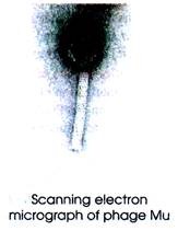
Phage Mu consists of an icosahedral head and a tail with helical symmetry and devoid of any envelope. The basic icosahedral symmetry of the head is derived by adding rows of capsomers to the capsid (T=13). The head is pro-late in shape and has a diameter of 54 nm. Capsids appear hexagonal in outline. Capsid consists of 152 capsomers.
The tail is a long, rigid and thick contractile tube with cross- bands and measuring 183 x 16-20 nm. Tail consists of a distinct axial canal. Tail has a base plate and fibers.
Sheath is composed of stacked rings. During contraction, sheath subunits of contractile tails slide over each other and the sheath becomes shorter and thicker. Upon contraction the sheath becomes 60-90 nm long. There are six long and terminal tail fibers attached to a large base plate.
Genetic Map of Phage Mu:
The genome is not segmented and contains a single molecule of linear dsDNA. The genome is completely sequenced which is 37,611 bp long. The genome has a guanine + cytosine content of 35 %. The genome contains unusual bases such as 5-hydroxy-methyl cytosine. Double stranded DNA is circularly permuted. The genome has terminally redundant sequences. Fig. 18.19 shows s simplified genetic map of the Mu.
ADVERTISEMENTS:
The E. coli temperate phage Mu lysogenizes its host by stable integration of its DNA at one of many possible sites in the host chromosome. The lytic cycle is initiated either by infection or by heat inactivation of a temperature-sensitive re-presser. Most of the genes of phage Mu have been characterized genetically.
The transcription rate revealed three defined phases of Mu transcription: early (0 to 9 min), intermediate (between 9 and the interval 14 to 17 min) and late (from the interval 14 to 17 min onward). Mu RNA is synthesized in two phases (early and late). Late and early mRNAs are mostly transcribed from one DNA strand from left to right (Fig. 18.19).
The leftmost transcription unit consists of the re-presser gene C which is transcribed to the left from a promoter initiating transcription at 1,063 to 1,064 bp or 1,066 bp from the left end of the Mu genome. A second, weaker promoter which initiates transcription at 884 to 885 bp is also present.
The early operon lies to the right of C, which consists of the genes involved in replication, integration, transposition, control of early gene expression and inhibition of cell division. Transcription of the early operon is initiated from a single promoter (Pe) at 1,028 bp and terminated approximately 8,700 bp from the left end.
Synthesis of the early transcript begins immediately after induction of a temperature-sensitive prophage is inhibited from the 4th min by her gene product (gpner), and after a reactivation phase (from the 9th min) it remains at low levels until the end of the lytic cycle.
The next transcription unit covers the C gene, which is located approximately 10 kb from the left end and is transcribed from the interval 15 to 20 min onward independently of the early operon.
Life Cycle of Phage Mu:
The transposable and temperate phage Mu reproduces by transposition. The mechanism of host recognition and penetration are the same as described earlier for the other viruses. Mu gets adsorbed on the surface of sensitive bacterial host and injects its linear DNA into the host cell (Fig. 18.20). The Mu DNA is inserted into the recipient genome through a non-replicative ‘cut and paste’ mechanism.
Fig. 18.20 : Life cycle of phage Mu.
After infection of the host E. coli, phage Mu either enters the lytic life cycle or remains as a repressed and integrated prophage. The re-presser protein Rep decides to initiate either lysis or lysogeny. Rep accumulates inside the cell where the prophage is completely de-repressed.
This accumulation is ClpX-dependent. Rep is a target of two protease systems. Inactivation of the clpP Ion gene results in stabilization of Rep. Such a reaction scheme explains that de-repression is correlated with high re-presser concentration. Under all conditions of phage induction the repressor is sequestered in a non-active form.
Wild type Mu lysogens are very stable. They cannot be induced by UV or other DNA damaging agents. But simply by transferring the lysogen to 42°C, one can induce the derivatives of Mu with a temperature sensitive repressor [e.g. Mu c(Ts)]. Upon inactivation of repressor, the A and B proteins are expressed.
ADVERTISEMENTS:
Then Mu transposes to 50, – 100 new sites on the chromosome by a replicative mechanism (Fig. 8.21). In the mean time, the phage genes express the late gene products i. e. the late proteins, such as phage heads, tails, lysis proteins, etc. The dsDNA of phage is cut in host sequences located about 100 bp from the left end of Mu, and the DNA is packaged by a headful mechanism.
The Mu DNA is about 37 kb long but about 39 kb long DNA is packaged into each head (Fig. 18.21). About 50-150 bp of host DNA is present at the left end of phage genome and a variable amount of host DNA on the right end. If part of the Mu genome is deleted, the length of host DNA on this end increases.
About 2 kb long host DNA is present on the right end of packaged wild type Mu DNA. The host DNA on the ends of Mu is unique in every different phage head. When phage assembly is completed, the host lysis occurs resulting in the release of about 50-100 phage particles per cell. The progeny phage restarts the life cycle in sensitive host.
The Targets of Transposase of Phage Mu:
ADVERTISEMENTS:
The two ends of Mu designated as atiL and atiR. (also called MuL and MuR) are involved in transposition (Fig. 18. 22). The atfL an attR of Mu genome represent the left end ancTngnt end, respectively. The genes A and B gene encode transposase. The A protein is involved for all events of transposition, but the B protein is required only for replicative transposition.
However, the c gene product can repress the expression of the transposase genes. The phage transposase recognizes and binds the enhancer sequence. Each att site is formed by three binding sites for Mu a replicative transposition. These structures are also recognized by phage repressor which shows overlapping binding specificity with the transposase. 


