ADVERTISEMENTS:
In this article we will discuss about the receptor organs of toad. This also help you to draw the structure and diagram of receptor organs of toad.
Associated with the nervous system are certain organs of special senses called receptor organs. Each receptor is affected by a particular kind of stimulus which is transmitted by sensory nerves to the central nervous system and produces its own peculiar kind of sensation.
The receptor organs themselves do not perceive anything but merely serve as a gateway to the central nervous system. The central nervous system is the seat of all sensation.
ADVERTISEMENTS:
There are receptors for:
(1) Touch,
(2) Pain,
(3) Heat,
ADVERTISEMENTS:
(4) Cold,
(5) Smell,
(6) Taste,
(7) Sight,
(8) Hearing, and
(9) Balancing (equilibration).
Our knowledge of the receptor organs of the toad is based upon a comprehensive knowledge of these structures in man. The receptors for touch, pain, heat, and cold are scattered microscopic structures situated beneath the epidermis of the skin.
They, therefore, are referred to as the cutaneous receptors. Some cutaneous receptors consist merely of free endings of sensory nerves in the deeper layers of the epidermis (Fig. 41, A). In others the nerve endings may be en-sheathed in special connective tissue capsules of various shapes (Fig. 41, C).
Receptors for smell are located in the nasal passages which open into exterior by the external nares and communicate with the buccal cavity by the internal nares. Each nasal passage is lined by mucous membrane containing peculiar sensory cells called olfactory cells and slender supporting cells (Fig. 41, E).
ADVERTISEMENTS:
The olfactory cells are the actual receptors of smell. The free ends of these are brush-like in appearance, each cell bearing several delicate hairs at its tip.
The other end of the olfactory cell is connected with a fibre of the olfactory nerve. The brush-like free ends of the olfactory cells are stimulated by chemical substances, and the impulse is conveyed to the brain via the olfactory nerve, but substances must be in solution before they can be detected.
Receptors for taste are microscopic structures distributed upon the mucous membrane of the mouth and tongue. These are called taste buds. A taste bud (Fig. 41, D) is composed of two kinds of cells: sensory cells or taste cells and supporting cells.
ADVERTISEMENTS:
Each taste cell bears a minute bristle at its tip, and its other end is connected with a sensory fibre of the glossopharyngeal nerve. Taste buds are particularly numerous on the tongue, in association with small elevations on the surface epithelium, the papillae.
The taste cells and the olfactory cells are recognised as chemo- receptors because they are stimulated by chemical substances in solution. Over and above the minute and scattered receptors just described there are two pairs of compact structures, the eyes for vision, and the ears for hearing and balancing.
Eye of Toad:
The eye is the organ of vision. Each eyeball is lodged in its own orbit. It can be turned and rotated within limits by the action of six extrinsic muscles which are attached on its outer surface.
These are:
ADVERTISEMENTS:
(1) Superior rectus,
(2) Inferior rectus,
(3) External rectus,
(4) Internal rectus,
ADVERTISEMENTS:
(5) Superior oblique, and
(6) Inferior oblique muscles.
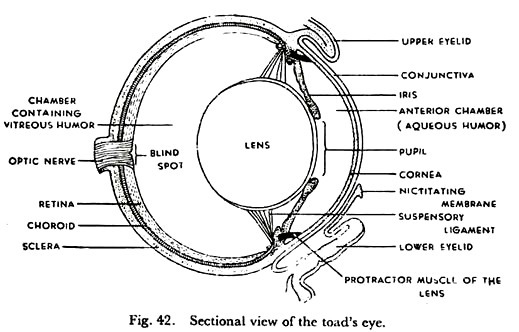
The eye can be thrust out by a muscle called levator bulbi and withdrawn into the socket by another muscle called retractor bulbi. The eye is a hollow spherical box fitted with a rounded lens in front and a light-sensitive screen behind. It is light-proof and works like a photographic camera. The eyeball is composed of three coats, which surround the transparent parts for transmitting light upon the sensory screen.
The outermost coat is thick and tough.
It consists of two portions:
(i) A transparent anterior portion, the cornea, and
ADVERTISEMENTS:
(ii) An opaque posterior portion, the sclera, the anterior part of which is commonly referred to as the ‘white’ of the eye. The cornea is practically circular in outline; it permits entry of light into the eyeball.
A thin membrane, the conjunctiva, which is continuous with the inner surface of the eyelids, covers the outer surface of the cornea. The sclera is composed of fibrous tissue and cartilage; it protects the soft parts and gives shape to the eyeball.
The middle coat is the choroid. It is a highly vascular black membrane forming an internal lining for the sclera. Anteriorly, the choroid turns sharply inward from the junction of the sclera with the cornea. This part of the choroid is modified to form a thin, circular, pigmented disc, the iris.
There is an opening in the centre of the iris which is spoken of as the pupil of the eye. The iris contains radial and circular muscle fibres which help the pupil to dilate or contract. Thus, a regulated amount of light is permitted to enter the eye through the pupil. Barring this, the eyeball is perfectly light-proof.
The innermost coat is the retina. It lies against the choroid and forms the light-sensitive screen of the eye. The retina contains peculiar cells, called rods and cones, which are the receptors of the visual impulse. The rods are sensitive to dim light, and the cones to bright light and colours. The reds and cones are connected to the fibres of the optic nerve.
ADVERTISEMENTS:
The crystalline lens is nearly rounded in shape. It lies just behind the pupil, and is held in position by the suspensory ligament. The cavity of the eye between the lens and the cornea is known as the anterior chamber. It is filled with a clear, watery fluid, the aqueous humor. The space between the black of the lens and the retina contains a transparent jelly-like substance, the vitreous humor.
The toad cannot change the shape of its lens. Two small muscles, one dorsal and the other ventral, join the suspensory ligament of the lens with the inner surface of the cornea. These are the protractor lentis muscles; when contracted, they draw the lens closer to the cornea.
Vision:
Light rays, entering the eye at the cornea, pass through the pupil, and being refracted by the crystalline lens, converge to a small part of the retina. A reduced and inverted image is caught on the retina in the same way as in a photographic camera.
Focusing of the image, that is accommodation, is effected by drawing the lens forward or backward through the protractor lends muscles. The retina contains rods and cones which are the actual photoreceptors. The reduced and inverted image caught by the rods and cones is transmitted to the brain via the optic nerves. During transmission the inverted image is mysteriously transformed into a corrected one.
Toad’s eyes are placed laterally and each eye covers a different visual field. Vision, therefore, is monocular. An animal which can focus both eyes on the same object is said to enjoy binocular vision.
Birds of prey, such as owls, have binocular vision which enables them to judge distance accurately when swooping down upon an object. Man too has binocular vision. Animals with laterally displaced eyes have monocular vision; they enjoy a wider visual field but are unable to judge distance.
Ear of Toad:
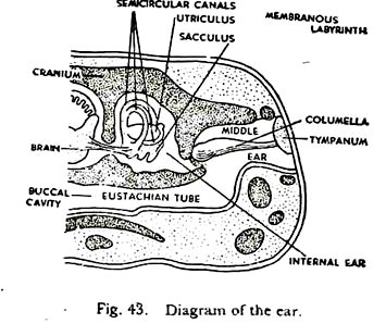
The ear serves as the organ of hearing and balancing. The toad’s ear is composed of three parts, the external ear represented by the tympanum or eardrum, the middle ear or tympanic cavity, and the internal ear including the membranous labyrinth. The ear is represented externally by the tympanum, one on each side of the head. This is a tightly stretched membrane covering the middle ear.
The middle ear is the funnel-shaped cavity beneath the tympanum. It is connected with the buccal cavity by the eustachian tube which acts as a safety valve by equalizing atmospheric pressure on the two sides of the eardrum. A small rod-shaped bone, the columella, extends across the middle ear, from the centre of the eardrum. It is joined to a membranous partition separating the middle ear from the internal ear.
The internal ear, including the membranous, labyrinth, is enclosed within the auditory capsule of the skull. The labyrinth floats in a fluid called perilymph which fills the cavity of the auditory capsule. The labyrinth itself is filled with another fluid called endolymph. The auditory capsule is sealed off from the middle ear by a membranous partition which is connected to the eardrum by the columella.
The membranous labyrinth consists mainly of two chambers, one placed above the other. The upper chamber is known as the utriculus, and the lower chamber as the sacculus. The two chambers are connected by a narrow canal.
The utriculus gives out three narrow tubes, the semicircular canals, all opening into the utriculus at both ends. Each canal bears an enlargement or ampulla at only one end. One of the canal is horizontal in position, but the other two are vertical and at right angles to one another. Thus the semicircular canals lie in three different planes. The sacculus is produced into a short lagena.
There are patches of sensory receptors in the inner wall of the membranous labyrinth. Each patch consists of supporting cells, and sensory cells with long hair-like bristles extending into the endolymph. The receptors are found within the ampullae of the semicircular canals, and within the utriculus, sacculus as well as lagena. Each receptor cell is connected with a fibre from-the auditory nerve.
Hearing:
Sound waves impinge directly upon the eardrum. Vibrations of the eardrum are conveyed to the internal ear by the columella. These vibrations are transmitted to the endolymph and stimulate the sensory cells.
The receptors receive the impulses by their bristles and transmit the same to the brain via the auditory nerves. The impulses are translated in the brain as sound. Sound impressions are recorded by the sensory receptors within the sacculus and lagena.
Balancing:
The semicircular canals are responsible for the maintenance of equilibrium. There are calcareous particles within the canals which impinge upon the bristles of the sensory receptors as soon as the animal loses its balance. Moreover, being arranged in three different planes and at right angles to one another, the canals can detect changes in the direction of the centre of gravity, when the toad is moving.


