ADVERTISEMENTS:
In this article we will discuss about the excretory system in toad with diagram. Also learn about: 1. Structure of Kidney in Toad 2. Mechanism of Urine Formation in Toad.
Various substances are manufactured within a toad’s body during its routine metabolic activities. Some of these are of no use and may cause harm if they are retained for a long time. Only these end products of metabolism are the waste products. Faeces is not a waste product, because it is not produced by metabolism in the tissues.
The principal waste products are urea, uric acid, carbon dioxide, excess of water and mineral salts. These are collected from the tissues by the blood and transported to the excretory organs, which promptly eliminate them from the toad’s body. The process by which metabolic waste products are separated and removed from the body is known as excretion.
ADVERTISEMENTS:
Excretory organs in Toad:
Of the waste products, CO2 and H2O are formed in all the tissues as by-products of internal respiration. These are expelled by the lungs and skin. Urea and uric acid are nitrogenous waste products.
Urea is formed in the liver by the decomposition of amino acids. The amino acids are first broken down to ammonia which is then converted into urea by the liver cells. Urea, uric acid, excess of water, and mineral salts are excreted in the form of urine.
The organs concerned with the removal of the urine are:
ADVERTISEMENTS:
(1) The kidneys,
(2) The ureters,
(3) The urinary bladder,
(4) The cloaca, and
(5) The vent.
The kidneys are a pair of flattened elongated bodies, reddish- brown in colour, attached to the dorsal wall of the body cavity, one on each side of the vertebral column. They lie behind and are completely covered by the peritoneum, that is, they are retro-peritoneal in position. The outer border of each kidney is somewhat convex, and the edges are indented by notches.
The toad’s kidney is recognised as the Wolffian body or mesonephros (mesos = middle; nephros = kidney), because it is more highly developed than the pronephros or the primitive kidney, but not as highly developed as the metanephros which is found in higher animals. The ureters or wolffian ducts are thin-walled whitish tubes found on the outer border of the kidneys.
The two ureters, one from each kidney, run backward. They join to form a single median tube at the posterior end of the trunk, and this tube finally opens into the dorsal wall of the cloaca. Arising opposite, from the ventral wall of the cloaca, there is a thin-walled bilobed sac called urinary bladder. Its narrow opening is guarded by a sphincter muscle. The cloaca communicates with the exterior by the vent.
Structure of the Kidney in Toad:
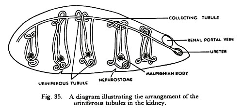
A kidney is composed of a large number of minute tubes called uriniferous tubules. Each tubule begins blindly in a two-walled cup with a narrow mouth. The cup is known as Bowman’s capsule.
ADVERTISEMENTS:
A small afferent vessel from the renal artery enters the capsule and breaks up into a bunch of capillaries which fits into the cup. This is the glomerulus (glomus = a ball). An efferent vessel emerges at the other end of the glomerulus and drains into the renal vein. A capsule with its glomerulus is recognised as the Malpighian body.
The other end of Bowman’s capsule is connected with the tubule proper, which extends as a much convoluted narrow channel. Ultimately the tubules join with one another to form larger tubes called collecting tubes and these in turn open into the ureter.
The convoluted part of each tubule is surrounded by capillary networks derived from the renal portal vein. On the ventral surface of the kidney, there are numerous microscopic openings called nephrostomes (nephrons= kidney; stoma = opening) which drain into the renal veins.
Mechanism of Urine Formation in Toad:
ADVERTISEMENTS:
Blood loaded with waste products is brought to the kidney by the renal artery and the renal portal vein. While passing through the glomerulus, nitrogenous waste products along with water and other substances are filtered out of the blood into the capsular space.
The filtrate enters the convoluted part of the tubule where useful substances are reabsorbed and sent back into the circulating blood. Oxygen that was brought by the renal artery is used up by the cells of the tubules, so that deoxygenated blood devoid of waste products now flows out of the kidney by the renal veins.
Probably the nephrostomes drain away excess of water and other wastes from the body cavity. The fluid which runs into the ureters via the uriniferous tubules is the urine. It drips into the cloaca continuously and gravitates into the urinary bladder for temporary storage. When filled with urine, the bladder contracts and the content is ejected through the vent.


