ADVERTISEMENTS:
The below mentioned article provides an outline of internal structure of stem of both dicotyledons and monocotyledons type.
The embryo develops into a plant with root-stem axis and the appendages. In fact, three important organs of a plant are the stem, the leaves and the root.
The continuity of the tissues, and particularly the vascular system, has been discussed in the preceding chapter. Apical meristems occurring at the tips of the axis, the stem tip and root tip divide and produce new cells.
ADVERTISEMENTS:
In course of time these tissues become permanent and the fundamental body of the plant is laid down. The tissues deriving their origin from the apical meristems are known as primary permanent tissues and body made of primary permanent tissues is designated as primary body.
The Stem:
In lowest vascular plants, as in Psilotales, a sharp demarcation between the stem and leaves is hardly possible. The boundary between the stem and leaves in the seed plants is, in fact, uncertain.
Both of them derive their origin from the same apical meristem—the shoot apex, and they are to a great extent interdependent in the process of growth and differentiation. For these reasons some workers prefer to put the stem and its foliar appendages (leaves) under the broader concept of shoot. However, it is a matter of practice to take up the three important vegetative organs separately.
An outline of the internal structure of the stems is given here. In view of the fact that wide diversities exist as regards the nature of the plants, a few common dicotyledons and monocotyledons have been selected for the study of anatomical structures.
Dicotyledons:
1. Young Stem of Sunflower:
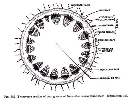
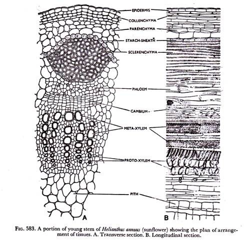
Transverse and longitudinal sections through the internode of a young sunflower stem (Helianthus annuus of family Compositae) should be taken and stained suitably for the study of internal structures. It would exhibit the following plan of arrangement of tissues from the periphery to the centre of the organ (Figs. 582 & 583).
I. Epidermis:
It is the outermost uniseriate zone, consisting of tabular cells attached end on end without leaving intercellular spaces. The cells are living with vacuolate protoplast. The chloroplasts are usually absent. The outer walls of the epidermal cells are cuticularised for checking loss of water. Stomata may be present here and there. A large number of multicellular hairy outgrowths develop on the epidermis.
II. Cortex:
It occurs next to epidermis and represents the extrastelar ground tissues. In sunflower stem cortex is differentiated into three zones. Just internal to epidermis there are a few layers of collenchyma, usually angular ones, forming a continuous band. This collenchymatous band meant for giving mechanical support to the growing stem, is called hypodermis.
Next to hypodermis a few layers of thin-walled parenchyma occur which have conspicuous intercellular spaces. A few glands, each having a hollow cavity surrounded by small densely protoplasmic epithelial cells, are present here and there in the parenchymatous portion.
Some authors used the term ‘general cortex’ for this part (parenchymatous part) of cortex; but that is rather misleading and hence has been abandoned. The last layer of cortex is a wavy band made of compactly-set barrel-shaped parenchyma cells. Starch grains are abundantly present in these cells.
This band is referred to as starch sheath. It is morphologically homologous to the endodermis commonly found in the roots. The starch sheath is the limiting layer of the extrastelar ground tissue, cortex.
ADVERTISEMENTS:
III. Stele:
All the tissues occurring internal to the starch sheath constitute the stele. It is a dissected siphonostele, consisting of vascular bundles arranged in form of a ring, and intrastelar ground tissues.
Vascular bundles:
The vascular bundles are typically collateral and open ones, with xylem and phloem on the same radius, xylem being internal and phloem external. A strip of lateral meristem, cambium, is present between xylem and phloem.
ADVERTISEMENTS:
The xylem is characteristically endarch, showing centrifugal mode of differentiation from the procambium. The protoxylem elements, tracheids and tracheae, with smaller cavities and annular and spiral thickenings occur towards the centre; and the metaxylem elements with pitted and other types of thickenings are present towards the circumference.
Xylem parenchyma cells are smaller than other parenchyma cells of the stele. The phloem is composed of sieve tubes, companion cells and phloem parenchyma. The cambium of the vascular bundle, what is called fascicular (fascicle =bundle), appears to be composed of two or three layers of fusiform cells, which look rectangular in cross- section. This tissue is responsible for growth in thickness.
Intrastelar ground tissues:
Against every vascular bundle there is a patch of sclerenchyma forming something like a cap. This has been called bundle cap. The patches of sclerenchyma and intervening parenchyma cells were considered to constitute the peri-cycle, the outermost portion of the stele.
ADVERTISEMENTS:
But modern workers are of opinion that the fibres associated with the vascular bundle in sunflower stem, and as a matter of fact, in many other plants, belong to phloem. So the term hard bast has also been attributed to this patch. A complete section (Fig. 582) shows that the bundles are located towards the periphery and the large central portion remains occupied by parenchyma cells with profuse intercellular spaces. This part is known as pith or medulla.
Continuations of the pith radiate, so to say, through the regions between the vascular bundles, the so-called interfascicular regions, and form the primary medullary rays. These are also composed of thin-walled parenchyma cells.
2. Young Stem of Cucurbita:
It (Cucurbita maxima of family Cucurbitaceae) is a weak plant climbing with the help of the tendrils. Transverse and longitudinal sections of the stem are taken, stained suitably and studied under the microscope. It is wavy in outline and thus shows distinct ridges and furrows (Fig. 584). The vascular bundles occur in two rings.
One ring usually consisting of five smaller vascular bundles is present against the ridges. These are leaf- trace bundles. Another ring of same number of bundles occurs against the furrows. The central part of the stem is hollow due to disintegration of pith.
ADVERTISEMENTS:
The Cucurbita stem shows the following plan of arrangement of tissues (Fig. 585):
I. Epidermis:
It is as usual the single-layered zone made of compactly-set tabular cells. The cells are living with vacuolate protoplast and cuticularised outer walls. A large, number of multicellular hairs develops from the epidermis. Stomata may be present in the young stem.
II. Cortex:
It is differentiated into three zones. The hypodermis is made of collenchyma cells forming conspicuous patches particularly at the ridges. Collenchyma cells do not form a continuous band, the number of layers become reduced towards the furrows and are interrupted by some parenchyma cells containing abundant chloroplasts.
These photosynthetic cells actually occur just beneath the epidermis where stomata are present. Chloroplasts may be present in the collenchyma cells as well. Next to hypodermis there are a few layers of parenchyma, larger in size and having lesser number of chloroplasts. The last layer of cortex is the starch sheath, a layer of barrel-shaped cells, larger than other cells of cortex. These cells contain abundant starch grains. The cortex naturally is more wide at the regions of ridges than at the furrows.
ADVERTISEMENTS:
III. Stele:
The central cylinder or stele containing the vascular bundles and intrastelar ground tissues remains enveloped by the cortex.
A few layers of sclerenchyma occur on the inner side of the starch sheath in form of a continuous band. Some parenchyma cells are usually present between the sclerenchymatous band and the vascular bundles. These tissues together form the pericycle. Pith, as already stated, disintegrates rather early in Cucurbita stem. The cells occurring in between the vascular bundles are referred to as internal parenchyma.
The vascular bundles are typically bicollateral, having two patches of phloem and two strips of cambium on either side of Xylem. So the sequence of tissues in the vascular bundle is outer phloem, outer cambium, xylem, inner cambium and inner phloem. The bundles, particularly those occurring against the furrows are fairly large and thus are quite suitable for study of detailed structure.
The outer phloem is more massive than the inner one and is composed of sieve tubes, companion cells and phloem parenchyma. The sieve tubes are quite conspicuous and sieve plates are noticed here and there. The Xylem has the characteristic elements. The metaxylem vessels are of very large size, what is characteristic of most climbers.
ADVERTISEMENTS:
They have pitted thickenings. A good number of proto- Xylem vessels with comparatively narrower cavities occur towards the centre. They have usually spiral and annular thickenings. Tracheids and fibres are rather few or lacking, whereas xylem parenchyma cells are abundant. The vessels do not remain arranged in radial rows. The cambium occurs on either side of xylem. The outer cambium is many-layered and is made of cells more or less rectangular in cross-section. The inner cambium is much smaller and is usually curved.
As formation of secondary tissues is confined to individual bundles in climbing plants like Cucurbita, the vascular bundles may contain some secondary tissues as well, in addition to the primary tissues. In that case outer phloem may be made of primary phloem occurring towards pericycle and secondary phloem against the cambium.
The elements are practically same, so that the two are hardly distinguishable, only the secondary phloem has larger elements. The internal phloem is solely primary. Secondary xylem similarly may occur internal to outer cambium. The elements, particularly the vessels, are much larger.
3. Young Stem of Leonurus:
A very young stem of Leonurus sibiricus of family Labiatae should be selected, because secondary growth commences unusually early in this plant. Transverse sections are taken and stained suitably for the internal structure. The stem is square in cross-section.
It shows the following plan of arrangement of tissues (Figs. 586 & 587):
I. Epidermis:
It is uniseriate zone composed of tabular cells attached end on end, without intercellular spaces. The cells are living with vacuolate protoplasts. The outer walls are strongly cuticularised. Many multicellular hairs develop on the epidermis. Stomata may be present here and there.
II. Cortex:
Like sunflower stem the cortex is differentiated in to three zones, though it is less massive. Next to epidermis occurs hypodermis composed of collenchyma cells.
These cells aggregate densely at the four corners of the stem, so that these patches serve as diagonally placed I-girders for withstanding flexion. Collenchyma cells extend beyond the corners, but do not form continuous bands. There are a few layers of thin-walled parenchyma just internal to collenchymatous hypodermis.
These cells contain abundant chloroplasts (chlorenchyma) and appear as a green belt under microscope. At the portions where collenchyma is absent, chlorenchyma cells occur next to the epidermis. These are usually the regions where stomata are present on the epidermis. The last layer of cortex is the starch sheath, composed of barrel-shaped compactly-arranged cells with abundant starch grains.
III. Stele:
The central cylinder or stele is limited by the starch sheath, and is made of vascular strands and intrastelar ground tissues. A few layers of sclerenchyma forming a continuous band occur next to the starch sheath. It may be called pericycle or perivascular tissue.
The vascular bundles are disposed more towards the periphery; a large parenchymatous pith is present at the central portion. As secondary growth in thickness is initiated unusually early, the primary medullary rays very soon lose their identity.
The vascular bundles are collateral starch sheath and open. The phloem is external, i.e., it occurs towards the periphery and has the usual elements—sieve tubes, companion cells, and phloem parenchyma. Next to phloem there is a strip of cambium, made of a few layers of fusiform cells appearing more or less rectangular in outline.
Cambium ring is formed rather early. Xylem occurs next. It has the usual tracheary elements—tracheids and tracheae, parenchyma and fibres. Xylem is endarch— protoxylem occurring towards centre and metaxylem towards circumference.
4. Stem of Calotropis:
A young stem of Calotropis procera of family Asclepiadaceae is selected. A transverse section through the internode would show the following plan of arrangement of tissues (Fig. 588).
I. Epidermis:
It is uniseriate—with tabular cells having cuticularised outer walls. Stomata are present here and there. Waxy depositions are quite common.
II. Cortex:
It is differentiated into two or three layers of lacunate collenchyma forming the hypodermis, large parenchyma cells and the starch sheath, made of barrel-shaped compactly-arranged cells with starch grains.
III. Stele:
The central cylinder includes the vascular bundles—compactly arranged with intervening ray cells and intrastelar ground tissues.
The pericycle is quite massive, made of parenchyma (a few layers) having patches of cellulosic fibres here and there. The central portion is occupied by a large parenchymatous pith.
The vascular bundles are made of internal xylem with endarch arrangement, external phloem and cambium ring in between -the two. Presence of patches of internal or intraxylary phloem is a distinctive character.
5. Stem of an Aquatic Dicotyledon:
Enhydra fluctuans of family Compositae is selected. It is an aquatic plant, and naturally the stem is soft with abundant air chambers and scanty mechanical tissues. A transverse section through the internode would show the following plan of arrangement of tissue (Fig. 589).
I. Epidermis:
It is a uniseriate layer consisting of comparatively small thin- balled cells. The outer walls have feeble cuticularisation.
II. Cortex:
It is less massive here than in other terrestrial dicotyledonous stems. The differentiation of the cortex into three zones is noticed. Next to epidermis, just a few layers of parenchyma with comparatively thicker walls occur as a continuous band forming the hypodermis. The absence of peripheral collenchyma is thus a notable feature.
The next zone consists of quite a few layers of parenchyma, larger in size and having abundant spaces and conspicuous air chambers. These are helpful in giving buoyancy to the plants, apart from normal aeration. Chloroplasts are present in the parenchyma cells. The last layer of cortex is as usual the starch sheath—a wavy layer composed of barrel-shaped compactly-arranged cells with starch grains.
III. Stele:
The central cylinder or stele includes the vascular bundles arranged in a ring and intrastelar ground tissues.
Against every vascular bundle there is a patch of sclerenchyma, like sunflower stem, but much smaller here, forming the so-called hard bast or bundle cap. The sclerenchyma patch together with intervening parenchyma has been called pericycle, though doubts prevail as regards the origin of the fibres. The pith disintegrates leaving a large cavity at the central region, so it may be called hollow pith. Some parenchyma cells occur in the interfascicular regions.
The vascular bundles remain arranged in a ring. They are collateral and open. Phloem is rather small and has the usual elements—sieve tubes, companion cells and phloem parenchyma. A strip of cambium occurs between xylem and phloem. Xylem is also small and is made of a few tracheary elements as protoxylem and metaxylem showing endarch arrangement, and a few parenchyma cells.
Monocotyledons:
A few typical monocotyledonous plants are selected for the study of internal structures. Grasses are undoubtedly the most suitable materials for the purpose. Transverse and longitudinal sections are taken, stained suitably and plan of arrangement of Tissues is noted.
1. Stem of Maize:
It (Zea mays of family Graminaceae) also shows the three tissue systems, but the number of vascular bundles is much larger and they remain irregularly scattered in the ground tissue (Figs. 590 & 591). The bundles occurring towards periphery are smaller in size and more crowded, whereas those at the central region are larger in size and more spaced.
All the bundles are common to the stem and leaves, the central ones from the median veins of the leaf blade and small peripheral ones form the marginal bundles. Unlike most of the grasses maize stem is not hollow in the internodal region.
I. Epidermis:
It is a uniseriate zone, composed of small tightly-set cells. The outer walls are cuticularised. The hairy outgrowths, so characteristic of dicotyledons, are usually absent.
II. Cortex:
The cortex and, in fact, the ground tissue system is not well-differentiated here. A few layers of sclerenchyma, rather parenchyma undergoing sclerosis, occur next to epidermis. This band usually continuous, but may be interrupted here and there by internal parenchyma, is referred to as hypodermis. All the ground tissues lying internal to hypodermis are composed by thin-walled parenchyma cells with profuse intercellular spaces. In fact, the bundles remain embedded in parenchymatous ground tissue.
III. Stele:
Due to irregular distribution of the bundles in the ground tissue the semblance of a stele is lost. As already stated, this type is called atactostele. The vascular bundles (Fig. 592) are collateral and closed. Xylem occurs in form of letter Y-—the two metaxylem vessels with wider cavities and pitted thickening at the two arms, and proto- xylem vessels usually one or two, with narrow cavities and spiral or annular thickening at the base.
A few tracheids occur near the protoxylem. In a mature bundle the Xylem elements undergo more and more lignification and the lowest protoxylem disintegrates forming a lacuna or cavity known as protoxylem cavity. Parenchyma cells also remain associated with other elements in xylem. Phloem is rather small. It is composed of only sieve tubes and companion cells, phloem parenchyma being absent.
In a mature bundle the protophloem often gets crushed due to pressure from internal tissues. The whole vascular bundle remains surrounded by sclerenchyma cells, forming what is known as bundle sheath. The sheaths of small peripherally located bundles and the hypodermal clarified cells often coalesce, so that those bundles appear to be embedded in the sheath.
2. Stem of Wheat:
The wheat stem (Triticum vulgar e of family Graminaceae) and, in fact, many grasses are hollow at the internodes with vascular bundles arranged in two rings.
A transverse section (Figs. 593 & 594) taken through the internode shows the following plan of arrangement of tissues:
I. Epidermis:
It is as usual single-layered, composed of compactly-arranged tabular cells with cuticularised outer walls. Stomata may be present here and there.
II. Ground tissues:
A few sclerenchyma cells occur as patches in the subepidermal region. They are not arranged in a continuous band, but are interrupted by chloroplast- containing parenchyma cells here and there. The latter occur at the regions where stomata are present on the epidermis. The remaining portion of the ground tissue is parenchymatous, the cells having conspicuous intercellular spaces. The central portion is hollow.
III. Vascular bundles:
Vascular bundles occur in two series. One ring of small bundles remain associated with subepidermal sclerenchyma, so that they appear to be embedded in sclerenchymatous patches. The second ring placed lower is composed of larger bundles. Like those of maize stem the bundles are collateral and closed ones. They usually remain surrounded by sclerenchymatous bundle sheath. The sheath of bundles of outer circle actually reaches the epidermis.
3. Stem Of an Aquatic Grass:
A transverse section of the stem of an aquatic grass (Leptochloa chinensis of family Graminaceae) is taken and stained suitably.
It would show the following plan of arrangement of the tissues (Fig. 595):
I. Epidermis:
It is as usual uniseriate, composed of a row of compactly-set cells with cuticularised outer walls.
II. Ground tissues:
Internal to epidermis there are two or three layers of sclerenchyma forming the hypodermis. Parenchyma cells with lots of intercellular spaces occur next to hypodermis. Another band of sclerenchyma is present—usually one- or two- layered in the ground tissue. The central portion is hollow.
III. Vascular bundles:
These are fairly large in number and remain scattered in the ground tissue. Those occurring towards periphery are smaller in size. The bundles resemble those of typical monocotyledon, maize. They are collateral and closed ones.
Xylem consists of two metaxylem vessels with pitted thickenings and one or two protoxylem vessels with spiral or annular thickening. Protoxylem cavity is present. Phloem is smaller, composed of sieve tubes and companion cells; phloem parenchyma is absent. The bundle remains surrounded by sclerenchymatous sheath.
4. Scape (Floral Axis) Of Canna:
It (Canna indica of subfamily Gannaceae) also shows monoootyledonous structure (Fig. 596), though a bit different from the maize and wheat stems.
I. Epidermis:
It is the uniseriate protective zone with cuticularised outer walls.
II. Ground tissues:
A few layers of parenchyma occur next to epidermis forming a small cortex. It is immediately followed by a band of chloroplast-containing cells.
This band is referred to as chlorophyllous tissue. Patches of sclerenchyma remain attached to the chlorophyllous tissue here and there. The remaining portion is parenchymatous ground tissue enclosing the scattered vascular bundles.
III. Vascular bundles:
The bundles are collateral and closed. Each of them has small amount of xylem and phloem, the latter with only sieve tubes and companion cells. Sclerenchyma cells do not form a sheath surrounding the whole bundle, as in maize and wheat stems, but remain in two patches on the outer and inner sides of the bundles. The patch on the outer side is larger forming something like a cap and that on the inner side is proportionately much smaller.
I. Stem of Asparagus:
A transverse section through the stem of Asparagus (A. racemosus of family Liliaceae) would show the following plan of arrangement of tissues (Figs. 597-599):
I. Epidermis:
It is single-layered composed of roundish cells with cuticularised outer walls.
II. Ground tissues:
Next to the epidermis there occur a few layers of parenchyma cells containing abundant chloroplasts, so that-they together form something like a green belt. This band made of parenchyma may be called cortex.
The layer internal to chlorophyll-containing parenchyma consists of a single row of compactly-set cells, forming the starch sheath. Next occur quite a few layers of sclerenchyma in form of a band. The 22 [one] central part of the stem is composed of thin-walled parenchyma with distinct intercellular spaces. Vascular bundles remain embedded in this central parenchymatous portion.
III. Vascular bundles:
As in other monocotyledons, bundles remain scattered; the central bundles are larger than the peripheral ones. The latter may touch the sclerenchymatous band. The bundles are collateral and closed.
Here xylem occurs in form of letter V (Fig. 599), the metaxylem vessels forming the arm and protoxylem, the base. Phloem with sieve tubes and companion cells is located between two arms of V. Sclerenchyma cells surrounding the bundle—-the so-called bundle sheath is absent.

