ADVERTISEMENTS:
Let us learn about Dicotyledonous Stems. After reading this article you will learn about: 1. Regions in Dicotyledonous Stems 2. Variations in Dicotyledonous Stem Structure 3. Types.
Regions in Dicotyledonous Stems:
Epidermis:
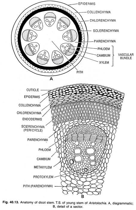
The epidermis consists of a single layer of cells and is the outermost layer of the stem. It contains stomata and produces various types of trichomes. The outer cell walls are greatly thickened and heavily cutinized.
The cells are compactly arranged and do not possess intercellular spaces. In transverse section the cells appear almost rectangular. It serves mainly for restricting the rate of transpiration and for protecting the
Cortex:
The region that lies next to the epidermis is the cortex. The innermost layer of the cortex is the endodermis, known also as the starch-sheath. It consists of a single layer of cells which surrounds the stele and contains numerous starch grains.
ADVERTISEMENTS:
Frequently it is most easily distinguishable from the surrounding tissue by the presence of these starch grains. The part of the cortex situated between the epidermis and the endodermis is generally divided into two regions, an outer zone of collenchyma cells and an inner zone of parenchyma cells.
Collenchyma:
On the inside of the epidermis there is usually a band of collenchyma. The cells of the collenchyma are modified parenchyma cells with cellulose walls thickened at the angles where three or more cells are in contact. The collenchyma resembles parenchyma in being alive and in having a moderate amount of protoplasm.
The chief function of collenchyma cells is to serve as strengthening material in succulent organs which do not develop much woody tissue, or in the soft young parts of woody plants before stronger tissues have been developed.
ADVERTISEMENTS:
They are especially fitted for giving strength to young, growing organs, since the thickened parts of the walls have considerable rigidity, while the thinner parts allow for an exchange of materials between the cells and for the stretching and growth of the cells. The collenchyma cells of stems sometimes contain chloroplasts and carry on photosynthesis.
Parenchyma:
The parenchyma cells are generally regular in shape, have comparatively thin walls, and are not greatly elongated in any direction. They are living cells and contain a moderate amount of protoplasm. When they are exposed to the light they develop chloroplasts and are known as chlorenchyma cells.
Chlorenchyma cells are thus only a special kind of parenchyma cells. The parenchyma cells in the cortex of a stem are near enough to the light so that some or all of them develop chloroplasts and perform photosynthesis. The turgid parenchyma cells frequently help in giving rigidity to an organ.
The function of parenchyma cells is important in succulent stems and in the young parts of the stems and woody plants before strong mechanical tissues have been developed. The parenchyma cells serve for the slow conduction of water and food.
In the case of the cortex of stem it becomes evident that the water which is received by the collenchyma and the epidermis must be conducted through the parenchyma. The parenchyma is the special storage tissue of plants.
Sclerenchyma:
The sclerenchyma cells are found in the cortex of some stems. There are two varieties of these sclerenchyma cells — short or irregularly shaped cells, known as stone cells, and sclerenchyma fibres. Sclerenchyma fibres are long, thick-walled dead cells and serve as strengthening material.
Stone cells give stiffness to the cortex. The sclereids have been reported from the cortex of many water plants (e.g., Limnanthemum, Nymphaea, etc.).
ADVERTISEMENTS:
Endodermis:
The innermost layer of the cortex is the endodermis consisting of barrel-shaped, elongated, compact cells, having no intercellular spaces among them. Usually the cells contain starch grains and thus the endodermis may be termed at starch sheath.
Stele:
The part of the stem inside of the cortex is known as the stele. The stele consists of three general regions — the pericycle, the vascular bundle region and the pith.
Pericycle:
ADVERTISEMENTS:
The region between the vascular bundles and the cortex is known as the pericycle. It is generally composed of parenchyma and sclerenchyma cells, but the sclerenchyma cells may be absent. The sclerenchyma may occur as separate patches or as a continuous ring in the outer part of the pericycle, forming a sharp line of demarcation between the stele and the cortex.
The sclerenchyma cells in the pericycle are like other sclerenchyma cells in being long, thick-walled dead cells which serve as strengthening material.
Vascular Bundles:
ADVERTISEMENTS:
The vascular bundles as seen in cross section are arranged in the general form of a broken ring. Each vascular bundle consists of three parts. That nearest the centre of the stem contained thick-walled cells and is known as xylem. The peripheral portion of the bundle is composed of thin-walled cells called phloem.
The xylem and phloem are separated by a cambium layer, which is composed of meristematic cells. By division the cambium layer increases the size of vascular bundles by forming xylem cells on the inner side and phloem cells on the outer side.
In some stems the bundles are separate and run the length of the internode. In others they are more or less united and form a hollow cylinder in which the medullary rays occur as radiating plates with slight vertical extension.
Xylem:
ADVERTISEMENTS:
The xylem which is formed before the activity of the cambium has begun to produce xylem and phloem cells is called primary xylem. It is composed of two parts. The xylem formed first is nearest the centre of the stem and is called protoxylem. The more peripheral part of the primary xylem is known as metaxylem.
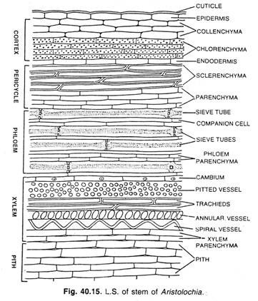
The xylem is composed of three different types of cells — tracheary cells, that include tracheids and vessels; wood fibres and wood parenchyma.
The tracheids are elongated dead cells, with walls that are thick in some places and thin in others. They serve both as water conducting and as strengthening cells. The walls of the tracheids are heavily impregnated with lignin.
The vessels are composed of rows of tracheary cells the cavities of which are connected by the total or partial disappearance of the cross walls. The diameter of vessels is usually much greater than that of tracheids. They form long tubes, and therefore, they constitute the principal water- conducting elements of the dicotyledonous stem.
The tracheary cells may be divided into several types according to the method by which the walls are thickened. Annular tracheary cells have thickenings in the form of rings, while spiral tracheary cells have spiral thickenings. Pitted tracheary cells have walls which are uniformly thickened except for thin places in the form of pits. When ladder-like thickenings are present, the vessel is said to be scalariform.
ADVERTISEMENTS:
The protoxylem is composed largely of annular and spiral vessels and parenchyma, while the tracheary elements of the secondary xylem are pitted.
The wood fibres are long, slender, pointed dead cells with greatly thickened walls and only comparatively few small pits. They serve as strengthening cells. The tracheids that have a structure approaching that of wood fibres are called fibre tracheids.
The parenchyma cells in the xylem are known as wood parenchyma. They serve mainly for the storage of food.
Phloem:
The primary phloem of the dicotyledonous stems consists of three types of cells — sieve tubes, companion cells and phloem parenchyma.
The sieve tubes consist of thin- walled, elongated cells arranged in vertical rows. The adjacent cells of a sieve tube are united by small holes in the cross walls. The areas on the walls of sieve tubes which contain such holes are called sieve plates. The mature sieve tubes do not contain any nuclei. The sieve tubes serve primarily for the conduction of food material.
The companion cells are small cells which are attached to the sieve tubes. Each companion cell is the sister cell of a sieve-tube cell, the two being formed by the division of a mother cell.
The phloem contains parenchyma cells whose structure is very similar to that of other parenchyma cells. These are known as phloem parenchyma.
Cambium:
There lies a layer of meristematic cells between the xylem and the phloem is known as the cambium. The cambium consists of a single layer of cells which, by division gives rise to xylem cells toward the centre of the stem and phloem cells toward the periphery.
At first the cambium is confined to the bundles, but later the parenchyma cells of the pith rays which lie between the edges of the cambium in the bundles divide and form a layer of cambium which reaches across the pith rays and connects that in the bundles, so that the cambium becomes a continuous cylinder.
Pith Rays:
The vascular bundles are separated from each other by radial rows of parenchyma cells known as pith rays. The pith-ray cells are usually elongated in a radial direction. They serve primarily for the conduction of food and water radially in the stem and for the storage of food.
Pith:
In a dicotyledonous plant the centre of the stem is composed of thin-walled parenchyma cells and is known as the pith. The cells have distinct intercellular spaces.
Variations in Dicotyledonous Stem Structure:
The above mentioned description of structure of the stems is applicable to the great majority of dicotyledonous plants, but there are a few which show minor variations. The relative development of the various parts, however, varies greatly in different species. In some cases the pith is wide, while in others it is narrow. It may be wide and transitory and its early disappearance results in a hollow stem.
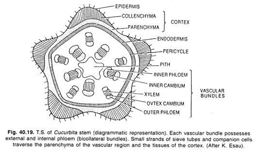
The vascular bundles vary considerably in number and size, while the pith rays and cortex vary in width. Bundles which have the phloem only on the outside of the xylem are called collateral bundles. The bundles of some plants have phloem on both the outside and the inside of the xylem (e.g., in the members of Cucurbitaceae) and known as bicollateral bundles.
Types of Dicotyledonous Stems:
Anatomy of Cucurbita Stem:
Epidermis:
This single outermost layer consists of compact barrel shaped cells having no intercellular spaces. The epidermis remains covered with a thin cuticle. Some of the epidermal cells possess multicellular epidermal hairs.
Cortex:
This region consists of external collenchyma, chlorenchyma (photosynthetic tissue) and endodermis.
(a) Collenchyma:
This lies immediately beneath the epidermis consisting of many layers of the cells in the ridges, whereas in furrows it is only two or three layered or sometimes altogether absent.
(b) Chlorenchyma:
Just below the collenchyma two or three layers of parenchyma containing chloroplasts (chlorenchyma—photosynthetic tissue) present which help in the process of assimilation.
(c) Endodermis:
It is innermost layer of the cortex, lying immediately outside the sclerenchymatous zone of pericycle. This layer is wavy and contains many starch grains.
Pericycle:
Just beneath the endodermis there is a multi-layered zone of sclerenchymatous pericycle. The cells are lignified and appear polygonal in cross section.
Ground tissue:
The vascular bundles are found lying embedded in the thin walled parenchyma cells of ground tissue. The ground tissue extends from just below the sclerenchymatous pericycle to the central medullary cavity.
Vascular bundles:
Generally vascular bundles are ten in number which are found to be arranged in two rows, those of the outer row corresponding to the ridges and those of the inner to the furrows.
The vascular bundles are bicollateral each consisting of xylem, two strips (inner and outer) of cambium and two strands of phloem (inner and outer):
(a) Xylem:
It occupies the central position of the vascular bundle, consisting of very wide, pitted vessels towards periphery of the metaxylem, and on the inner side of narrow vessels which form the protoxylem. In the xylem certain tracheids, wood fibres and xylem parenchyma are also present. The xylem vessels are not arranged in radial rows.
(b) Cambium:
In each vascular bundle two strips of cambium are found. The cambial activity remains confined within the vascular bundles. The cambial strip is found between xylem and phloem on either side of the bundle. Of the two strips of cambium it is only the external one which divides and causes growth in thickness.
The cells of cambium are thin walled, rectangular and arranged in radial rows. Usually the outer cambium is many layered and flat while the inner cambium is few layered and somewhat curved. Only fascicular cambium is found. The stem is not woody, and therefore, the periderm and lenticels are not formed.
(c) Phloem:
On the extreme ends of the vascular bundle the phloem occurs in two patches, towards the periphery, the outer phloem, and towards pith, the inner phloem. Each strand of phloem consists of sieve tubes, companion cells and phloem parenchyma. Sieve tubes are very well developed. The sieve plates with perforations are also visible. Fibres and ray cells are absent.
Special Structure:
The bicollateral open vascular bundles are found each consisting of xylem (central position), two strips of cambium (outer and inner) and two patches of phloem (outer and inner). (See figs. 40.19 and 40.20).
Anatomy of Bryonia Stem:
Epidermis:
The single outermost layer consists of compact barrel shaped cells having no intercellular spaces. Usually the epidermis is covered with a thin cuticle. At certain places stomata are also found.
Cortex:
This consists of collenchyma, loose chlorenchyma and inconspicuous endodermis.
(a) Collenchyma:
This lies immediately beneath the epidermis consisting of rounded or oval cells (in cross section) usually thickened laterally due to the presence of cellulose. The collenchymatous region is multi-layered.
(b) Chlorenchyma:
Below the collenchyma few layers of chlorenchyma are found. The cells are thin walled, rounded or oval, with chloroplasts and having intercellular spaces among them.
ADVERTISEMENTS:
(c) Endodermis:
The single layered endodermis is inconspicuous. The endodermal cells may contain abundant starch grains in them.
Pericycle:
Immediately beneath the endodermis multi-layered sclerenchymatous pericycle is found. The cells are lignified and polygonal as seen in cross section.
Ground tissue:
The ground tissue extend from immediately beneath the pericycle to the central pith cavity. The vascular bundles are found lying embedded in it.
Vascular system:
Usually the vascular bundles are found to be arranged in two rows. They are open and bicollateral. Each bundle consists of xylem, cambium and two strands (inner and outer) of phloem.
Xylem occupies the central position of the vascular bundle consisting of bigger vessels (pitted) of metaxylem outwards and narrower vessels (annular and spiral) of protoxylem towards pith. Certain tracheids, wood fibres and xylem parenchyma are also present.
In between the strands of outer phloem and xylem a strip of cambium is present. The cells of cambium are thin walled, rectangular and arranged in radial rows.
The phloem strands are found on extreme ends of the vascular bundle. Each strand consists of sieve tubes, companion cells and phloem parenchyma. Each sieve tube is accompanied by a conspicuous companion cell. Sieve tubes are well developed. Sieve plates with perforations are also visible in cross section.
Pith:
It consists of thin-walled rounded or oval parenchymatous cells having well defined intercellular spaces among them.
Special Structure:
The bicollateral and open vascular bundles are present. Each bundle consists of central xylem, a cambial strip and two strands (inner and outer) of phloem.


