ADVERTISEMENTS:
The following points highlight the top three theories of root apical meristem in plants. The theories are: 1. Apical Cell Theory 2. Histogen Theory 3. Korper-Kappe Theory.
1. Apical Cell Theory:
This theory was proposed by Nageli who drew the attention to the occurrence of a single apical cell or apical initial that composes the root meristem. A single apical cell is present only in vascular cryptogams, e.g. Equisetum, Adiantum and Polypodium etc. The apical initial is tetrahedral in shape and generates root cap from one side.
The other three sides donate cells to form epidermis, cortex and vascular cylinder. In other words all tissues that compose a mature root including root cap are the derivatives of a single apical cell. Apical cell theory is confined to vascular cryptogams only as the root apical meristem of flowering plants does not have a single apical cell.
2. Histogen Theory:
ADVERTISEMENTS:
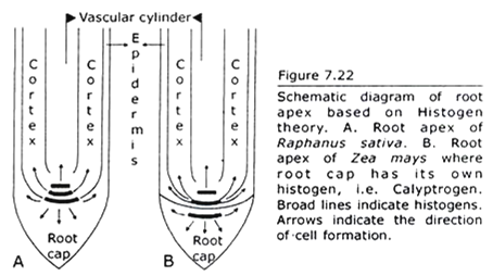
Hanstein in 1868 advocated the theory. According to Hanstein root apical meristem consists of three cell-initiating regions called histogens (Fig. 7.22). The histogens are called dermatogen, periblem and plerome that respectfully form epidermis, cortex and vascular cylinder that are present in a mature root.
The derivatives of dermatogen vary. In Zea mays (monocot) dermatogen generates root cap only and this histogen is referred to as calyptrogen. In Brassica (dicot) dermatogen generates both protoderm and root cap and this histogen is referred to as dermatocalyptrogen.
Histogen theory explains both root and shoot apical meristem. This theory attributes specific destinies to the derivatives of the three histogens. Though histogen theory is abandoned to explain shoot apex, Eames and MacDaniels illustrated the root apical meristem on the basis of histogen concept.
3. Korper-Kappe Theory:
ADVERTISEMENTS:
This theory of root meristem was proposed in 1917 by Schiiepp who regarded the occurrence of two systems of cell seriation that characterize the root apex with reference to planes of cell division in its parts.
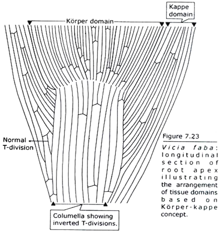
Korper-kappe concept is also referred to as body-cap concept (Korper = body and kappe = cap) and the concept illustrates distinct type of cell wall pattern formation during cell division. The body-cap concept is illustrated below on analyzing the divisions in the derivatives of apical cell (Fig. 7.23).
The root meristem exhibits multicellular structure. It consists of conspicuous longitudinal files of cells. During growth the root changes in diameter. This happens due to cell divisions that occur in such a way that a single longitudinal file of cells becomes double files. The initial cell divides transversely. The two cells thus formed one has the capability of cell division. This cell divides longitudinally and both the daughter cells inherit the property of cell division.
The daughter cells are parallel in arrangement, share a common wall and divide by transverse partition followed by longitudinal partition in one cell. The sequences of wall formation when viewed together appear to form a configuration resembling the letter ‘T’ or ‘Y’. Such divisions are described as T-divisions. Continuous T-divisions result in the formation of double-rowed region over a single rowed region.
It is the T-division that characterizes korper and kappe. In the kappe the initial cell first divides transversely and forms two cells. The daughter cell that faces the root apex inherits the initial function. It divides longitudinally. The two cells thus formed have the capability of cell division.
When transverse and longitudinal partition are viewed together the combined cell walls appear as ‘T’ that is right-way-up. When such division continues it is observed that a single rowed region is left behind over the double-rowed region. This occurs in downwardly pointed roots.
In the korper the initial cell first divides by transverse partition and forms two cells. The daughter cell that faces the base of root, i.e. away from the apex inherits the initial function.
It divides longitudinally and the two daughter cells thus formed have the potentiality of cell division. The daughter cells divide by transverse partitions followed by longitudinal partitions. When transverse and longitudinal partitions are viewed together the cell walls form a configuration resembling an inverted ‘T’.
ADVERTISEMENTS:
Korper and kappe-these two zones of root are delimited by planes of cell division. The zones exhibit clear boundary when they originate from separate initials, e.g. root with calyptrogen. The zones do not exhibit sharp demarcation line when they are the derivatives of same apical cell. In root with dermatocalyptrogen the cap extends into protoderm.
The central part of root cap is the columella where the cells are arranged in longitudinal files. These cells seldom divide. When division occurs the partition walls form the configuration of an inverted ‘T’ that is observed in the korper. The ‘T’ has normal configuration in the peripheral region of root cap.
The korper-kappe theory of root apex is comparable with tunica-corpus theory of shoot apex. The body-cap concept and tunica-corpus concept both are based solely on the planes of cell division. Anticlinal division is the characteristic of tunica whereas corpus exhibits both anticlinal and periclinal division.
On the other hand the inverted ‘T’ – and normal ‘T’ pattern of cell wall formation are the characteristic of korper and kappe respectively. The boundaries between korper and kappe, and between tunica and corpus are not always sharply demarcated.


