ADVERTISEMENTS:
In this article we will discuss about the the structure of larger and smaller subunits of ribosomes.
Ribosomes are ribonucleoprotein particles present in all types of cells. They were first observed in EM by Claude in the cytoplasm of cells, and later on the surface of endoplasmic reticulum by Porter and Palade.
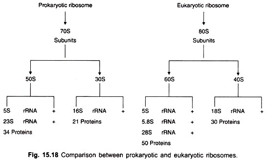
Ribosomes occur in 3 sizes: 70S in bacteria and chloroplasts, 60S in mitochondria, 80S in cytoplasm of eukaryotes. All ribosomes consist of two unequal subunits each containing RNA and protein in the ratio of 63: 37. In bacteria the 70S ribosomes have 50S and 30S subunits and a diameter of 18 nm.
ADVERTISEMENTS:
An E. coli cell contains about 15,000 ribosomes constituting about 25 per cent of the dry weight of the cell. The cytoplasmic ribosomes of eukaryotic cells are larger having a diameter of 20-22 nm (Fig. 15.18), and have a higher protein content and larger RNA molecules.
They either lie free in the cytoplasm or are bound to the endoplasmic reticulum. In eukaryotes ribosomes are also present in the nucleus (they are assembled in the nucleolus) and in organelles like mitochondria and chloroplasts. In cells that are synthesizing proteins, electron micrographs show ribosomes associated in bead-like strings called polyribosomes or polysomes formed by attachment of ribosomes to a single molecule of mRNA.
The 70S ribosomes of prokaryotes have in the larger 50S subunit, 23S and 5S types of rRNA and 34 different proteins. The 30S subunit contains a molecule of 16S rRNA and 21 different proteins. The eukaryotic cytoplasmic ribosomes show variation in sizes of subunits in different plants and animals and in size of rRNA.
ADVERTISEMENTS:
Plant ribosomes contain 25S and 16S or 18S rRNA whereas animal ribosomes contain 28S and 18S rRNA; 5S RNA is present in the larger subunit of plants and animals. The 28S RNA of eukaryotes has a small rRNA called 5.8S rRNA. It has about 160 bases and is bound to the 28S rRNA.
In 1964 Watson had suggested that ribosomes dissociate into their subunits and again reassociate during protein synthesis. This was later confirmed by Kaempfer in E. coli. It is necessary for the subunits to dissociate because mRNA and the initiating aminoacyl tRNA cannot bind directly to 70S ribosome, but are first bound to the 30S subunit.
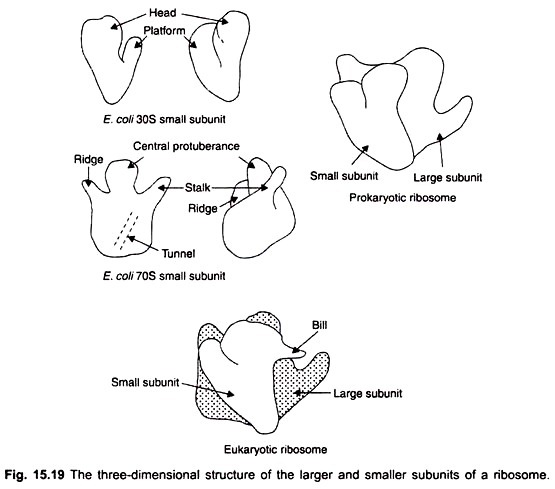
Recently, high resolution studies on ribosomes have revealed new features in their fine structure. The three-dimensional structure of the large and small subunits, as well as the locations of their proteins have been determined by the techniques of immune electron microscopy and neutron diffraction.
The first of these two techniques consists in preparing antibodies that bind to specific subunit proteins thus revealing their locations. The procedure involves purification of the ribosomal protein and injecting it into the bloodstream of an animal such as rabbit, to elicit the formation of antibodies (immunoglobulin G, IgG).
The purified antibodies are mixed with whole ribosomes allowing them to bind to specific proteins that are exposed on the surface of the ribosome. As there are two binding sites in the antibody, therefore one antibody becomes bound to the subunit protein of two ribosomes. The subunits become associated in pairs and are visible in the electron microscope.
The analysis not only reveals the positions of the proteins, but also the three-dimensional structure of the ribosome, In the neutron diffraction technique, the subunits of the ribosomes are made to include a pair of proteins containing heavy hydrogen, deuterium.
From the interference pattern made by a beam of neutrons which traverses the subunit, the distance between the two proteins (which contain deuterium) can be estimated. A number of such observations lead to mapping of the proteins.
In the three-dimensional model of an E. coli ribosome the smaller subunit is elongated and indented and fits into the concave surface of the larger subunit. The small subunit consists of 3 regions, a head, a base and a platform which is separated from the head by a cleft (Fig. 15.19). The larger subunit has a central protuberance and two projections, the larger called stalk, the shorter one ridge. The depression between the ridge and the central protuberance is called the valley.


