ADVERTISEMENTS:
Quick Notes on Ribosomes!
Structure of Ribosomes in Cell:
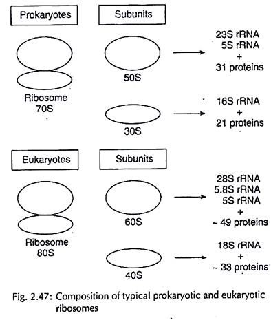
Ribosomes are cytoplasmic non- membranous ribonucleoprotein granules of 150 -200 A diameter. They have a typical binary and constricted structure with the two units being unequal in size. The prokaryotic and eukaryotic ribosomes are differentiated on the basis of the sedimentation coefficient.
These are of two basic types, 70S and BOS ribosomes found in prokaryotes and eukaryotes respectively. The ‘S’ (Svedberg units) refers to sedimentation coefficient which shows how fast a cell organelle sediments in an ultracentrifuge. The heavier a structure the more is its sedimentation coefficient.
ADVERTISEMENTS:
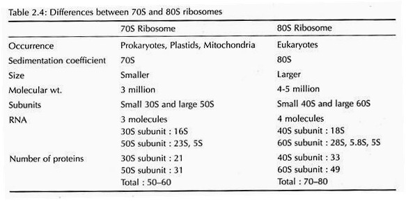
The 70S ribosome of prokaryotes is relatively smaller and consists of a large 50S subunit and a small 30S subunit. The SOS ribosomes of eukaryotes are heavier and made up of large 60S subunit and small 40S subunit (Fig. 2.47). The differences between 70S and SOS ribosomes are given in Table 2.4.
Chemically ribosome is made up of rRNA, proteins and some divalent metallic ions. The subunits of ribosome can dissociate and associate on the basis of Mg++ concentration. Ribosomes may occur in the free form in prokaryotes, and are then called monosomes, or may be associated with mRNA to form polysomes as in eukaryote.
ADVERTISEMENTS:
Ribosomes reported in plastids and mitochondria have a sedimentation coefficient of 70S and similar to prokaryotes in size and are different from cytoribosomes.
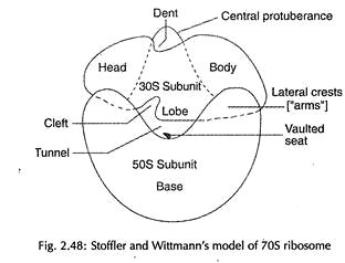
The structural model of ribosomes, as proposed by Stoffier and Wittmann, has the frontal face of the 30S subunit with its hollow facing the vaulted seat of the SOS subunit. The long axis of the BOS subunit is oriented transversely to the central protuberance of the SOS subunit. A tunnel is formed between the hollow of the small sub- unit and the vaulted seat of the large subunit (Fig. 2.48).
Functions of Ribosomes in Cell:
Ribosomes are the site of protein synthesis and are string together by mRNA to form polysomes or polyribosomes. Interaction of the tRNA-amino acid complex with mRNA, which brings about translation of the genetic code, is coordinated by the ribosomes.
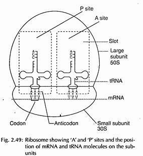
During protein synthesis, the messenger RNA moves through a channel between two subunits of ribosome. Each ribosome has two functional sites – amino acyl or acceptor (A) site and the peptidyl or donor (P) site (Fig. 2.49). The acceptor sites receive the tRNA amino acid complex, and the donor site binds the growing polypeptide tRNA. As such, they perform most important function in protein synthesis.


