ADVERTISEMENTS:
In this article we will discuss about:- 1. Habit and Habitat of Marsilea 2. External Features of Marsilea 3. Internal Structure 4. Reproduction 5. Life Cycle Patterns.
Habit and Habitat of Marsilea:
Marsilea is commonly known as “pepper wort” or “water fern” (although it is a fern but hardly resembles a true fern). It is represented by about 53 species which are cosmopolitan in distribution but abundantly found in tropical countries like Africa and Australia. About 9 species have been reported from India.
Either the species are hydrophytic or amphibious i.e., they grow rooted in mud or marshes and shallow pools or are completely submerged or partially or entirely out of water in wet habitats. M. hirsuta is an Australian xerophytic species. M. hirsuta and M. quadrifolia are two most common Indian species usually found growing in marshy places, wet soil or near muddy margins of ponds and are commonly found in U.P., Punjab, Bihar, Delhi etc.
External Features of Marsilea:
The mature sporophyte is an herbaceous plant. Its underground rhizome spreads in a diameter of 25 meter or more. The plant body is distinctly differentiated into rhizome, leaves and roots (Fig. 1 A).
1. Rhizome:
All the species possess a rhizome which creeps on or just beneath the soil surface. It is slender, dichotomously branched with distinct nodes and internodes and is capable of indefinite growth in all directions as a result of which it occupies an area of 25 metre or more in diameter.
In aquatic species the internodes are long while in sub-terrestrial species they are short. Usually from the upper side at nodes, the leaves are given out while from their lower side, the roots.
ADVERTISEMENTS:
2. Leaves:
They are borne alternately on upper side of rhizome at nodes, in two rows. Young leaves show circinate vernation (like ferns) (Fig. 1 A). In some species young leaves are covered with multicellular hairs. The leaves are compound, with basal petiole and terminal lamina.
In submerged plants the petiole is a long and flexible structure and the lamina floats over the surface of water but in muddy or marshy plants the petiole of the leaf is short and rigid with short lamina spreading in the air.
The lamina consists of 4 leaflets (pinnae) which are present at the apex of petiole. The 4 leaflets arise as a result of 3 dichotomies of the lamina in close succession to each other i.e., 2 leaflets arise slightly higher than other two (Fig. 1B).
Puri and Garg (1953) suggested that the leaf consists of single pinna consisting of 4 pinnules. Pinnae have got a dichotomously branched vein system with cross connections (Fig. 1C). The veinlets at the margin are connected with loops thus forming a reticulum. The shape of pinna varies from obovate to obcuneate and margin also varies from entire to crenate or crenate to lobed.
Sometimes the pinnas are once or twice deeply dichotomously lobed (M. biloba) or toothed (M. minuta). At night the pinna are folded upwardly. This is known as sleeping movement of pinna. Near the base of petiole the stalked bean-shaped sporocarps are borne.
3. Roots:
ADVERTISEMENTS:
The roots are adventitious, arising from the underside of the node of rhizome, either singly or in groups. In certain cases the roots are given out even from the internodes (M. aegyptiaca).
Internal Structure of Marsilea:
1. T. S. Rhizome (stem):
A T. S. of the young rhizome shows a protostelic structure i.e., pith is absent and xylem is completely surrounded by phloem but in the old stem pith is developed in the centre and the stele is amphiphloic siphonostelic type.
ADVERTISEMENTS:
A. T. S. of the old stem is somewhat circular in outline and shows the following structures:
(i) Epidermis:
ADVERTISEMENTS:
It is the outermost limiting layer of single celled thick parenchymatous cells. The stomata are absent.
(ii) Cortex:
It is differentiated into three regions – the outer cortex, the middle cortex and the inner cortex.
(a) Outer cortex:
ADVERTISEMENTS:
It is present just below the epidermis (also called hypodermis). It is parenchymatous and may be one to several cells thick. Some of its cells contain tannin.
(b) Middle cortex:
It is also called aerenchyma. It lies below the hypodermis. It consists of large air spaces (chambers) separated by one cell thick parenchymatous septa. In the xerophytic species e.g., aegyptiaca the air chambers are obliterated.
(c) Inner cortex:
It is a solid tissue of several cells thickness. The outer layers are thick walled (sclerenchymatous) while the inner layer of cells is thin walled (parenchymatous) and compactly arranged. Some of these cells are filled with starch or tannin.
(iii) Stele:
ADVERTISEMENTS:
Stele is amphiphloicsiphonostele i.e., in the centre there is a pith which may be either parenchymatous (aquatic species) or sclerenchymatous (terrestrial muddy species). Xylem is present in the form of a complete ring which is surrounded on both sides by a complete ring of inner and outer phloem, pericycle and endodermis.
In this way the continuation of different tissues in the form of complete ring in stele is as follows—outer endodermis, outer pericycle, outer phloem, xylem, inner phloem, inner pericycle and inner endodermis. The protoxylem may be well defined exarch (M.vestita) or mesarch (M.aegyptiaca) or ill defined (M.quadrifolia).
A T. S. of the nodal region shows an amphiphloicsolenostelic condition and is provided with one leaf gap.
2. T. S. of Petiole:
A T. S. of the petiole is somewhat circular in outline and is differentiated into epidermis, cortex and stele.
(i) Epidermis:
ADVERTISEMENTS:
It is the outermost layer of single cell thickness. The cells are parenchymatous and slightly elongated.
(ii) Cortex:
It is differentiated into three regions: The outer cortex, the middle cortex and the inner cortex.
(a) Outer cortex:
It is present just below the epidermis, (also called hypodermis). It is made of thin walled cells (parenchymatous).
(b) Middle cortex:
It lies below the hypodermis and called aerenchyma. It consists a ring of air chambers. The air chambers are separated by single layered partitions of thin-walled parenchymatous cells.
(c) Inner cortex:
It is a solid tissue of several cells thickness. The cell layers are parenchymatous and contain starch and tannin filled cells. In M.minuta few sclerenchymatous layers are also present just inner to middle cotex.
(iii) Stele:
It is somewhat triangular in outline and is of protostelic type i.e. pith is absent. Xylem is “V” shaped with 2 distinct arms. Each arm is provided with metaxylem elements in the centre and protoxylem is situated at both the margins i. e., protoxylem is exarch.
The xylem is surrounded on all sides by phloem. Phloem is externally surrounded by a single layer of parenchymatouspericycle which, in turn, is bounded by a single layered endodermis.
3. Transverse Section of Leaflet:
A. T. S. of the leaflet shows epidermis, mesophyll and vascular bundles.
(i) Epidermis:
It is the outermost surrounding layer and is only one cell in thickness. It is differentiated into upper and lower epidermis. In floating leaflets the stomata are present on the upper epidermis but in case of plants growing in mud or moist soil where the leaves are aerial, the stomata are present both on upper as well as lower epidermis.
(ii) Mesophyll:
It occupies a wide space between upper and lower epidermis. It is usually differentiated into an upper palisade tissue and lower spongy parenchyma. The palisade tissue is made up of elongated cells provided with chloroplast. The spongy tissue is made up of loosely arranged parenchymatous cells with large air spaces separated by single layered septa. In submerged species, however, the mesophyll is not differentiated into palisade and spongy parenchyma.
(iii) Vascular bundles:
In between the mesophyll tissue are present several vascular bundles. Each vascular bundle is concentric and amphicribal type i. e., made up of a centrally situated xylem, surrounded on all sides by phloem. The phloem is enclosed by a single layered thick endodermis.
4. T. S. Root:
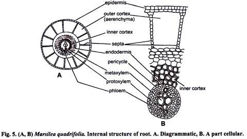
A T. S. of root is somewhat circular in outline and can be differentiated into epidermis or piliferous layer, cortex and stele (Fig. 5A, B).
(i) Epidermis:
It is the outermost, parenchymatous, single layered covering.
(ii) Cortex:
It can be differentiated into two parts: outer cortex and inner cortex. The outer cortex consists of large air chambers arranged in the form of a ring (parenchymatous). These chambers are separated from each other by longitudinal septa. The inner cortex is differentiated into outer parenchymatous and inner sclerenchymatous regions. The inner cortex is delimited by single layered thick endodermis.
(iii) Stele:
It is of protostelic type and occupies the central position. It is devoid of pith. Xylem is situated in the centre which is diarch and exarch. It is surrounded by phloem. The phloem is bounded externally by a single layer of pericycle.
Reproduction in Marsilea:
Marsilea reproduces by two methods:
(i) Vegetative reproduction
(ii) Sexual reproduction.
Vegetative reproduction:
It takes place by means of tubers which are produced in dry conditions from the rhizome. First a branch is given out from the rhizome, which later on swells up due to the accumulation of food material. The structure is termed as tuber and is capable of tiding over the unfavourable conditions. On the return of favourable conditions it germinates to produce a new sporophytic plant, e.g. ,M. hirsuta, M. quadrifolia.
(ii) Sexual Reproduction:
1. Sporophytic Phase:
Spore producing organs:
Marsilea is heterosporous i. e., it produce two types of spores—microspores and megaspores. These spores are produced in microsporangia and megasporangia, respectively. These sporangia are borne in special type of spore producing organ called sporocarp. The sporocarp are born laterally on the short and lateral branches of the (called the peduncles or pedials) petiole of leaf either near the base or a little higher up.
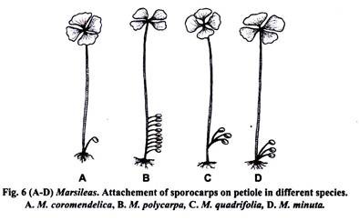
They arise solitary or in clusters. The peduncle is usually unbranched but it may be branched also. Number of sporocarp differs in different species and varies from 1 to 20 or more. In M. vestitasporocarparises single, in M. quadrifolia the peduncle is dichotomously branched bearing 2-4 sporocarps, in M. polycarpa several sporocarps arise in a linear row. The attachment of the pedicel sporocarp varies in different species.
Mainly it is of three types (Gupta 1962):
(i) Pedicel of the solitary sporocarp is directly attached to the base of the petiole (e.g., M. coromendelica Fig. 6A) or pedicels of many sporocarps are attached on the petiole in a linear sequence on the same side (e.g.,M. polycarpa; Fig. 6B).
(ii) Pedicels first become united themselves with one another and then are attached to the petiole by a common stalk e.g., M. quadrifolia; (Fig. 6C).
(iii) Pedicels are free or slightly united and attached to the petiole by a single point (e.g.,M. minuta; Fig. 6D).
External Morphology of Sporocarp:
Each sporocarp is an oval or bean shaped biconvex, flattened structure. It is green and soft when it is young but at maturity it becomes very hard and brown in colour. It is made up of a short stalk like structure known as peduncle and the body.
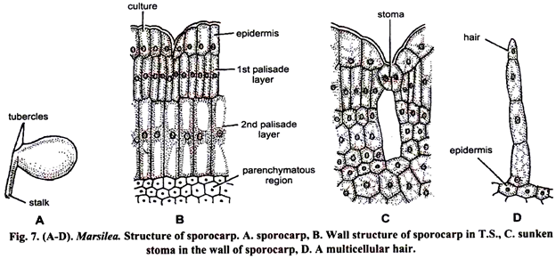
The point of attachment of peduncle with the body is called raphe (Fig. 7A). Slightly above the raphe in a median plane are present 1 or 2 protuberances called tubercles. They are unequal in size and lower one is stouter than the upper one. In some cases the tubercles are absent e.g.,M. polycarpa.
Internal Structure of Mature Sporocarp:
The sporocarp is a bivalved structure. It can be split open in the dorsiventral plane into two halves (valves).
If we split open the sporocarp, we can see the following structures:
Wall of sporocarp:
It is very hard, thick and highly resistant to mechanical injury. It can be differentiated into three zones—outer epidermis, middle hypodermis and inner parenchymatous zone. Epidermis is single layered made up of broad and columnar cells. Its continuity is broken by the presence of sunken stomata (Fig. 7C).
Some of the epidermal cells develop into multicellular hairs (Fig. 7D). Hypodermis consists of two layers of radially elongated palisade like cells. Both the layers are without intercellular spaces and have chloroplast in their cells. Next to hypodermal layers is the parenchymatous zone (Fig. 7B). In mature sporocarp the cells of this zone gelatinise and form a gelatinous ring which helps in the dehiscence of the sporocarp.
Cavity of sporocarp:
The alternating rows of sori (sing, sorus, a group of sporangia is called sorus), one along each side lies transversely-dorsiventrally to the long axis of the sporocarp. The sori on either side alternate with each other. The number of sori inside the sporocarp varies from species to species. It may be from two (e.g., M. aegyptiaca) to twenty (e.g.,M. vestita). Each sorus bears both microsporangia and megasporangia.
Their number also varies from species to species. In M. minuta a sorus has 4-8 megasporangia and 8-13 microsporangia. In M. aegyptiaca each sorus has 5-16 megasporangia and 9-19 microsporangia.
In M. minuta, M. vestita, M. rajasthanensis, sometimes megasporangia are absent in sorus. Each sorus arises on a ridge like placenta or receptacle formed on the sporocarp wall. Each sorus is surrounded by a thin, membranous two layered true indusium. The indusia of adjacent sori are partially fused.
Vascular supply of the sporocarp:
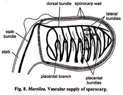
It is supplied by a main dorsal vein which runs along the narrow side facing the peduncle. From the dorsal vein, lateral branches are given alternatively right and left, at right angle to the dorsal vein which supplies laterally (Fig. 8). These lateral veins at their middle divide dichotomously. In the region here lateral vein forks, arises a placental bundle which too branches dichotomously. The first and the last lateral veins do not possess placental bundles.
The entire internal structure of the sporocarp can be best seen in section cut in three plains:
(i) Horizontal Longitudinal Section (H.L.S.): Section is cut horizontally but the sporocarp is cut longitudinally.
(ii) Vertical Longitudinal Section (V.L.S.): Section is cut vertically but the sporocarp is cut longitudinally.
(iii) Vertical Transverse Section (V.T.S.): Section is cut vertically but the sporocarp is cut transversely.
(i) Horizontal Longitudinal Section (H.L.S.):
The section passes through the peduncle and cuts it transversely. Peduncle shows characteristic ‘V’ shaped xylem (Fig. 9 A, B). A H.L.S. of sporocarp shows the usual wall layers. The gelatinous ring is cut transversely and it appears in the form of dorsal and ventral mass at its proximal and distal ends.
The dorsal mass is more prominent than transversely along with their two layered inducia. Sori show their alternate arrangement in the two rows. Each sorus has a receptacle which has a central terminal megasporangium and two lateral microsporangia, one on either side. The lateral bundle is also cut transversely below each sorus.
(ii) Vertical Longitudinal Section (V.L.S.):
A V.L.S. of the sporocarp shows the usual wall layers. The peduncle along its vascular bundle is cut longitudinally. The entire gelatinous ring is cut vertically and it appears as a complete ring around the sori. The section cut the sori longitudinally, which are arranged in many vertical rows.
If the section passes strictly through the median plane of sporocarp, only megasporangia are visible (Fig. 10A, B) but if it passes slightly away from the median line, only microsporangia are visible (Fig. 10C).
(iii) Vertical Transverse Section (V.T.S.):
A V.T.S. of the sporocarp shows the usual wall layers. Peduncle is not cut in the section. The gelatinous ring appears in the form of dorsal and ventral mass (as in H.L.S.). The gelatinous mass on the dorsal side is much more prominent.
In V.T.S. only two sori covered with indusia are visible on the inner side and attached to the placental ridge on the outer side (Fig. 11 A, B). The sori reveal many megasporangia and only two or three microsporangia at the sides. The dorsal bundles, the lateral bundles and the placental bundles are clearly visible.
Morphological Nature of the Sporocarp:
Two main views have been put forward by different morphologists to explain the morphological nature of the sporocarp of Marsilea which are discussed below:
(1) The laminar or leaf segment hypothesis.
(2) Petiolar or whole leaf hypothesis.
The laminar or leaf segment hypothesis. According to the supporters of this hypothesis the sporocarp is a lateral modified fertile segment of the leaf but their interpretations are different which are given below:
(i) Russow (1872) and Busgen (1890) regarded the sporocarp to be made up of 2 leaflets with ventral surface facing each other (Fig. 121).
(ii) Goebel (1882, 1905, 1930) considered the sporocarp as a fertile leaflet (pinna), comparable with one or more leaflets.
(iii) Campbell (1893, 1928, 1940) regarded the sporocarp as a folded pinnate leaf formed by the fusion of several pairs of pinnae.
(iv) Bower (1926) stated “the hypothesis seems to be tenable that the sporocarp consists of rachis, bearing 2 rows of pinnules, this is indicated by the veination (Fig. 12 G, H).”
(v) Eames (1936) compared it to the tip of the leaf with 4 leaflets. He was of the opinion that the body of the sporocarp represents the two distal leaf-lefts and the region of 1st and 2nd protuberance represent the remaining 2 proximal leaflets (Fig. 12 D, E).
(vi) Smith (1938, 1955) considered the sporocarp as a modified pinna with one midrib and several lateral branches (Fig. 12 A-C).
(vii) Takhtajan (1953) considered the sporocarp as a more specialised fertile segment of a leaf as in Schizaeaceae.
(viii) Puri and Garg (1953) regarded the sporocarp equivalent to a single leaflet which consists of as many pinnules as the number of lateral bundles (Fig. 12F).
(ix) Gupta K. M. (1962) regarded the sporocarp to be leaflet with as many lobes as the number of lateral bundles. The sori are situated marignally in the depressions of the lobes.
The following evidences support the above hypothesis:
(1) The sporocarp initial and the leaf initial both are two sided apical cells and they behave in the same way during early segmentation.
(2) In M. polycarpa, 10-15 sporocarps arise acropetally on one side of the petiole.
(3) In M. quadrifolia, branching of peduncle is similar to the formation of pinnules.
(4) The vascular supply of the sporocarp is similar to that of the sterile leaflets.
(5) Presence of epidermis with stomata, hypodermis and parenchyma with air spaces also supports the laminar hypothesis of sporocarp.
Petiolar or whole leaf hypothesis:
Johnson (1898, 1933) proposed this hypothesis and regarded the sporocarp as homologous to the swollen end of the petiole. The marginal cells here develop sporangia instead of lamina. The evidence in support of this theory is that sometimes the primary sporocarp gives rise to secondary sporocarp which develops from the initial arising from the marginal cells of the stalk of primary sporocarp.
Development of Sporocarp:
The development of sporocarp is described by Johnson (1898) in M. quadrifolia. According to him the leaf arises from a bifacial apical leaf cell. It cuts off a pair of segments. When the leaf primordium is 6-7 cells high, the sporocarp initial develops at its base (Fig. 13 A, B).
This cell behaves as a two sided apical leaf cell and cuts off segments on either side. The second sporocarp initial appears in the same way as the first sporocarp initial appears. Similarly, the third sporocarp initial arises. This means that these sporocarps are the branches of the leaf.
The sporocarp initial forms a mass of undifferentiated cells by cutting segments on both lateral sides. It grows into sporocarp. Two rows of soral mother cells appears on ventral side of the young sporocarp.
Formation of inoculations takes place due to undifferentiated growth of soral mother cells and their derivatives. These soral mother cells form two alternating rows of canals. These are called soral canals (Fig. 13 C-E). Each soral canal is lined on its inner surface by a two layered tissue called indusium (Fig. 13 D, E). The receptacle of the sorus faces the indusium in each canal and the sporangia arise upon the receptacle in basipetal manner (Fig. 13 F).
The first sporangial initial appears at the tip of the receptacle and develops into megasporangia. Subsequently the two initials develop on the lateral side of the megasporangium. These are microsporangial initials and develop into microsporangia (Fig. 14A).
Structure of Microsporangium:
It is somewhat oval structure with a long stalk and is present laterally on the receptacle. It is smaller in size. It has a single layered jacket followed by two layers of tapetal cells. In the centre is present a cavity filled with microspore mother cells (Fig. 14H).
At maturity the tapetal cells disintegrate and each microspore mother cell divides reductionally forming 4 haploid microspores (Fig. 141). The microspores are usually 32-64 in number and are liberated by the disintegration of the microsporangial wall (Fig. 14J).
Development of Microsporangium:
It takes place from a superficial cell situated laterally on the receptacle. This cell is called as sporangial initial. It divides transversely into an outer and inner cell (Fig. 14 B, C). The outer cell later on gives rise to the whole of the sporangium i. e., stalk, wall, tapetum and microspores. It divides by three successive diagonal divisions to form a tetrahedral apical cell (Fig. 14D) with three cutting faces.
This apical cell cuts off two cells from its each face which helps in the formation of stalk. Now a periclinal wall is formed towards the outer face of the apical cells forming an outer smaller primary jacket cell and an inner archesporial cell (Fig. 14J). The primary jacket cell divides only anticlinally to form a single layered jacket.
The archesporial cell again divides periclinally to form an outer primary tapetal cell and inner primary sporogenous cell (Fig. 14F, G). The primary tapetal cells divide periclinally as well as anticlinally to form a two layered tapetum. The primary sporogenous cell divides to form 8-16 microspores mother cells (Fig. 14H). Now each microspore mother cell divides reductionally to form a tetrad of spores as a result of which 32-64 microspores are produced (Fig. 14J).
Structure of Megasporangium:
It is a spherical structure with a short stalk and is present on the top of the receptacle (Fig. 14A). It is bigger in size than the microsporangium (Fig. 14A). Its structure is similar to microsporangium except that only one megaspore is present per megasporangium at maturity. The megaspore is liberated by the disintegration of the megasporangial wall.
Development of megasporangium:
The development of megasporangium is exactly in the same way as that of microsporangium except that out of the total number of megaspores formed, all degenerate leaving except one which behaves as a functional megaspore (x). It increases in size.
Dehiscence of Sporocarp and Liberation of Spores:
The decaying of the wall of the sporocarp takes place due to bacterial action and thus the sporangia and spores are liberated. The sporocarp bursts open only in water in valvecular manner along the ventral side and apex. The gelatinuous ring absorbs water and extends greatly through the open margins of the sporocarp thus dragging out sori along with it.
It straightens and behaves as sporophore. The gelatinous ring bears two alternating rows of sori. The delicate mucilage wall of the sporangia (micro-or mega) opens in water and the spores (micro-or mega are liberated which germinate soon (Fig. 15 A, E).
2. Gametophytic Phase:
The microspores and the megaspores are the unit of male and female gametophytes respectively.
They germinate to produce the respective gametophyte in the following ways:
Development of male gametophyte:
The microspore is the initial stage in the development of male gametophyte. Each microspore is a unicellular, uninucleate, thick walled globose and haploid structure, ranging from 0- 060 to 0- 075 mm in diameter.
The cytoplasm is surrounded by inner wall called endosporium and outer wall called exine or exosporium. The microspore germinates just after its liberation. The first division is in a lenticular plane to form a small lens shaped prothallial cell and a apical cell (Fig. 16A, B).
The apical cell divides transversely to form 2 equal antheridial initials (Fig. 16C). Each antheridial initial divides periclinally to form an outer first jacket cells (initial) and inner wedge-shaped sister cells (3-3) (Fig. 16D). The inner cell further divides by periclinal division to form a second smaller jacket cell and a large outer cell (Fig. 16E).
The large cell again divides by a periclinal wall to form an outer or peripheral third jacket layer and a central cell or the primary androgonial cell (Fig. 16F). Each primary androgonial cell divides to form 16 androcytes (Sperm mother cells) and finally metamorphosises into antherozoids. In all 32 antherozoids are produced (Fig. 16 G-I).
The male gametophyte is developed inside the microspore and produces 32 antherozoids with usually one prothallial cell. Sometimes 2 prothallial cells are also produced. By breaking of the jacket cells and disintegration of male gametophytic tissue the antherozoids are liberated.
Each antherozoid is a cork screw shaped, spirally coiled multiflagellate, structure (Fig. 16 J, K). It is characterised by the presence of a prominent terminal vesicle. The cilia are attached only to the posterior coils. The coiling looses at the time of fertilization.
Development of female gametophyte:
The megaspore is the initial stage in the development of female gametophyte. Each megaspore is a unicellular, uninucleate, ellipsoidal structure with an apical papilla (Fig. 17A). The mucilaginous wall is a thick structure except in the papillate region.
The wall has 2 covering layers, outer one is known as exospore and inner one as endospore. The nucleus lies in the apical papilla (Fig. 17A). The rest of the basal portion of the megaspore contains granular starch, oil globules and albuminous substances. The first division is in a transverse plane at the base of papilla, thus forming an upper small cell and a basal bigger prothallial cell (Fig. 17B).
The prothallial cell serves as a nutritive cell and provides nutrition to the developing gametophyte while the apical cell forms the female gametophyte. The upper smaller cell again divides transversely to form an upper apical cell and lower basal cell (Fig. 17C). The apical cell divides by three vertical divisions so as to form 3 lateral cells surrounding a central cell or archegonial initial (Fig. 17D).
The archegonial initial divides by a periclinal wall forming an outer primary cover cell and an inner central cell (Fig. 17E). The 3 lateral cells and a basal cell by further horizontal and vertical divisions form a jacket round the archegonium. The cover cell divides vertically by 2 divisions at right angle to each other to form 4 neck initials (Fig. 17F) which by transverse division forms a neck of 2 tiers of 4 cells each.
The inner cells first divides transversely to form an upper primary- neck canal cell and lower primary venter cell. The former may or may not divide to form 1 or 2 neck canal cells. The latter divides to form an upper ventral canal cell and a basal egg. Thus, at maturity the mature archegonium has 8 celled neck, 1 neck canal cell, a small venter canal cell and a large egg. Each megaspore produces a single archegonium (Fig. 17G).
Fertilization:
The free swimming antherozoids are attracted chemotactically towards the neck of a mature archegonium but only one enters the neck and reaches the egg. The male and female nuclei fuse to form a diploid structure called oospore or zygote. Thus the gametophytic generation ends and the unit of sporophytic generation is formed. In some species e.g.,M. drummondii (Strasburger 1907), parthenogenesis has been observed.
Development of embryo:
Oospore is the initial stage of sporophytic generation. The first division of the oospore is in a vertical plane (parallel to the long axis of archegonium) to form 2 unequal cells. The bigger one is known as epibasal cell and the smaller one as hypobasal cell (Fig. 18 A, B). This is followed by a second transverse division to form 4 cells (quadrant stage) (Fig. 18C).
The epibasal half gives rise to shoot and leaf whereas the hypobasal half gives rise to root and foot. The cell of epibasal half near the neck gives rise to cotyledon and other away from the neck, to the stem.
In the same way the cell of the hypobasal half near the neck gives rise to root and other away from the neck, to the foot. Simultaneously, the tissue surrounding the archegonium divides to form a 2 or 3 celled thick calyptra which protects the embryo in young stage. The embryo later on gives rise to an adult plant.
Life Cycle Patterns of Marsilea:
Mature plant of Marsilea is diploid. Marsilea is a heterosporous fern because it produces 2 different types of spores i. e., microspores and megaspores. Micro- and megaspore mother cells are produced inside micro- and megasporangium respectively which represents the late stage of sporophytic generation. After reduction division microspores and megaspores are produced which represent the initial stage of gamestophytic generation.
Microspore gives rise to make gametophyte which, in turn, produces archegonium and egg. Both antherozoid and egg fuse to from a diploid oospore (2x).The oospore is the initial states of sporophytic generation. Hence, in the way the sporpytic andgametophytic generation alternate with each other although the sporophytic phase is dominant over gametophytic phase (fig 19,20).


