ADVERTISEMENTS:
In this article we will discuss about:- 1. Occurrence of Isoetales 2. Sporophyte of Isoetales 3. Gametophyte 4. Phylogeny.
Occurrence of Isoetales:
The genus Isoeles has about 65 species reported from many parts of the world. In our country also Isoeles is represented by many species.
Panigrahi (1981) has reported the occurrence of nine species of Isoeles from India. Some of the common Indian species are I. coramandaliana, I. dixitii, I. panchananii, I. sahyadrii etc.
ADVERTISEMENTS:
Some of the species that grow in Karnataka are I. sahyadrensis, I. dixtii, I. coromandaliana, I. sampathkumarini etc. Of these I. coromandalina occurs in Amruthoor (Tumkur district) I. sampathkumarini has been reported from Lal bagh (Bangalore) gardens.
Of all the genera of Lycopsida, Isoetes is the most interesting and problematic. With its shortened axis and long cylindrical leaves, it resembles a monocotyledons plant (in particular, Garlic plant). The morphological similarity tempted many people to propose “Isoetes monocotyledon theory” to account for the origin of monocotyledons from Isoetes. The resemblance between Isoetes and monocots however is superficial.
Isoetes is popularly called ‘Quill wort’ or ‘Merllyn’s grass’. The former name is due to the quill like nature of the leaves. The plant body usually grows in swampy regions. Sometimes the plant may grow in aquatic or amphibious habitats. Certain species like I. butleri grow on dry soil.
Sporophyte of Isoetales:
Morophology of the Plant:
ADVERTISEMENTS:
The plant body consists of a condensed, lobed (two or three) structure called the axis or corm (Fig.81). The morphological nature of the corm is debatable. It has been variously interpreted as an erect rhizome, a stalk, stem, rhizophore and an upper leaf bearing part-the stem and a lower root bearing part-the rhizomorph.
The axis or corm is a fleshy structure having complex morphology and anatomy. The corm on its upper surface bears a number of long, quill like, ligulate leaves. The leaves are two to several centimetres long and are crowded in a closed spiral forming a sort of a fascicle. Sometimes the leaves may be as long as 0.5 metre (I. cormanandaliana) to 1 metre (j. japanica).
The leaves have broad spoon shaped bases and tapering tips. The outermost leaves are sterile; successively within them are found megasporophylls, micro-sporophylls and sporophylls with immature sporangia. From the lower surface of the corm are produced a number of roots.
The roots usually take their origin from the grooves of the lobed corm. They are arranged in vertical rows (at right angles to the groove). In a two lobed corm there are four series of roots, two on each side of the corm. Roots are produced in such a definite plan that the number of roots produced may be calculated as per the formula proposed by West and Takeda (1915).
Therefore,
x = yz(z + 1)
Where x = approximate total number of roots produced by a plant.
ADVERTISEMENTS:
y = number of lobes.
z = number of series on the flank of a lobe.
The roots branch dichotomously. In some species of Isoetes swellings are known to occur in root tips. The occurrence of proximal swellings and connectives in the roots of Isoetes have been reported in a comparative study of stigmarian appendages and Isoetes roots.
Internal Structure:
ADVERTISEMENTS:
Corm: Anatomically the corm exhibits many interesting features, the notable being the peculiar type of secondary growth. A vertical section of the axis shows a central vascular cylinder surrounded by a broad parenchymatous cortex (Fig.82).
The vascular cylinder in its shape resembles an anchor or a vegetable chopper. In the lower portion it is spade shaped, flattened in the plane of the grooves of the corm. In a transverse section the lower shade shaped portion appears triradiate even though there are four radiating arms.
The stele is protostelic, the central region of the vasculature consists of xylem, en-sheathed by phloem. The xylem consists of parenchyma intermixed with tracheids. Leaf traces depart from the stele but there are no leaf gaps. The cortex is parenchymatous and has starch filled cells.
ADVERTISEMENTS:
As has already been said, a peculiar type of secondary growth occurs in Isoetes. This is brought about by the activity of a cambium that arises external to the phloem (The normal position of a fascicular cambium is between xylem and phloem). There is difference of opinion as to the functioning of this cambium and the tissues derived from it.
The cambium according to some cuts off tissues only internally. But there are evidences to show that the cambium produces tissues on either side. The outer derivatives of cambium cut off parenchyma cells (not phloem) constituting the secondary cortex.
The inner derivatives are of a complex nature and are referred to as Prismatic layer. The composition of this layer has been variously interpreted. Stockey (1909) considers it secondary xylem, West and Takeda; (1915) regard it secondary phloem while Scott and Hill (1890) interpret it as a mixture or tracheid’s and sieve elements. Among the inner derivatives, the first formed one mature into tracheids while all the later ones differentiate into sieve elements.
In a corm, after the secondary growth has taken place the following tissues are seen (Fig.83). In the centre is a mass of primary xylem. Next to this is the primary phloem. Outlining the phloem is the prismatic tissue. External to the prismatic tissue is the cambium; following cambium is the secondary cortex and then is found the primary cortex. The cortex is very broad.
The growing point of the corm lies in a shallow depression at the distal end of the corm. Growth is initiated by a group of meristematic cells. Vertical elongation of the corm is very little.
Root:
The root grows by an apical meristem; sometimes the apical meristem divides, as a result there is branching of the root. New sets of roots are formed every year from the growing point and the older roots are pushed further back from the growing point.
The roots may persist more than a year or may be sloughed off by an abscission layer. Karrfalt (1984) has studied the origin and early development of the root producing meristem in Isoetes andicola. According to him the rhizogenic meristem differentiates between the first and the second root after the emergence of the second root.
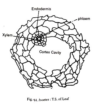
A transverse section of the root shows an outer epidermis middle cortex and central stele. The epidermis is single layered. Cortex is parenchymatous and has a large ‘C’ shaped cavity (Fig.84). This cavity results due to the breakdown of the cortical cells.
The presence of this cavity recalls a similar thing found in the stigmarian rootlets of Lepidodendrales. The stele is a monarch protostele. The xylem and phloem are collateral and are arranged in such a way that the phloem is on the side away from the rhizome. Surrounding the vasculature is a well defined endodermis.
Leaf:
The leaves develop from the apical meristem. Each leaf bears a single ligule at the junction of the cylindrical and the basal wider portion of the leaf. Like roots the leaves are also produced every year. Internally, the leaf shows a single vascular bundle surrounded by an undifferentiated mesophyll.
The outermost layer is the epidermis in which stomata are found (except in submerged species). The mesophyll may consist of only parenchyma, sometimes sclerenchyma may also be located. There are four cylindrical air chambers in the mesophyll separated by parenchymatous partitions.
Here and there, the air chambers are closed by means of a diaphragm to offer mechanical support to the leaf. Reports of Goswami (1985) indicate the presence of internal hairy outgrowths protruding into the lacunae in the leaves of Isoetes pantii. The vasculature bundle of the vein is mostly collateral but may become concentric towards the lip of the leaf.
Reproduction:
The main method of reproduction is by the formation of spores. Rarely however gemmae or vegetative buds may be produced.
ADVERTISEMENTS:
Spore Production:
Isoeles is heterosporous. The micro and megasporophylls are arranged spirally on the corm. There is also no distinction between the foliage leaves and sporophylls. Those leaves which bear the sporangia constitute the sporophylls.
There is no strobilar organisation in Isoetes. The sporophylls bear a single flattened sporangium between the ligule and leaf base on their adaxial surface. The sporangium lies within a cavity (fovea) in the leaf base. Completely or incompletely covering the sporangium will be a membranous outgrowth called Velum that arises just below the ligule.
Structure and Development of the Sporangia:
The micro and mega-sporangia are similar in their development up to the spore mother cell stage. A group of initial cells arise a little below the ligule. These are the sporangial initials. A periclinal division in these cells results in forming an upper layer of jacket cells and an inner layer of archesporial cells.
At the same time, some of the cells between the sporangial cells and the ligule divide and project out forming the velum. Subsequently the velum grows downwards completely or incompletely overarching the sporangium. The jacket initials divide and constitute the single layered jacket of the sporangium. At later stages the jacket may become 3-4 layered.
The archesporial cells divide in all the planes and produce a mass of sporogenous tissue. At the time of the formation of spore mother cells bands of sterile cells are differentiated separating the fertile spore mother cells. These bands constitute the trabeculae that divide the sporangium completely or incompletely. The sporogenous cells adjacent to the trabeculae and to the sides of the sporangium differentiate into a two layered tapetum.
In a sporangium that is destined to become a microsporangium, most of the spore mother cells function, undergo reduction division and produce tetrads of haploid spores. The spore output is enormous and varies from one to three lakhs per sporangium.
In a sporangium that is to become a mega-sporangium, all the spore mother cells except about 40-80, degenerate. The surviving ones undergo reduction division and produce haploid megaspores. Each mega-sporangium of Isoetes indica is known lo contain megaspores ranging in number between 703-2345.
A peculiar type of sporangium called mixed sporangium has been described in some individuals of Isoeles coromandaliana. A mixed sporangium is defined as “a sporangium possessing monolete microspores, trilete megaspores and alete sterile spores”.
Dehiscence of the Sporangia:
The sporangia remain indehiscent for a long time. Liberation of the spores takes place only upon the decay of the sporophyll. Spore dissemination is mostly by wind. According to a report, worms may also help in the dispersal of spores. On liberation, the spores develop into gametophytes.
Gametophyte of Isoetales:
As Isoetes is heterosporous, two types of gametophytes are produced. Both the gametophytes are endosporic and highly reduced.
Structure of the Microspore and Development of the Male Gametophyte:
The microspores are bilateral or tetrahedral in shape, very minute, brownish in colour and have a diameter ranging from 20 to 45µ. The wall is two layered. As the spore liberation is delayed for quite a long time, the germination is immediate and a mature gametophyte is formed within a few days.
The first sign of germination is the migration of the microspore nucleus to one side, when it divides asymmetrically to give rise to a small prothallial cell and large antheridial initial (Fig.87a, 87b).
The antheridial initial first divides diagonally to form two cells (Fig.87c); of these, the cell nearer to the prothallial cell will not divide but forms the first jacket cell of the antheridium; the other cell divides at right angles to the previous plane of division (Fig.87d).
Of the two cells thus formed, the one away from the prothallial cell forms the second jacket cell. The other cell divides in a plane almost parallel to the preceding plane of division. Of the two cells, one forms the third jacket cell and the other one divides periclinally to form the fourth jacket cell and the primary androgonial cell (Fig.87e, 87f, 87g).
The primary androgonial cell divides twice to form four androcytes which metamorphose into cork screw shaped, multi-flagellate (nearly fifteen flagella attached to one pole) antherozoids. When the antherozoids are mature, the microspore wall disintegrates liberating the antherozoids.
Structure of the Megaspore and Development of the Female Gametophyte:
The megaspores are very huge (in comparison with the microspores) having a diameter of 250-900µ. They are tetrahedral in shape with prominent triradiate ridges. The colour of the spore is white, gray or black. The spore wall is three layered with the outer layer marked with crusts, spines or ridges.
The first sign of germination is the migration of the nucleus towards one side where it undergoes a series of free nuclear divisions to produce about 40-50 nuclei distributed towards the periphery. However, there is no central vacuole as in Selaginella. During the later stages many nuclei accumulate towards the apical region where wall formation sets in, first. Subsequently the lower portion also becomes cellular. According to La Motte (1933), cell formation may be delayed until embryogeny.
After the formation of a cellular gametophyte the megaspore wall breaks open along the triradiate ridge to expose the pro-thallus. The prothallial tissue is devoid of chlorophyll but may be develop a number of rhizoids.
Archegonia develop from the apical tissue of the pro-thallus. Generally two or three archegonia are formed at first. If these are not fertilized, a few more archegonia are produced from the apical tissue. Formation of archegonia continues until their fertilization or the exhaustion of reserve food in the gametophyte. An archegonial initial periclinally divides to form an upper primary cover cell and a lower central cell (Fig.88c).
The primary cover cell divides twice to form four neck initials which in turn divide to form a three to four celled high neck. The central cell divides to form a primary canal cell and a primary venter cell. The primary canal cell may divide, but wall formation may not take place. The division of the primary venter cell produces a venter canal cell (V.C.C.) and an egg cell.
Fertilization:
In a mature archegonium, the neck opens apart and the mucilage formed by the disintegrating neck canal cells and ventre canal cells comes out. Many antherozoids enter the archegonium, but one succeeds in fusing with the egg.
Embryogeny:
Embryogeny in Isoetes differs from that in other lycopods in the lack of a suspensor and in early differentiation of the embryonal parts. The first division of zygote is obliquely vertical (about 20°) to the long axis of the archegonium. Both these cells divide transversely to form four cells (Fig.89a, 89b).
In the four celled embryo, root and stem tip are formed from the epibasal half and cotyledon and foot are formed from the hypo basal half (Fig.89c). This type of embryogeny is similar to what is seen in some ferns, but differs from them in having the epibasal half giving rise to stem and root instead of cotyledon and root.
Cell divisions taking place in the embryo result in its longitudinal differentiation with cotyledon at one pole and the primary root at the other.
In the beginning, the embryo is diagonal to the archegonium but later becomes perpendicular to it
ADVERTISEMENTS:
Further development is very rapid. Within about a week after fertilization, the cotyledon comes out of the gametophyte with the primary root growing downwards.
The growth of the stem is slow in the beginning. It forms a ligule like structure called the “cotyledonary sheath” (Fig.89d).
With the formation of root, the young sporophyte becomes independent.
Some experiments conducted on the influence of gravity on the four celled embryo have shown that gravitational force has no influence on the parts into which the four initials of the embryo grow.
Chromosome Number:
Considerable variation exists in Isoetes with regard to the basic chromosome number usually it is n-10. The chromosomes are very small.
Phylogeny of Isoetales:
Isoletes is a queer and enigmatic genus with several unique characters which have made its systematic position a little uncertain. The axis or corm is something special in Isoetes. It is not certain whether it is, a stem or a rhizome or a rhizophore. Eames (1964) regards it, a part stem and a part root comparable to the storage organ of Beta vulgaris.
The crowded leaves and roots, stunted axis with no external differentiation into root and stem and rare branching suggest that originally the axis was extended but later telescoped during the course of evolution.
The peculiar type of secondary growth resulting in the formation of a prismatic tissue has perhaps no parallel in the plant kingdom.
There are two views regarding the affinities of Isoetes. According to Campbell (1939) there is some (remote) relationship between eusporangiate ferns and Isoetales. He states that the general morphology and anatomy of Isoetes is much more like that of Ophioglossaceae and Marattiaceae than Lycopodiales. The aquatic habit which is typical of heterosporous ferns is also seen in Isoetes.
According to another opinion Isoetales are related to Lycopodiales and Selaginellales. In habit and general structure Isoetes is very much like Phylloglossum. Isoetes resembles Selaginella in being heterosporous, ligulate, in having highly reduced endosporic gametophytes and in the early differentiation of the embryo (though there is no suspensor in Isoetes).
The presence of ‘C’ shaped cavity in the roots recalls the stigmarian rootlets of Lepidodendrales.
Eames (1964) observes “Habit and structure suggest that the quillworts are a group that have long lived as amphibious plants, and that the terrestrial forms represent the secondary adaptation”.
Though Campbell (1939) regards that Isoetales have affinities with eusporangiate ferns, it seems highly likely that the close relatives of Isoeles are to be found among Selagincellales and Lycopodiales.
Between Lycopodiales and Selaginellales, Isoetes is more related to the latter than to the former. Very often Selaginella and Isoetes are included under the group “Ligulatae” standing apart from “Eligulatae” (Lycopodiales).


