ADVERTISEMENTS:
In this article, we propose to discuss about the mechanical tissues of plant cell and their distribution.
Some tissues are specially meant for providing mechanical strength or support to the plant members which are frequently subjected to various kinds of strains and stresses.
The problem is not so acute in case of small herbaceous land plants and plants adapted to grow in water. The cell wall and the turgidity of the cells may be adequate to keep them in proper position. But comparatively larger plants, and, in fact, their main organs have to encounter considerable, often vigorous, strains operating from outside. They possess conspicuous mechanical tissues for withstanding them.
ADVERTISEMENTS:
The most important mechanical tissues are sclerenchyma fibres with highly lignified walls and peculiarly interlocked ends, sclereids, with massive lignified walls, and collenchyma with unevenly thickened cellulose walls. The sclerenchyma fibres, as already stated, may be present in cortex, in pericycle, with vascular elements and even in pith. They are undoubtedly the most effective mechanical tissues. Sclereids may occur in different parts of the plants for the same purpose.
Collenchyma forms either continuous strands or isolated patches in the superficial regions of the aerial organs of the dicotyledons. They provide sufficient strength to the growing organs.
These tissues giving mechanical strength had been put under a system, known as stereome, by some workers in the last century. The tracheary elements of xylem—the tracheids and tracheae, are primarily meant for the conduction of water and solutes. But as they possess thick lignified walls with different types of localised thickenings, they possibly can give mechanical support as well.
The principle of distribution of mechanical tissues is, curiously enough, similar to the practice followed in the construction of a house or bridge. The primary consideration of an engineer or architect is to secure maximum strength with minimum expenditure of materials.
ADVERTISEMENTS:
Thus the guiding principle is to ‘economise materials’, as Haberlandt has suggested in his classical book, without in any way impairing the strength.
In the later part of the nineteenth century Schwendener made comprehensive studies on the distribution of mechanical tissues in plants and asserted that the principle was in conformity with the practice followed by the engineers in constructions. Without in any way underestimating the classical importance of his work it should be said that engineering practices have considerably advanced in the meantime, so that one demerit of his contention cannot be ignored now.
He treated sclerenchyma as an isolated system and disregarded the soft tissues in which they remain embedded.
An analogy with ferroconcrete structure will be helpful in understanding that the soft tissues are to the sclerenchyma what the concrete is to the iron framework. So the soft tissues support the framework of sclerenchyma and take up a part of the strain.
An account of the distribution of mechanical tissues in accordance with the principle stated above is being given:
Inflexibility:
If a load is put at the middle portion of a straight girder supported at the ends, the result would be a curvature when the upper surface would be shortened and the lower lengthened. That shows that the upper surface is subjected to compression and the lower to tension, while at the middle portion tension will come to zero.
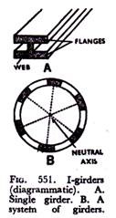
So requisite materials should be concentrated on the two surfaces, which are regions of greatest tension. The typical girders are constructed accordingly, so that they appear as I in cross-section (Fig. 551). Here the top and bottom portions, called flanges, are really stronger and the upright middle portion, known as web, connecting the two flanges may be made of lighter materials. In fact, web represents the neutral line or null-line where no strain operates.
Many plant organs, particularly the cylindrical ones like stems, etc., are frequently subjected to bending stress or flexion involving compression on one side and tension on the other.
ADVERTISEMENTS:
The mechanical tissues are distributed at the peripheral region here, so that they approach the I-girders in construction, the web being made of parenchyma or other tissues. Typical dicotyledons like sunflower possess mechanical tissues in hypodermis, in the region of pericycle and in association with vascular tissues (Fig. 552A) where the large pith at the central region serves as the web.
Thus it resembles a group of composite I-girders (Fig. 551B). In the square stems of members of family Labiatae like Leonurus, extra patches of collenchyma occur at the four corners, so that they appear like diagonally-placed I-girders (Fig. 552B).
In a typical monocotyledon like maize sclerenchyma is present as a band in hypodermal region and the bundles with sclerenchymatous sheaths remain more crowded towards the periphery (Fig. 552G).
Some members of sedge family, Gyperaceae possess patches of mechanical tissues just internal to epidermis and corresponding semilunar patches of the same on the lower side of the bundles, the two patches constituting the flanges of a girder (Fig. 552D).
ADVERTISEMENTS:
The central portion being hollow (web) the vascular bundles with associated mechanical tissues constitute composite I-girders. In some monocotyledons, as in the onion family, Liliaceae, the vascular bundles remain completely embedded in a peripherally located cylinder of mechanical tissues.
Bilaterally symmetrical organs like the foliage leaves have mechanical tissues arranged in form of I-girders parallel to one another and at right angles to the surface.
In the leaves of many grasses and sedges subepidermal I-girders extend from one surface to another (Fig. 553A), the web being composed of the bundle and parenchyma.
ADVERTISEMENTS:
In case of long leaves the upper surface is subjected to more vigorous tension, and so patches of subepidermal mechanical tissues occur at the outer surface to prevent tension, and the small I-girders present at the lower surface are meant for withstanding compression (Fig. 553B).
Inextensibility:
The roots and other organs which attach the plants to the soil or other substratum suffer from longitudinal pull or tension. Mechanical tissues are advantageously put in the central region in form of a compact mass in these organs. The degree of resistance of course depends on the cross-sectional area of the mechanical elements. Thus the roots have mechanical tissues associated with the vascular elements inside the stele (Fig. 554B).
The underground rhizome has also centrally located hollow or solid mechanical strand (Fig. 554A). The aquatic plants, particularly those which are adapted to grow in rapidly running water, e.g., Potamogeton of family Potamogetonaceae are inextetisible and naturally possess mechanical tissues at the central region.
ADVERTISEMENTS:
The same principle is applicable to the inflorescence axis and pendulous fruit-stalks. The stilt roots of the members of grass family, e.g., maize, have to encounter both flexion and longitudinal pull.
So here in addition to aggregation of mechanical tissues at the central region forming a massive stele, a peripheral band is also present (Fig. 554C).
Incompressibility:
The axis of a spreading tree with its arrays of branches and leaves has to bear the weight of the heavy crown, which may be compared to putting a load at the top of a cylindrical axis. Here the axis is subjected to longitudinal compression. The mechanical tissues are effectively aggregated at the central portion which serves as a solid column for withstanding longitudinal compression.
The underground and aquatic organs are subjected to radial or crushing pressure by the surrounding medium. The aquatic plants have loosely-arranged cortical cells which give requisite protection.
In plants like Sagittaria, Alisma, etc., the epidermis also becomes associated with the outer layers of cortex for the purpose. The roots of grasses in particular develop tubular sheaths of cells, often with suberised walls.
Shearing Stresses:
ADVERTISEMENTS:
The flat organs like the leaves are often subjected to violent shearing stresses due to movement of surrounding air or water. The wind currents work at right angles to the surface of the leaves and cause considerable laceration. To stand against this stress the I-girders present for securing inflexibility are firmly held together by a large number of cross-ties in form of veins which often form a network.
The I-girder arrangement is more pronounced in monocotyledonous leaves, usually having parallel venation (Fig. 553). The margins of the leaves are particularly exposed to shearing stresses. They have special arrangement for protection by increased thickness of the epidermis, and frequent occurrence of thick-walled collenchyma in the subepidermal region.


