ADVERTISEMENTS:
The following points highlight the top five types of Staining. The types are: 1. Simple Staining 2. Differential Staining 3. Gram Staining 4. Acid Fast Staining 5. Endospore Staining.
Staining Type # 1. Simple Staining:
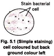
Colouration of microorganisms by applying single dye to a fixed smear is termed simple staining. One covers the fixed smear with stain for specific period, after which this solution is washed off with water and slide blotted dry. Basic dyes like crystal violet, methylene blue and carbolfuchsin are frequently used in simple staining to determine the size, shape and arrangement of prokaryotic cells. (Fig 5.1)
Staining Type # 2. Differential Staining:
ADVERTISEMENTS:
These staining procedures are used to distinguish organisms based on staining properties. They are slightly more elaborate than simple staining techniques that the cells may be exposed to more than one dye or stain, for instance use of Gram staining which divides bacteria into two classes-Gram negative and Gram positive.
Staining Type # 3. Gram Staining:
It is one of the most important and widely used differential staining techniques in microbiology. This technique was introduced in 1884 by Danish Physician Christian Gram. Gram staining procedure is illustrated in fig 5.2.
In the first step the smear is stained with basic dye crystal violet (Primary stain) followed by treatment with iodine solution functioning as mordant.
ADVERTISEMENTS:
Iodine increases the interaction between cell & dye so that cell stains strongly. The smear is next decolourized by washing with ethanol or acetone. This step generates the differential aspect of Gram stains. Gram positive bacteria retain crystal violet and become colourless.
Finally smear is counter-stained with a simple basic dye different in color from Crystal violet. Safranin is the most common counter stain which colours Gram negative bacteria pink to red and leaves Gram positive bacteria dark purple. (Fig 5.2)
The differences in staining responses to the Gram stain can be related to chemical and physical difference of cell walls. The Gram-negative bacterial cell wall is thin, complex multilayered structure and contains relatively high lipid contents in addition to protein and mucopeptide.
The higher amount of lipid is readily dissolved by alcohol, resulting information of large pore in the cell wall, thus facilitate leakage of crystal- violet – iodine (CV-I) complex which results in decolorization of the bacterial cell.
Which later take counter stain and appears red. In contrast the cell wall of gram+ve bacteria is thick and chemically simple, composed mainly of mucopeptides. When treated with alcohol, it causes dehydration and closure of cell wall pore, thereby does not allow the loss of (CV-I) complex and cell remain purple.
Staining Type # 4. Acid Fast Staining:
It is another important differential staining procedure. It is most commonly used to identify Mycobacterium spp. These bacteria have cell wall with high lipid content such as mycolic acid -a group of branched chain hydroxy lipids, which prevent dyes from readily binding to cells.
They can be stained by Ziehl-Nulsen method, which uses heat and phenol to derive basic fuchsin into the cells. Mycrobacterium spp. were penetrated with basic fuchsin, not easily decolourized by acidified alcohol (acid alcohol) and thus are said to be acid fast.
Non arid fast bacteria are decolourized by arid alcohol and thus are stained blue by methylene blue counter stain.
Staining Type # 5. Endospore Staining:
ADVERTISEMENTS:
Spore formation takes place in some bacterial genera to withstand unfavourable conditions. All bacteria cannot form spores, only few bacterial genera including Bacillus, Clostridium, Desulfotomaculum produce sporulating structure inside vegetative cells called endospore.
Endospore morphology and location vary with species and are valuable for identification Endospores are not stained well by most dyes, but once stained, they strongly resist decolorization.
In the Schaffer-Fulton procedure, endospores are first stained by heating bacteria with malachite green, which is very strong stain that can penetrate endospores. After malachite green treatment, the rest of the cell is washed free of dye with water and is counter-stained with safranin. This technique yields a green endospore with red vegetative cell. (Fig. 5.3)


