ADVERTISEMENTS:
In this article we will discuss about:- 1. Discovery of Lysosomes 2. Types of Lysosomes 3. Functions.
Discovery of Lysosomes:
They were discovered accidently by a Belgian scientist, Christian de Duve, in 1955 through fractionation technique. The organelles were observed under electron microscope by Novikoff (1956).
He also coined the term, lysosomes. Lysosomes (Gk. lysis- digestive or loose, soma- body) are small vesicles which are bounded by a single membrane and contain hydrolytic enzymes in the form of minute crystalline or semi crystalline granules of 5-8 nm.
ADVERTISEMENTS:
About 50 enzymes have been recorded to occur in them. All the enzymes do not occur in the same lysosome but there are different sets of enzymes in different types of lysosomes.
The important enzymes are acid phosphatases, sulphatases, proteases, peptidases, nucleases, lipases and carbohydrates. They are also called acid hydrolases because these digestive enzymes usually function in acidic medium or pH of 4—5. Acidic conditions are maintained inside the lysosomes by pumping of H+ or protons into them.
The covering membrane of lysosomes keeps the hydrolytic enzymes out of contact from the cellular contents. It is itself protected from them by high glycosylation of its proteins and lipids.
The covering membrane becomes fragile in the absence of the oxygen, or the presence of excess of vitamins A and E, male and female hormones, bile salts, carcinogens, silica, asbestos particles, heat, many drugs, X-rays and ultra-violet rays.
ADVERTISEMENTS:
The membrane is protected from these agencies by cortisone, cortisol, chloroquine and a type of cholesterol. Lysosomes are called suicide bags because of the presence of a large number of digestive enzymes or acid hydrolases in them. Only a thin membrane separates the destructive enzymes from the rest of the cell. If the membrane happens to get broken, the various cellular constituents would undergo lysis.
Lysosomes are generally rounded but can be irregular (e.g., root tip cells) in outline. The diameter varies from 0.2-0.8 µm but sometimes it may grow to a very large size (up to 5 µm in leucocytes, kidney cells, etc.). The interior may be almost solid or differentiated into outer denser region and a central less dense mass with granular content.
Lysosomes occur in all animal cells with the exception of red blood corpuscles. In plants and fungi, their function is taken over by vacuoles. In animals, lysosomes are abundant in leucocytes, macrophages, Kupffer’s cells and similar cells with phagocytic activity.
Lysosomes are believed to be formed by the joint activity of endoplasmic reticulum endosomes and Golgi complex (GERL system). The precursors of hydrolytic enzymes are mostly synthesised at the rough endoplasmic reticulum.
The latter transfers those to the forming face of Golgi complex either directly or from smooth endoplasmic reticulum through its vesicles. In Golgi complex the precursors are changed to enzymes. The enzymes are then packed in larger vesicles which are pinched off from the maturing face. Golgian vesicles are joined by endosomes to produce lysosomes.
Lysosomes do not normally burst in the cytoplasm. All materials which are to be acted upon by lysosome enzymes must enter them. Rather the materials are usually enclosed inside vacuoles and the vacuoles fuse with the lysosomes for digestion of materials.
Lysosomes take part in intracellular digestion of various types of materials of endogenous or exogenous origin. Extracellular digestion can be performed by them under certain conditions. They help in removing various toxic substances including carcinogens.
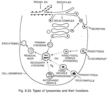
Lysosomes pass through various stages in the same cell. The phenomenon is called polymorphism or existence of more than one morphological form. Depending upon their morphology and function, there are four types of lysosomes— primary, secondary, residual bodies and auto-phagic vacuoles (Fig. 8.33).
Types of Lysosomes:
1. Primary Lysosomes:
ADVERTISEMENTS:
They are newly pinched off vesicles from the Golgi apparatus which generally fuse with some endosomes to become fully functional. The primary lysosomes are small in size. They contain hydrolytic enzymes in the form of granules.
2. Secondary Lysosomes:
They are also called heterophagosomes or digestive vacuoles. A secondary lysosome is formed by the fusion of food containing phagosome with lysosome (having hydrolytic or digestive enzymes). Digestion occurs. The digested food passes out into the cytoplasm. Finally, the secondary lysosome is left with undigested food.
ADVERTISEMENTS:
3. Residual Bodies (Residual or Tertiary Lysosomes):
They are those lysosomes in which only indigestible food materials have been left. The residual bodies or lysosomes pass outwardly and fuse with the plasma membrane to throw out the debris into external environment by exocytosis or ephagy.
Sometimes, residual bodies remain inside the cells due to failure of exocytosis and absence of some hydrolytic enzymes. This leads to pathological diseases (storage diseases) like hepatitis, Pompe’s disease, Hurler’s disease, Tay-Sachs disease and polynephritis. Ageing is also due to them. Lipofuschin pigment granules are actually residual bodies.
4. Autophagic Vacuoles (Auto-phagosomes, Auto-lysosomes):
ADVERTISEMENTS:
They are produced by the fusion of a number of primary lysosomes around worn out or degenerate intracellular organelles. The latter are first wrapped over by one or two membranes from endoplasmic reticulum (Dunn, 1990) before being recognised by lysosomes.
The cell debris is digested the phenomenon is also called autophagy or auto digestion. It helps in disposal of cell debris. The worn out, aged or injured cells are also disposed of similarly (apoptosis). Therefore, lysosomes are also called disposal bags or disposal units.
The digested products are made available to the cell for new synthesis. Lysosomes are, therefore, also known as recycling centres. Besides removing worn out organelles, old or diseased cells, the auto-phagic vacuoles are also used in removing internal obstructions. Autophagic vacuoles provide nourishment during starvation.
Autolysis:
ADVERTISEMENTS:
It is self destruction of a cell, tissue or organ with the help of lysosomes. Lysosomes performing autolysis do not enclose the structures to be broken down. Instead, they themselves burst to release the digestive enzymes. Autolysis occurs in ageing, dead or diseased cells. The disappearance of larval organs during metamorphosis (e.g. tail in frog) is due to autolysis.
Functions of Lysosomes:
1. Intracellular Digestion:
Individual cells may obtain food through phagocytosis. The same is digested with the help of lysosomes.
2. Extracellular Digestion:
For this the lysosomes release enzymes in the external environment through exocytosis.
3. Body Defence:
ADVERTISEMENTS:
Lysosomes of leucocytes devour foreign proteins, toxic substances, bacteria and other microorganisms. They thus take part in natural defence of the body.
4. Autophagy:
In the metamorphosis of many animals (e.g. amphibians, tunicates) certain embryonic parts like tail, gills, etc. are digested through the agency of lysosomes. The digested food is used in the growth of other parts.
5. Removal of Obstructions:
Obstructing structures are destroyed by lysosomes.
6. Mobilisation of Reserves:
ADVERTISEMENTS:
During periods of starvation, lysosomes provide nourishment by rapidly hydrolysing the organic foods stored in the cells (carbohydrates, fats and proteins). Mobilisation of reserve food during germination of seeds is also accomplished by lysosomes. Extra nourishment may also be got by digesting some organelles and cells.
7. Intracellular Scavenging:
In long lived cells the lysosomes perform intracellular scavenging by removing old or useless organelles.
8. Sperm Lysins:
They are lysosomal enzymes which are used for breaking limiting membrane of eggs.
9. Disposal of Useless Cells:
They cause breakdown of ageing and dead cells.
ADVERTISEMENTS:
10. Storage Diseases:
In certain regions due to some malfunction, the residual bodies do not undergo exocytosis. Instead, they remain inside the cells and cause disease, e.g., hepatitis, polynephritis.
11. Formation of Thyroxine:
In thyroid, active hormone thyroxine is formed through hydrolysis of thyroglobulin by the agency of lysosomes.
12. Cell Division:
Lysosomes seem to be essential for cell division perhaps by overcoming agents that cause repression of mitotic cycle.
13. Genetic Changes:
They may harm genetic material through the release of nucleases.
It may result in mutations, breakage of chromosomes and other abnormalities. Blood cancer may be result of such an activity.
14. Carcinogenesis:
Lysosomes remove carcinogens by engulfing and separating them. However, when the carcinogen is in excess, lysosome may harm the living cells as in case of lung fibrosis caused by silicosis or asbestosis.
15. Leucocyte Granules:
Leucocyte granules are derived from lysosomes.
16. Osteogenesis:
At the time of formation of bones from cartilage and during re-modelling of the bone, lysosomes of the osteoclasts cause breakdown of existing matrix so that it may be replaced by the new one.

