ADVERTISEMENTS:
In this article we will discuss about Amoeba Proteus:- 1. Habit, Habitat and Culture of Amoeba Proteus 2. Structure of Amoeba Proteus 3. Locomotion 4. Nutrition 5. Respiration and Excretion 6. Metabolism 7. Behaviour 8. Reproduction 9. Immortality 10. Biological Significance.
Contents:
- Habit, Habitat and Culture of Amoeba Proteus
- Structure of Amoeba Proteus
- Locomotion of Amoeba Proteus
- Nutrition in Amoeba Proteus
- Respiration and Excretion in Amoeba Proteus
- Metabolism in Amoeba Proteus
- Behaviour of Amoeba Proteus
- Reproduction in Amoeba Proteus
- Immortality of Amoeba Proteus
- Biological Significance of Amoeba Proteus
ADVERTISEMENTS:
1. Habitat and Culture of Amoeba Proteus:
Amoeba Proteus is widely distributed. It is commonly found in the ooze or bottom mud in freshwater pools, ponds, ditches, lakes and slow streams, often in shallow water on the underside of aquatic vegetation. It is also found in damp soils. The sides of lotus ponds and the water troughs are excellent places for the collection of amoebae.
It occurs in abundance in the water which contains bacteria and organic substances, such as leaves, twigs and other aquatic vegetation in abundance.
Culture of Amoeba Proteus:
Amoeba Proteus may be obtained for laboratory use from a variety of places such as organic ooze from decaying vegetation or the lower surface of the lily pads. To make a culture of Amoeba put some pond water, mud and leaves in 100 ml of water containing a few grains of wheat.
ADVERTISEMENTS:
Amoebae will appear after a few days, this shows the presence of cysts in the pond water. To make a pure culture, boil four or five grains of wheat in 100 ml of distilled water for 10 minutes and cool for a few days; to this add some Amoebae from the first culture and cover with glass plate; in ten days many Amoebae will be formed in the pure culture.
2. Structure of Amoeba Proteus:
(i) Size and Shape:
Amoeba proteus is a one-celled microscopic animal about 0.25 mm (250 microns) in diameter and so transparent that it is invisible to the naked eyes. Under the compound microscope, it appears as an irregular, colourless, translucent mass of living animal-like jelly or protoplasm that is constantly changing its shape by sending out and withdrawing finger-like processes, the pseudopodia.
The name Amoeba is derived from a Greek word amoibe which means change. The specific name proteus is based on the name of a mythological Greek sea-god who constantly changes his shape. Although it possesses no cell wall, it has a thin delicate outer membrane called the plasma lemma.
Just beneath this is a non-granular layer, the ectoplasm which encloses the granular endoplasm. However, there is no line of demarcation between the ectoplasm and endoplasm.
(ii) Pseudopodia:
Pseudopodia (Gr., pseudos = false; podos = foot) are temporary finger-like and blunt extensions which are constantly being given out or withdrawn by the body. They are broad to cylindrical with blunt rounded tips and are composed of both ectoplasm and endoplasm.
Such pseudopodia are called lobopodia. These are formed as a result of liquefaction and flowing forward of the cytoplasm. As many pseudopodia are formed simultaneously, Amoeba proteus is a polypodial species.
(iii) Plasma-Lemma:
ADVERTISEMENTS:
The plasma lemma is very thin, delicate, invisible, elastic external cell membrane. The thickness of plasma lemma may be from 1/4 micron (0.00025 mm) to 2 microns.
It is composed of a double layer of lipid and protein molecules. According to Schneider and Wohifarth Batterman (1959), the plasma lemma consists of two darkly staining layers, about 200 A0 thick separated by a clear layer. Other filaments, about 80 A0 in diameter, extend 1100 to 1700 A0 out into the medium from the plasma lemma.
The outer layer of plasma lemma is supposed to contain mucoprotein. Plasma lemma can regenerate itself when broken. This membrane is selectively permeable and it regulates an exchange of substances such as water, O2 and CO2 between the animal and the surrounding medium.
It also retains the protoplasm within the cell. Numerous fine, ridge-like projections from the outer surface of plasma lemma are supposed to be adhesive and binding the organism to its substratum.
(iv) Cytoplasm:
Inside the plasma lemma, there is a dense mass of cytoplasm containing several organelles. It is differentiated fairly into two district zones, an outer ectoplasm and an inner endoplasm.
(v) Ectoplasm:
ADVERTISEMENTS:
The ectoplasm forms the outer and relatively firm layer lying just beneath the plasma lemma. It is a thin, clear (non-granular) and hyaline layer. It is thickened into a hyaline cap at the advancing end at the tips of pseudopodia. The ectoplasm has a number of conspicuous longitudinal ridges. Due to the presence of longitudinal ridges in the ectoplasm, it is considered as a supporting layer. Ectoplasm lends form to the cell body.
(vi) Endoplasm:
The endoplasm forms the main body mass completely surrounded by the ectoplasm. It is granular heterogeneous fluid containing bi-pyramidal crystals. According to Mast, the endoplasm is made up of an outer, relatively stiff plasmagel and a more fluid inner plasmasol.
The plasmagel is granular and more solid but its granules show no movement. The plasmasol is highly granular fluid having various inclusions and it shows streaming movements. Besides granules, endoplasm contains a number of important inclusions such as nucleus, contractile vacuole, food vacuoles, mitochondria, Golgi apparatus, fat globules and plate-like or bi-pyramidal crystals.
ADVERTISEMENTS:
(vii) Endoplasmic Organelles:
Endoplasm contains a number of organelles or structures suspended in it. These organelles are nucleus, contractile vacuole, food vacuoles and water globules.
1. Nucleus:
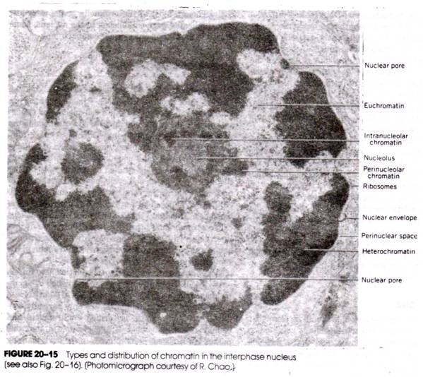
In Amoeba proteus, there is a single conspicuous nucleus. The nucleus appears as a biconcave disc in young specimens but it is often folded and convoluted in older specimens. The nucleus has a firm nuclear membrane or nuclear envelope and contains a clear achromatic substance with minute chromatin granules or chromidia distributed uniformly near the surface.
The nucleoplasm is small in quantity. Such a nucleus is called massive or granular nucleus. Electron microscopic studies show a honeycomb lattice under double layered nuclear envelope. Within the nucleus are many small spherical chromosomes said to number over 500. The position of nucleus in the endoplasm is not definite, but changes during the movement of Amoeba.
During the life of an Amoeba, before the period of reproduction, the nucleus plays an important role in the metabolic activities of the cell. This has been proved by experiments in which the animal was cut in two. The streaming of cytoplasm ceases within a few minutes in the piece without a nucleus but is resumed after a few hours.
ADVERTISEMENTS:
The enucleated Amoeba may attach itself to the substratum and exhibits irritability although its Reponses to stimuli are modified. Food bodies are engulfed and digested in an apparently normal manner but death finally ensues. The part with the nucleus continues its life as a normal Amoeba. An isolated nucleus will not survive.
Thus, the nucleus and cytoplasm are interdependent and their separation ultimately results in the death of both.
2. Contractile Vacuole:
The outer part of the endoplasm near the posterior end contains a clear, rounded and pulsating vacuole filled with a watery fluid. This vacuole, called the contractile vacuole, is enclosed by a unit membrane. It is not fixed but circulates in the endoplasm. It arises near the hind end of the animal and it grows in size probably by fusion of a number of smaller vacuoles.
As it continues to grow, it comes to lie in the peripheral plasmagel layer where it remains as the endoplasm flows onwards, so that it is left at the posterior end where it bursts by contraction of its wall and the contents are forced out through no obvious pore. It appears near the point of its disappearance, then it travels towards the nucleus and finally moves backwards.
The vacuole fills rhythmically with fluid and then discharges it to the exterior. In protozoans, the contractile vacuole is surrounded by a crowd of mitochondria, close to which tiny vacuoles of water appear, which then coalesce to form a larger vacuole.
The mitochondria provide the energy for actual formation and working of the vacuole. It expands (diastole) and contracts (systole) rhythmically and serves for excretion and osmoregulation.
It removes CO2 and waste substances from the animal, it is not only excretory and respiratory but mostly it is a hydrostatic organ because it constantly removes the water which the Amoeba absorbs, thus, it regulates the osmotic pressure,and it harmonizes the tension between the protoplasm and the surrounding water, consequently it regulates the weight of the animal also.
In many marine and parasitic amoebae, there is no contractile vacuole, this is because the osmotic pressure of their protoplasm is about the same as that of the surrounding medium.
3. Food Vacuoles:
Numerous food vacuoles are scattered in the endoplasm. These are non-contractile and of different size. Each food vacuole contains a morsel of food under digestion. The food vacuoles are carried about by the movements of the endoplasm. Digestion of food takes place inside the food vacuole. The endoplasm also contains some waste substances and grains of sand.
4. Water Globules:
These are several small, spherical, colourless and non-contractile vacuoles filled with water.
Ultrastructure or Structure by Electron Microscope:
The electron microscopic studies have not revealed the occurrence of two colloidal phases, sol and gel, in the endoplasm. It is believed that it is the ectoplasm which is in the gel state, while endoplasm is in the sol state.
De Bruyn has proposed that the protoplasm can be presumed as a “three dimensional network” of protein chains, linked together by cross linkages of side chains. The gel state is due to the fully extended protein chains and the sol state is due to the contraction of such chains.
The structure of nucleus of Amoeba proteus is like that of a honeycomb-lattice. The nuclear membrane is double-layered having pores. The nucleoplasm contains a few nucleoli and a large number of chromosomes. Various organelles, characteristic of an animal cell, are also seen. The mitochondria are more or less oval with tubular cristae.
The Golgi apparatus appear as groups of sac-like tubules. The lysosomes are found scattered as minute membrane-bound spherical bodies. The endoplasmic reticulum forms a network of tubules as well as vesicles. The endoplasm contains reserve food material in the form of plate-like or bi-pyramidal crystals. Contractile vacuole is seen surrounded by several mitochondria and vesicles.
3. Locomotion of Amoeba Proteus:
Amoeba Proteus exhibits characteristic amoeboid movement by the formation of finger-like temporary processes, the pseudopodia. The pseudopodia of Amoeba are known as lobo-podia due to their blunt, finger-like appearance and rounded tips. The ectoplasm forms a blunt projection into which the endoplasm flows to form a pseudopodium at the advancing end which is then spoken as the anterior end.
Usually several small pseudopodia are formed at first, one of these enlarges and the others disappear. The protoplasm which enters pseudopodia is naturally withdrawn from other parts, so that the animal not only changes its body shape but also its position, thus, pseudopodia bring about change of shape and position of the animal. These are called amoeboid movements which occur not only in Amoeba but in other Protozoa and some amoeboid cells of Metazoa.
4. Nutrition in Amoeba Proteus:
An Amoeba is unable to form its food from simple substances, but it requires ready-made organic substances for food; such a mode of nutrition in which solid organic particles are ingested is called zoo trophic or holozoic.
The food of Amoeba consists of algal cells and filaments, bacteria, other protozoans, small metazoans such as rotifers and nematodes and organic matter. Smaller flagellates and ciliates appear to constitute the favorite food of Amoeba.
Amoeba proteus does not feed on diatoms, as is often claimed. Amoeba exhibits a choice for food and it can discriminate between inorganic particles and organic food. If a particle of carbon is fixed to the food, the animal will ingest the food and leave the carbon particle out.
There is no mouth but food is ingested at any point which is generally at the anterior advancing end. The nutrition involves a number of processes, viz., ingestion, digestion, assimilation, dissimilation and egestion.
(a) Ingestion:
No definite regions or organelles for food intake are present. The food is captured by pseudopodia, usually by the formation of food cup, in which a pseudopodium embraces the prey from each side while a thin sheet advances over it from above pinning it to the substratum.
The cup is then completed below and the food is enclosed. Food particles may be engulfed at any point on the surface of the body, but is usually taken in at what may be called the temporary anterior end, i.e., the part of the body toward the direction of locomotion. According to Rhumbler (1930), ingestion in Amoeba takes place in many ways depending upon the nature of the food.
The following methods of ingestion are employed:
(i) Circumvallation:
When an Amoeba comes near its food the part immediately in line with it stops moving, and pseudopodia are formed above, below and on the sides of the food to form a food cup, the food cup does not touch the food, but edges of the food cup fuse around the food to form a non-contractile food vacuole into which some water is also taken.
The walls of the food vacuole were made of ectoplasm which becomes internal and changes into endoplasm. This method of ingestion is used for capturing live prey. It is, however, not understood how Amoeba perceives that the particle is suitable for food and puts forward the pseudopodia so as to engulf it.
(ii) Circumfluence:
When the food is less active or immobile, a pseudopodium comes in contact with the food to form a food cup above it and pinning it down to the substratum, the cup is then completed below to enclose the food in a food vacuole. By repeating this process, the Amoeba can ingest and roll up long filaments of algae. In other species of Amoeba, food is ingested by import and invagination.
(iii) Import:
In Amoeba verrucosa, food comes into contact with the animal and sinks passively into the body. It absorbs and devours filamentous algae in this way.
(iv) Invagination:
Amoeba verrucosa comes in contact with food and adheres to it, the ectoplasm along with food is invaginated as a tube into the endoplasm, and the food particle is sucked in, the plasma lemma disappears and ectoplasm changes into endoplasm.
The newly ingested organism may remain active for a time in the large primary food vacuole. Within an hour, the primary food vacuoles break down into smaller secondary vacuoles, these subdivide into numerous minute vacuoles which form a large portion of the endoplasmic content.
(v) Pinocytosis:
When the ingestion of fluid material in bulk takes place by the cell through the plasma membrane, the process is known as pinocytosis (Gr., pinein = to drink). The process of pinocytosis was first of all observed by Mast and Doyle (1934) in Amoeba proteus. By pinocytosis, the animalcule (amoebae) absorb the high molecular compounds from the outer environment.
Pinocytosis does not take place through the whole surface of the body of the amoeba. It is understood that plasma lemma along with the colloidal food material forms pinocytosis channels which run from the surface deep into the endoplasm.
The internal ends of the channels then break off forming pinocytosis vesicle or pinosomes containing engulfed food material. The pinosomes, later on, are transported to the interior of the cell where they are fused with the lysosomes. It is yet to be confirmed whether pinocytosis is a normal means of ingestion in Amoeba.
(b) Digestion:
Digestion takes place in a primary food vacuole after it gets embedded in the endoplasm. The contents of the food vacuole are at first acidic due to HCI, but later they become alkaline, living food dies in the acid phase. The protoplasm secretes enzymes into the vacuoles which convert the proteins into amino acids, starch into soluble sugar, and fats into fatty acids and glycerol.
The presence of some enzymes, e.g., proteases (protein-splitting enzymes), amylases (starch-digesting enzymes) and lipases (fat-splitting enzymes) have been demonstrated in Amoeba. When the digested food is reduced to molecular form, then the food vacuole buds off smaller and smaller secondary food vacuoles which carry away the digested food.
The digestion of food in Amoeba is said to be intracellular in contrast to the extracellular digestion in higher animals, like earthworm and frog, taking place the cells in the cavity of an alimentary canal.
(c) Assimilation:
The digested food, water and minerals are absorbed by the surrounding protoplasm by a simple process of diffusion. In Amoeba, the food vacuoles constantly move about in streaming endoplasm by cyclosis and directly supply nourishment to all parts of the cell. In the protoplasm, the digested food gets assimilated to build new protoplasm.
The amino acids are built up to form living protoplasm; sugar, fatty acids and glycerol provide energy. This ability to form living protoplasm from simple substances is a fundamental property of living matter.
(d) Dissimilation:
The living protoplasm is broken down constantly by oxidation to produce heat, kinetic energy and waste products. Complex molecules of protoplasm are broken down by dissimilation to produce energy for various activities of the animal.
(e) Egestion:
The undigested residue of food vacuoles is waste which is heavier than protoplasm, hence, it gravitates towards the posterior end from where it is dropped out by the Amoeba moving away from it. Egestion of undigested particles occurs at no fixed point, they pass out at any point on the surface through no special opening.
The process of egestion is not so simple in Amoeba verrucosa, which possesses an ectoplasmic pellicle that is a thick and tough membrane. Waste pellets are extruded as shown in Fig. 14.14. A new pellicle is formed at the point of exit to prevent the outflow of the endoplasm.
5. Respiration and Excretion in Amoeba Proteus:
Amoeba has no respiratory organs and no respiratory pigments. Respiration in Amoeba occurs by diffusion through the general body surface (plasma lemma). Amoeba is aerobic, takes in oxygen and gives off carbon dioxide like other animals. The oxygen dissolved in the surrounding water passes into the cytoplasm of Amoeba by diffusion.
Since the concentration of oxygen in the water is higher than that in the Amoeba’s cytoplasm, oxygen constantly enters and is immediately used up in the burning of foods.
Thus, the concentration of oxygen within the animal always remains lower than that in the outside water, and oxygen continuously enters the animal and is available for energy requirements. During metabolic activities, the oxygen burns or oxidizes the living matter or cytoplasm of Amoeba and breaks it into simpler compounds.
As a result, water, carbon dioxide and urea are formed and energy is liberated which is stored in the high energy bonds of ATP and used in the life activities of the organism. Carbon dioxide diffuses to the outside because it is always at a high concentration within the body of Amoeba than in the surrounding water.
If an Amoeba is placed in hydrogen instead of oxygen, then movements cease and death results, if carbon dioxide is introduced in place of oxygen then the Amoeba first encysts but finally dies.
Excretion in Amoeba Proteus:
The oxidation of carbohydrates and fats results in the production of metabolic wastes, carbon dioxide and water. In Amoeba, the by-product of oxidation of proteins is ammonia and carbon dioxide and less often urea. Carbon dioxide and ammonia are soluble in water and these are excreted out through the plasma lemma by diffusion in the surrounding water or in the water discharged by the contractile vacuole.
Osmoregulation:
Osmoregulation is a process in which the water contents of the protoplasm are controlled. The regulation of water contents in the protoplasm of Amoeba is performed by the contractile vacuole. The protoplasm of Amoeba is more concentrated than the surrounding water, so that a regular water current enters its body by osmosis through the semi-permeable plasma lemma. If the excess of water is not expelled, would lead to the rupture of the animal. This excess of water is collected by the contractile vacuole and expelled out of the protoplasm.
The disappearance of one vacuole is followed by the production of a new one. The regulation of water in the protoplasm maintains an osmotic equilibrium with the surrounding water. The absence of contractile vacuole in most marine amoebae may be due to the fact that the salt concentration of the protoplasm is almost equivalent to that of the surrounding medium so that water does not accumulate in the protoplasm.
Marine amoebae develop contractile vacuole when they are placed in freshwater. On the other hand, if freshwater amoebae are transferred to salt water their contractile vacuoles decrease and finally disappear altogether. Thus, it is probable that the chief function of the contractile vacuole is to regulate the water contents of Amoeba.
6. Metabolism in Amoeba Proteus:
Amoeba Proteus takes in food and O2, from which it makes protoplasm, then the protoplasm is broken down into waste products and kinetic energy is produced; these processes involve many complex chemical reactions, the sum total of which is called metabolism.
The processes which use energy and build up protoplasm are known as anabolism and those which break down protoplasm to release energy and produce waste products are called katabolism.
The waste products of katabolism are urea, CO2, H2O and minerals. In metabolism, the nucleus controls the assimilation of food, and the cytoplasm carries on the kataholic phase. The metabolic processes exhibited by Amoeba (Fig. 14.16) are ingestion, digestion, egestion, absorption, circulation, assimilation, dissimilation, secretion, excretion and respiration.
7. Behaviour of Amoeba Proteus:
There are no special structures for the reception of stimuli in Amoeba Proteus still it responds to various kinds of stimuli. The responses of Amoeba to stimuli or changes in its environment, either internal or external, constitute its behaviour. The responses or reactions of Amoeba Proteus to stimuli are due to the fundamental property of protoplasm called irritability.
All changes in the environmental conditions are termed as stimuli or irritations and the property of response to stimuli is called irritability or sensibility. The behaviour of Amoeba involves change in shape, locomotion, food getting (ingestion of food), avoiding of un-favourable environment, hunger and so on. Its responses to different forms of sitmuli vary.
The responses to stimuli are called taxes (singular, taxis). A taxis may be either positive, in which the organism moves towards the stimulus, or negative, in which the organism moves away from the stimulus. Amoeba proteus exhibits both types of taxes, positive as well as negative, specifically to different stimuli.
With respect to the kinds of stimuli, the taxes are classified as follows:
(i) Thigmotaxis (response to contact or touch):
The response of Amoeba Proteus to contact is varied. Amoeba Proteus reacts negatively when touched at any point with a solid object, the part affected contracts and the animal moves away. A floating Amoeba with spread pseudopodia, responds positively to contact with the solid object by fastening to it.
Contact with the food also results positive reactions. The creeping Amoeba touches lightly with a needle responds negatively by drawing back and moving away. Amoeba, therefore, reacts negatively to a strong mechanical stimulus and positively to a weak one.
(ii) Chaemotaxis (response to chemicals):
Amoeba Proteus reacts negatively to many chemicals and changes in the culture water. It also avoids sand particles. It responds positively to the food organisms.
(iii) Thermotaxis (response to heat):
Negative reactions result if Amoeba is locally affected by heat, since the animal will move away from heat stimuli. Amoeba’s rate of locomotion is lessened by colder temperatures and may cease entirely near the freezing point. Its rate increases up to 30°C but it ceases to move at temperatures higher than this.
(iv) Phototaxis (response to light):
Amoeba moves away from strong light and may change its direction a number of times to avoid it, but it may react positively to a weak light.
(v) Galvanotaxis (response to electric current):
When an electric current is passed through the water containing Amoeba, it stops moving, withdraws its pseudopodia and becomes globular. In the weak electric current, it moves towards the negative pole (cathode) and, thus, avoids positive pole (anode).
(vi) Rheotaxis (response to water current):
Amoeba Proteus shows positive rheotaxis as it tends to move in line with the water current.
(vii) Geotaxis (response to gravity):
Amoeba Proteus exhibits positive geotaxis since it moves toward the centre of gravity like other animals.
8. Reproduction in Amoeba Proteus:
Reproduction in Amoeba Proteus is a periodic process taking place at intervals. The duration between successive phases of reproduction essentially depends on the rate of growth of Amoeba. When Amoeba attains a maximum size, i.e., 0.25 mm, it starts to reproduce. Reproduction in Amoeba chiefly occurs by asexual method, i.e., by binary fission, multiple fission and sporulation. Amoeba does not reproduce sexually by mating, i.e., by the fusion of cells or gametes.
(i) Binary Fission:
Binary fission is the most common mode of reproduction. It results in the division of parent amoeba into two daughter amoebae. The division involves the nuclear division followed by cytoplasmic division. Amoeba undergoes binary fission during favourable conditions of food and temperature.
Binary fission occurs when the organism reaches a maximum limit of size, it becomes sluggish and spherical with its surface covered with small radially arranged pseudopodia.
In binary fission, the contractile vacuole ceases to function, the nucleus divides mitotically, then the cell constricts in the middle to form two daughter cells. There is a correlation between nuclear division and changes in external characters. The amoeba divides by mitosis and involves the prophase, metaphase, anaphase and telophase.
In the prophase stage, the nucleus becomes oval and numerous fine pseudopodia are formed radiating in all the directions. The cytoplasm loses its transparency to a large extent and the contractile vacuole disappears. The honeycomb-like lattice just below the nuclear membrane first fragments and then disappears. The nucleoli disintegrate. The chromosomes emerge in the central nucleoplasm.
The metaphase stage is marked by the arrangement of the chromosomes at the equator. Each chromosome splits longitudinally and becomes paired. The chromosomes, on each side, become attached to the spindle fibres arising from multiple poles, situated within the nuclear membrane. Externally the pseudopodia begin to thicken.
In the anaphase stage, the daughter chromosomes move towards opposite poles and the constriction of the nuclear membrane begins in the middle. The pseudopodia become thick and coarse. In the telophase stage, the transverse constriction of the nuclear membrane is completed and the nucleus is finally divided into two daughter nuclei.
In each daughter nucleus, the lattice is formed just below the nuclear membrane and the nucleoli reappear. Next follows cytokinesis. Amoeba stretches and constricts in the middle. Numerous large pseudopodia are formed at opposite poles, drawing both the daughter amoebae in opposite directions.
Ultimately the amoeba divides into two daughter amoebae. The pseudopodia become normal, then each daughter amoeba acquires a contractile vacuole and begins to grow. At about 24°C, the process takes about 20 to 30 minutes.
A very interesting feature of binary fission, observed in the division of nucleus, is the existence of multipolar nuclear spindle in the metaphase, which is reduced to tri-polar nuclear spindle in the mid-anaphase stage and finally reduced to unipolar nuclear spindle at the end of late anaphase.
In Amoeba reproducing by binary fission, the parent becomes completely merged in the offspring. Thus, there exists a continuity of life, so that Amoeba is potentially immortal. However, death may be due to starvation, accident or some other misfortune.
(ii) Multiple Fission:
Amoeba Proteus reproduces by multiple fission during un-favourable conditions of food and temperature (i.e., scarcity of food and rise and fall in temperature). Pseudopodia are withdrawn, the animal becomes rounded, streaming movements of endoplasm cease, larger granules dissolve and protoplasm becomes minutely granular, distinction between ectoplasm and endoplasm is lost.
The animal begins to rotate and secretes a cyst inside to which two new layers are added to complete a three-layered cyst, then the rotation of the animal stops. The cyst is a resting stage and it protects the animal, it also brings about dispersal of the animal when the pond dries up.
On return of favourable conditions or the cyst being blown into another pond, the cyst bursts and the protoplasm flows out to reform the Amoeba. It has been reported that reproduction occurs in the cyst by multiple fission.
The nucleus divides amitotically into 500 to 600 nuclei which move towards the periphery of the cell. Each nucleus acquires some cytoplasm around it to form pseudopodiospores or amoebulae. When favourable conditions return, the cyst wall absorbs water and bursts, the pseudopodiospores escape and each grows into Amoeba.
The segmentation of cytoplasm does not extend to the centre of the cyst and some residual cytoplasm is left. Multiple fission in the cyst has been described but not established, the modern view is that no multiple fission occurs in the cyst, in fact only cyst formation occurs.
(iii) Sporulation:
Recently Taylor has described that Amoeba proteus multiplies by a process of sporulation without encystment, during un-favourable conditions. In A. proteus, spores are formed internally. The nuclear membrane ruptures and the nucleus breaks into several small chromatin blocks which are liberated into the streaming endoplasm. Each chromatin block acquires a nuclear membrane to form a new nucleus.
The new nuclei get surrounded by some cytoplasm to form amoebulae within the parent body. Each amoebula is surrounded by a spore case to form a spore. About 200 such spores may be formed within a single parent. Finally the parent body disintegrates and the spores are set free which remain inactive for some time.
On the return of favourable conditions, each spore forms a young Amoeba which soon grows to the adult size.
According to Johnson (1930) and Hasley (1936), multiple fission does not occur in larger free living species such as Amoeba proteus and A. dubia but it may occur in smaller forms or parasitic amoebae.
Encystment:
Amoeba Proteus tides over un-favourable conditions by secreting a protective covering or cyst around it.
This process of cyst formation is known as encystment. In extremes of coolness or hotness, or when the pond dries up or in the scarcity of food and in other un-favourable conditions, Amoeba encysts. During encystment, pseudopodia are withdrawn and body becomes round.
The food particles are either absorbed or thrown out and the contractile vacuole disappears. The ectoplasm secretes a tough double-walled cyst around the body.
The cyst is a resting stage and it protects the animal. It may be blown off with the wind and facilitates the dispersal of Amoeba to long distances. On return of favourable conditions, cyst breaks and the Amoeba emerges out of it to lead an active life. The cyst in Amoeba is protective and not reproductive.
Evidences in favour of Amoeba undergoing nuclear division in encysted condition are very rare. It is to be noted that one Amoeba comes out of one cyst.
Conjugation:
It has also been observed that two amoebae come closer and unite together temporarily and get separated after some time. However, its significance is not fully known.
Regeneration:
Bruno and Hoger have described that if Amoeba is cut into pieces having a part of nucleus, each piece grows into a new Amoeba, but the pieces without nuclear part degenerate.
9. Immortality of Amoeba Proteus:
Hertman (1928) first suggested that the natural death did not occur in Amoeba, hence, it is immortal.
Weismann emphasised that the body of multicellular animals is formed of two types of cells:
(i) The somatic cells
(ii) The germ cells.
The somatic cells are related with the general maintenance of the body. Hence, these cells undergo wear and tear during life activities and finally they are subjected to death. The germ cells are related with reproduction, i.e., the production of new individuals. Hence, these cells give something to their new individuals before their death. Therefore, the germ cells may be called immortal in contrast to the somatic cells.
Amoeba is an a cellular animal without differentiation into somatic and germ cells. So, during different reproductive processes the whole body of Amoeba gets divided into daughter cells. Thus, the parent body is replaced by the daughter Amoeba. Hence, Amoeba is said to be immortal.
Thus, we see that Amoeba remains as such though in the form of daughter amoebae. However, only accidental death may occur in Amoeba.
10. Biological Significance of Amoeba Proteus:
Amoeba Proteus exhibits the following biological significance:
1. Amoeba depicts organisation of protoplasmic mass or a single cell into a complete organism.
2. Binary fission of Amoeba provides a clear-cut understanding of the mitotic division of a cell.
3. The responses or taxes of Amoeba Proteus represent the early beginning of sensitivity in animals.
4. The various organelles of Amoeba Proteus provide the first indication of division of labour concerning the vital activities.
5. The large number of chromosomes present in the nucleus of Amoeba suggests the occurrence of isolated genes, which in higher animals are located in chromosomes.
6. Amoeba provides a faint idea regarding the anatomical structures of higher animals. For example, the food cup is comparable to the buccal cavity, food vacuole to gut, pseudopodia to legs, contractile vacuole to urinary bladder, and so on.


