ADVERTISEMENTS:
In this article we will discuss about Platyhelminthes:- 1. Habit and Habitat of Platyhelminthes 2. Structure of Platyhelminthes 3. Organs of Adhesion 4. Body Wall 5. Digestive System 6. Excretory System 7. Respiratory System 8. Nervous System 9. Reproductive System 10. Development 11. Phylogenetic Considerations.
Habit and Habitat of Platyhelminthes:
The platyhelminthes are mostly ecto or endoparasitic and few are free-living. The free-living ones belong to the class Turbellaria and live in freshwater, ponds, lakes, streams and springs. Some of them are found in shores in tropical and subtropical regions. Trematoda and cestoda are total parasites. In adult stage they parasitise vertebrates and in larval stages they occur as parasites of the invertebrate animals.
Structure of Platyhelminthes:
ADVERTISEMENTS:
The members of the phylum are usually elongated in appearance. The Polyclads are broad leaf-like while the tapeworms are flat and ribbon-like. The contour of the body is simple in general but some trematodes offer bizzare contour. The bodies of the tapeworms are made up of a number of squarish or rectangular segments called proglottids.
The anterior proglottid is smallest and the posterior-most proglottid is largest. That means, the size of the proglottid increases in the anteroposterior direction. The presence of proglottids imparts in the tapeworms a segmented condition. Some sort of pseudometamerism is encountered in Procerodes lobata in which some of the internal organs are repeated.
Anterior and posterior ends as well as dorsal and ventral surfaces are easily recognisable. Often the anterior end is marked off from the rest of the body by the presence of a ‘head’ followed by a constricted ‘neck’.
In some forms definite head is lacking but the anterior end can be detected by the sense organs or by its movement being directed forward during locomotion. Ventral surface bears mouth and genital apertures, when present.
ADVERTISEMENTS:
The sizes of platyhelminthes range from microscopic to extreme elongated forms as long as 10-15 metres in length (Tapeworms). Most of the members are of small to moderate dimensions. The platyhelminthes in general are colourless or white in colour, Free- living forms are white, brown, grey or black in colour. Some polyclads and land planarians are with bright colours arranged in patterns.
Organs of Adhesion or Suckers in Platyhelminthes:
The platyhelminthes possess a variety of organs of adhesion and attachment. Acetabulum or sucking organ in the form of ‘sucker’ is very common in adult flat worms. In flukes, there are two suckers on the ventral side of the body. One of these suckers is located in the anterior side of the body and its position is more or less fixed.
While the other one called the posterior sucker is not constant in position. In Paramphistomum the sucker is posterior-most in position, in Echinostoma and Fasciola the position is shifted more anteriorly.
In tapeworms the adhesive organs are present in the form of grooves or cups and are situated at the cephalic end. Often hooks are present near these grooves or in the eversible buccal chamber to aid in anchorage. Suckers also occur in some free-living Planaria.
Body Wall of Platyhelminthes:
Platyhelminthes lacks exo- or endoskeleton and as such the body is soft. The epidermis is single-layered and in some cases it is syncytial. The epidermis may be ciliated in whole or in part. The sub-epidermal layer consists of musculature of circular, longitudinal and oblique fibres. The muscle fibres are smooth. All the spaces between the organs are filled in with packing cells, called parenchyma.
The hard parts in Platyhelminthes are the hooks and spines. Many earlier workers regarded that the body of monogeneans, digeneans and cestodes is covered by ‘cuticle’.
Non-ciliation in these forms gives the false impression of cuticle under light microscopy. But electron microscopic studies have revealed the outer layer of the body to be epidermis. The cells in this layer contain microchondria and remain continuous with the underlying cells.
Digestive System of Platyhelminthes:
Digestive system is absent in the turbellarian order Acoela and in the cestodes. In Turbellaria and Trematoda alimentary system is represented by mouth, pharynx and intestine which ends blindly and as such the entire disposition of digestive organs closely resembles that of anthozoans and ctenophores.
The mouth in primitive condition is situated at about the middle of the ventral line but in many flatworms the position is shifted anteriorly along the mid- ventral line.
ADVERTISEMENTS:
Mouth is absent in endoparasitic rhabdocoels. The pharynx is stomodaeal in nature and is a strong muscular tube. It shows variation in the phylum. The intestine shows wide variation in form. It may be a simple sac or may have complicated branching’s and sub-branching’s. Anal openings are rarely present.
Excretory System of Platyhelminthes:
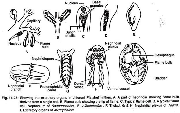
Excretory system of platyhelminthes consists mainly of longitudinal water vessels or longitudinal excretory canals having a number of specialized excretory cells, called flame cells or flame bulbs, collectively called protonephridia. The arrangement of water vessels inside the body offers wide range of variation.
In triclads there are two longitudinal vessels (canals) which open to the outside by numerous pores (Fig. 14.28G). The rhabdocoels have two lateral or a single median vessel (Fig. 14.28E). The median vessel opens to the outside by a single pore situated at the posterior end of the body.
In forms where two vessels are present the openings are either on the ventral side or in the pharynx. In some forms the two vessels unite at the posterior region and open to the outside by a single median aperture. The main nephridial canal is extensively branched and there are accessory excretory pores besides the nephridiopore (Fig. 14.28F).
In monogenic trematodes there are two longitudinal excretory ducts and the two ducts open separately on the dorsal surface. The ducts dilate to form excretory sacs at their terminal ends.
In other trematodes the excretory ducts are connected posteriorly to one another by a transverse duct and open to the outside by a single median aperture. The transverse duct dialates and in some cases forms an excretory vesicle. The two longitudinal ducts may unite with each other in the posterior region and may have a single opening.
In cestodes there are four longitudinal excretory verssels. The excretory vessels are connected by a transverse vessel situated in the scolex to form nephridinal plexus (Fig. 14.28H) and open in a contractile excretory vesicle situated in the last proglottid.
Numerous protonephridia and large number of nephridiopores are present in freshwater turbellarians because influx of excessive water is prevented in the body.
Respiratory and Circulatory Systems of Platyhelminthes:
ADVERTISEMENTS:
These two systems are totally absent in the phylum. In some trematodes the presence of a system of tubes, called lymphatic system, has been described. The tubes are considered to be of uncertain functions.
Nervous System of Platyhelminthes:
The main nervous centre or brain is located in the head as a pair of cerebral ganglia. Several ganglionated longitudinal cords arise from the brain. Of these longitudinal cords a pair becomes most conspicuous and the rest becomes insignificant. Numerous transverse connections occur between the longitudinal cords and the whole nervous system becomes ladder-like in appearance.
Sensory organs as ocelli or eyes are present in turbellarians and monogenic trematodes. They are numerous in polyclads and two to four in Rhabdocoels and monogenic trematodes. The eyes are present either as pigment cells or in the form of cups containing pigmented and sensory cells. Chemo and tangoreceptors are present widely and statocysts occur in Acoela and few other forms.
Reproductive System of Platyhelminthes:
Excepting a few Turbellaria and Trematoda, the platyhelminthes are hermaphrodites. Male and female reproductive organs in each individual are separate and open externally by their own pores or by a common genital aperture. In some cases the gonoducts open into the digestive tract and the sex cells are liberated through the mouth.
ADVERTISEMENTS:
In some flatworms an extra female pore or vaginal pore which serves during copulation is present. The gonopores are usually ventral in position but are occasionally dorsal. In tapeworms the gonopore is lateral in position.
Male reproductive organs consist of testes which in primitive condition are numerous and scattered. The number of testes are reduced to one or two in many and in Acoela definite gonads and ducts are lacking. The vasa efferentia, when present, correspond to the number of testes present. In general, there is a pair of vasa deferentia which unite and open into the complicated copulatory apparatus.
The copulatory apparatus consists of an eversible cirrus or a protrusible penis armed with spines or hook-lets. Various glands are associated with it. Vesicle in single or paired condition is often present and acts as the reservoir for storing sperms. The female reproductive organs consist of one or two ovaries. The oviducts when paired fuse distally and form a common oviduct which opens into the copulatory apparatus.
The copulatory apparatus consists of a sac, the seminal vesicle or called seminal bursa or copulatory bursa. Various glands which help in the formation of egg-shells and production of secretory substances are also associated with the copulatory apparatus. A long tubular or branched uterus for the purpose of accumulating ripe eggs forms a conspicuous part of the reproductive system.
The female gonads in platyhelminthes is peculiar as it is distinctly differentiated in two zones, the ovary proper and the yolk or vitelline glands.
The yolk is never incorporated into the structure of the egg as in the case of other animals but it is produced as abortive eggs to be included inside the egg capsule or shell to provide food for the developing embryo. In cestodes the reproductive organs are repeated in each proglottid. In young proglottids the organs are rudimentary while these are highly developed in the gravid ones.
Development of Platyhelminthes:
ADVERTISEMENTS:
Though hermaphrodite, the practice of cross-fertilization is the rule. Fertilization is internal and fertilized, shelled eggs containing embryos are shed to the exterior. Acoela and Polyclads lack yolk glands and development is direct. In Polyclads a free-swimming larva is produced which is supposed to foreshadow the Trochophore.
The larva is called Muller’s larva or Protrochula (Fig. 14.29). It is oval in shape and bears eight prominent arms which are beset with long cilia forming one continuous band. General body surface is covered with small cilia.
The mouth aperture is located in the mid-ventral line and three eyes exist in the anterior part of the dorsal surface. During development the ciliated arms are absorbed. In other groups the embryonic development is greatly modified and complicated. 
Phylogenetic Considerations of Platyhelminthes:
There is no unanimous view among zoologists regarding the origin of flatworms. There are several views for the origin of flatworms.
1. Lang (1881) proposed ctenophore- polyclad theory. This theory states that polyclad turbellarians might have evolved from ctenophores through Platyctenea. The Muller’s larva of polyclad bears some resemblances with ctenophores.
The resembling features are:
ADVERTISEMENTS:
(i) Similarities of the symmetries between the ctenophores and larva.
(ii) 8 ectodermal cilialed ridges of Muller’s larva can be compared with the meridional combplates of ctenophores.
(iii) Statocyst of turbellarians can be compared with the ctenophores.
This theory does not favour much support among zoologists.
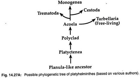
2. Planula larva theory states that turbellarians bear some resemblances with the planula larva of cnidarians. So many zoologists such as Hyman (1951), Jagersten (1955) and Hadzi (1963), consider that flatworms have evolved from a planula-like ancestral stock and Acoela is the most primitive group among platyhelminths (Fig. 14.27A).
Molecular data and cladistic analyses have been suggested a monophyletic origin for the three parasitic groups. The three parasitic classes—Trematoda, Monogenea and Cestoda, have evolved from free-living turbellarians.
3. Again the enterocoel theory states that all bilateral animals are basically coelomate, and acoelomate flatworms have evolved from coelmate ancestors by loss of the cavity secondarily. This theory is not acceptable to the zoologists because it is difficult to explain some aspects, such as the change from coelomate to acoelomate condition and from bilateral to radial symmetry.


