ADVERTISEMENTS:
In this article we will discuss about Sepia:- 1. Habit and Habitat of Sepia 2. External Structures of Sepia 3. Coelom 4. Digestive System 5. Locomotion 6. Respiratory System 7. Circulatory System 8. Excretory System 9. Nervous System 10. Reproductive System 11. Development.
Habit and Habitat of Sepia:
Sepia is a typical representative of the class Cephalopoda. Members of this class show highly specialised and complex structural organisation amongst the molluscs. Description of Sepia will give a general idea about the cephalopod organisation. Sepia is popularly known as cuttle fish.
Sepia is exclusively marine and usually remains near the surface of water. They are good swimmers. They usually swim at night and rest flat on bottom during daytime. The lateral fins, funnel and the arms help in swimming. Their flattened bodies indicate their sand and mud-dwelling habit.
ADVERTISEMENTS:
They can make burrow by using the funnel and use fins as shovel to cover the body with sand. They usually move in groups (Gregarious). They are carnivorous and live on small fishes, crustaceans and other small animals.
External Structures of Sepia:
The body is divided into a distinct head and an elongated dorso-ventrally flattened trunk. A narrow neck connects the head with the trunk. The head bears two well-formed eyes and ten oral arms.
The mouth is surrounded by the bases of the ten long oral arms (Fig. 16.38A). Excepting the fourth pair, all the oral arms have convex outer and flat inner surfaces. The inner surface possesses four longitudinal rows of suckers which help to grasp the prey or foreign bodies.
The fourth pair of the arms are comparatively longer and are called the tentacles. The tentacles have suckers restricted to the thickened tips. Each sucker is a muscular cup-like body. The rim of the sucker has small horny teeth and the whole sucker rests on a short stalk, called pedicel (Fig. 16.38B).
During breeding season the fifth arm on the left side (only in males), becomes modified as a copulatory organ and this modified arm is called hectocotylus or hectocotylized arm which helps in copulation and such a change is known as hectocotylization. The oral arms are modified foot and are innervated by pedal ganglion.
The trunk is covered by a thick mantle. Communicating with the mantle cavity a large tube known as funnel is present on the mid-ventral side of the oral end. The margin of the body has lateral, thin muscular folds called the fins.
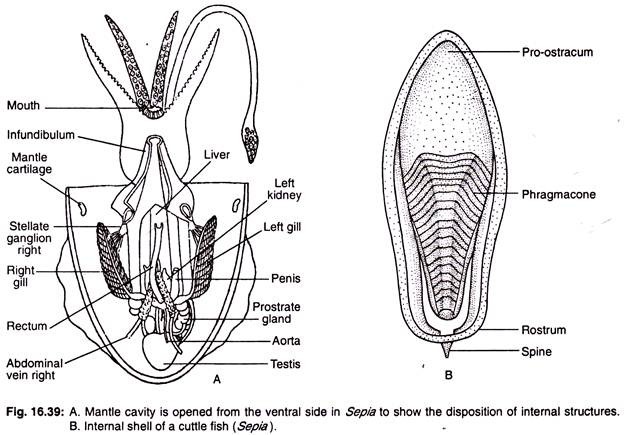
The shell is internal and is bilaterally symmetrical. The shell is a flat ovoidal structure and is composed mostly of calcareous matter. It supports the body as an endoskeleton and is also known as cuttle bone (= sepion). It functions in buoyancy regulation.
Living Sepia can change its colour due to the presence of chromatophores in the integument and matches the colour of sand and rock it passes over.
Sepia has a complicated structural organisation. A longitudinal incision of the mantle through the mid-ventral line will show the disposition of internal structures. The internal organs occupy the major portion of the mantle cavity (Fig. 16.39).
Coelom of Sepia:
The true coelom is represented by the viscero-pericardial coelom and the cavities in the kidney. The viscero-pericardial coelom is divided into two parts—the anterior is the pericardial cavity and the posterior is the gonocoel.
Digestive System of Sepia:
Sepia feeds on small fish and crustacea, specially prawn. The circlet of eight short arms help in capturing the prey and bringing it to the mouth. The mouth is bounded by a circular lip within which a pair of powerful jaws are lodged. The jaws help in cutting the food into small bits.
The circular lip is provided with many papillae. Mouth leads into the buccal cavity possessing an odontophore and the radula. The radula is relatively weak. The buccal cavity descends as a straight narrow tube, called oesophagus, which in turn opens into a round stomach. The stomach is represented by two separate chambers communicating by a sphincter.
ADVERTISEMENTS:
The first chamber is called gizzard. It is muscular and strongly-built. The other chamber is the caecum. It is thin-walled. The caecum leads into the intestine. The intestine runs paralled to the oesophagus, continues towards the oral end as rectum and ultimately terminates into the mantle cavity by anus (Figs. 16.40 and 16.41). Two pairs of salivary glands are present.
The anterior pair of salivary glands produce mucus. The posterior pair may show the tendency towards fusion and produce mucus, digestive enzyme (protease) and a number of poisons (indolic and phenolic amines). Two sets of digestive glands are present. The ducts of the digestive glands open at the junction of the two chambers of the stomach.
ADVERTISEMENTS:
Of the two sets of digestive glands, the larger one is formed by the fusion of a pair of brownish glandular lobes. The ducts from the larger one enter into the stomach near the point of origin of caecum. These glands are called liver. The other gland is clustered round the paired ducts of the liver. It is small, wedge-shaped, creamy in colour and follicular in appearance. It is called the pancreas and it opens into the gizzard.
A cylindrical duct coming from the ink- sac opens into the rectum. The ink-sac is a pear-shaped body (Fig. 16.42). The inner glandular mass secretes blackish ink. The ink is forced out to produce a coloured surrounding under the cover of which the animal can escape the-sight of enemies.
Locomotion of Sepia:
Sepia is a good swimmer. It swims by the undulatory movement of the lateral fins. During rapid movement, sepia moves backward by sudden forceful expulsion of water like a jet from the mantle cavity through the siphon.
Respiratory System of Sepia:
ADVERTISEMENTS:
The respiratory organs are the ctenidia. They are two in number. They are plume- shaped structures having numerous paired, delicately folded lamellae (see Fig. 16.39). Such a type of ctenidium is called bipectinate type. They are attached throughout the greater part of the mantle wall. The blood flows into the ctenidium by afferent branchial vessels and returns to the auricle by efferent branchial vessels.
Circulatory System of Sepia:
The circulatory system is highly developed in Sepia (Fig. 16.43). The heart consists of two auricles and one ventricle which is constricted into two lobes. The ventricle gives off anteriorly an oral or anterior aorta and a smaller aboral or posterior aorta in the posterior side.
The arteries arising from the two aortae supply the various parts of the body. Blood from the different parts of the body is collected into a large vena cava. The vena cava bifurcates anteriorly into precavals which pass through the substance of the kidney and continue as afferent branchial veins supplying the ctenidia.
The afferent branchial veins at the base of ctenidium dilate to form a contractile branchial heart. Attached to the branchial heart there exists a rounded glandular appendage. From the ctenidia blood returns to the heart by efferent branchial veins.
Excretory System of Sepia:
The excretory system consists of a pair of thin-walled renal sacs which communicate with the mantle cavity by two separate apertures, one in each side. The right and left sacs communicate with each other both anteriorly and posteriorly.
Through each sac runs the corresponding branchial vein. Surrounding the branchial vein there are masses of glandular tissue of the renal sac. This glandular tissue extracts the nitrogenous waste from the blood in the form of guanin.
Nervous System of Sepia:
The nervous system is highly developed in Sepia. The cerebral, pedal, pleural and visceral ganglia are large in size and are closely aggregated around the oesophagus by shortening of their connectives (Fig. 16.44).
They are all protected by cranial cartilaginous box. The cerebral ganglia are fused to form a single rounded mass and give off laterally the optic nerves which become expanded into optic ganglia. Another pair of nerves emerging from the cerebral ganglia are connected with the superior buccal ganglia. The superior buccal ganglia are also connected with the inferior buccal ganglia located on the posterior side of oesophagus.
The pedal ganglia, like the cerebral, are also fused into a single mass which is differentiated anteriorly into a brachial portion giving origin to ten brachial nerves supplying the arms and posteriorly into the infundibular portion supplying nerves to the funnel and statocysts. The brachial nerves are connected with one another by ring commissure.
ADVERTISEMENTS:
The pleuro-visceral ganglia are also united to form a single ganglionic mass and remain in immediate contact with the pedal. The pleuro- visceral mass gives off a pair of visceral nerves supplying the viscera and also gives a branchial nerve to the ctenidium. The branchial nerve possesses a branchial ganglion at its base. The pleuro-visceral ganglion also gives two very stout nerves posteriorly.
These two nerves are called the pallial nerves containing a large stellate ganglion (Fig. 16.68E) on each nerve (for diagram see Fig. 16.68E in the general notes on the phylum). The inferior buccal ganglion gives off sympathetic nerve which terminates into a gastric ganglion located on the dorsal side of the stomach.
The cuttle fish and squids possess two long nerves containing giant nerve fibers, situated close to the fused visceral ganglia whose function is to co-ordinate the contraction of mantle musculature and reponse of rapid escape from the place.
Sense Organs of Sepia:
The sense organs are highly developed in Sepia.
ADVERTISEMENTS:
(i) Eyes:
They are highly developed. The eye ball has stout outer wall called sclerotic layer, which is provided with sclerotic plates. Other structural elements, viz., the cornea, lens, aqueous and vitreous humours, retina are present in the eye of Sepia (Fig. 16.45). Cuttle fish is the only animal in the world which contains ‘W’-shaped pupils.
(ii) Statocyst:
The statocysts are two in number and are situated very close to the pleuro-visceral ganglion. They have complicated structures comprising of a crista statica and a macula statica. The crista statica and macula statica are rounded elevations on the inner cavity of the statocyst.
The crista statica is lined by flattened epithelium and the macula statica is composed of elongated cylindrical cells with hair-like processes at the free ends. A very large statolith is present in the cavity which is attached to the macula.
(iii) Olfactory pits:
In addition to eyes and statocysts, a pair of ciliated pits located behind the eyes, are supposed to be olfactory in function.
(iv) Gustatory organ:
This organ for taste comprises of an elevation in front of the odontophore.
Besides these, the arms and tentacles are regarded as tactile organs.
Reproductive System of Sepia:
The sexes are separate. Sexual dimorphism is present. The males are distinguishable by the presence of hectacotylized arm.
Male reproductive system:
The male reproductive system consists of a testis which is enclosed in a capsule. The capsule leads into an elongated and greatly convoluted vas deferens (Fig. 16.46A). The vas deferens opens into an elongated seminal vesicle. The seminal vesicle at the other end expands into a wide sperm sac which opens into the mantle cavity.
The prostate is glandular and opens into the seminal vesicle at this end. The gonopore lies on an elongated papilla, called penis. Inside the seminal vesicle sperms are rolled up and become enclosed by a chitinoid capsule which is called spermatophore (Fig. 16.46B).
Each spermatophore is elongated and about 16 mm in length. Two-thirds of it house the viscous mass of sperm and the anterior one-third contains a spring apparatus. The outer wall of the spermatophore is made up of chitinised capsule and the inner wall forms the tunic.
All the spermatophores remain stored in a pouch, called Needham’s sac. The hectacotylized arm takes out the spermatophores, keeps them for some time in the suckers and after the ritual of courtship places them on the body of the female.
Female reproductive system:
Female reproductive system consists of a single ovary. It is enclosed in a capsule which leads into a wide tubular oviduct. The oviduct opens into the mantle cavity near the rectum (Fig. 16.47A). A pair of nidamental glands are present on the sides of the ink-duct.
They are oval in shape and their secretion helps the eggs to adhere together like a bunch of grapes (Fig 16.47B). Another accessory nidamental gland of unknown function is present around the anterior ends of the nidamental glands proper.
Development of Sepia:
The development of Sepia is direct, because the young emerges out of the egg and looks like the adult. The fertilized egg is enveloped by a protective gelatinous covering. The eggs are very large in size. They are pear-shaped and are heavily yolked. The yolk provides nourishment for the developing embryo.


