ADVERTISEMENTS:
In this article we will discuss about Antedon:- 1. Habit and Habitat of Antedon 2. External Structures of Antedon 3. Body Wall 4. Coelom 5. Digestive System 6. Respiratory System 7. Water Vascular System 8. Haemal System or Blood Lacunar System 9. Locomotion 10. Excretory System 11. Nervous System 12. Reproductive System 13. Fertilization and Development.
Contents:
- Habit and Habitat of Antedon
- External Structures of Antedon
- Body Wall of Antedon
- Coelom of Antedon
- Digestive System of Antedon
- Respiratory System of Antedon
- Water Vascular System of Antedon
- Haemal System or Blood Lacunar System of Antedon
- Locomotion of Antedon
- Excretory System of Antedon
- Nervous System of Antedon
- Reproductive System of Antedon
- Fertilization and Development of Antedon
1. Habit and Habitat of Antedon:
Antedon is an inhabitant of the shallow water. It is a stalked form at the beginning and with the attainment of adulthood it breaks off from its stalk and swims about actively by the arms. They can also cling to the substratum by the cirri. They are of gregarious forms and feed on microscopic living organisms.
2. External Structures of Antedon:
ADVERTISEMENTS:
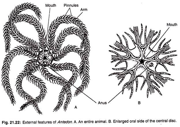
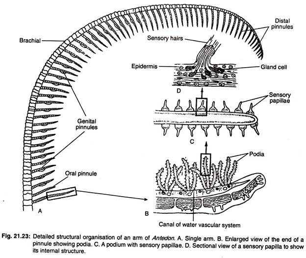
The body of Antedon is composed of small central part, called calyx from which elongated slender arms radiate (Fig. 21.22). The arms are actually five in number and each arm is bifurcated at the basal end. So the number apparently comes to ten. Each arm bears double rows of alternate small branches, called the pinnules (Figs. 21.22A, 21.23A).
The aboral side is flat. On the aboral side the calyx bears a knob-like structure which is the stung of the stalk. The knob is composed of centrodorsal ossicle which is provided with many-jointed cirri (Fig. 21.24). Each cirrus terminates in a small hook-like claw which help in temporary attachment.
ADVERTISEMENTS:
The oral surface is lined by a leathery tegmen and bears the mouth at the centre. The mouth is surrounded by tube-feet or podia which are sensory in function. The podia are beset with sensory papillae. The anus is situated on a papilliform projection in one of the inter- radii on the oral surface. Radiating from the mouth there are five ambulacral grooves across the tegmen.
In the mouth region the ambulacral grooves are separated by five oral valves. Each groove runs into one of the pairs of arms and gives branch to each pinnule. There are, in all, ten grooves— supplying ten arm-branches. The grooves throughout the course bear finger-like non- prehensile tube-feet or podia. The ambulacral grooves are actually the food grooves in Antedon.
The ambulacral grooves and the podia are ciliated. They help to convey food particles to the mouth which are gathered from the water current set by the cilia.
3. Body Wall of Antedon:
The ectoderm is rudimentary except at the ambulacral grooves. The basement membrane is absent. The dermis is leathery at the oral side. The body is covered by a delicate cuticle.
Skeleton:
The skeletal elements, specially on the aboral side, are well-formed. The aboral skeleton comprises of the large pentagonal centrodorsal ossicle, a thin plate, called the rosette (formed by the coalescence of the larval pieces, called basals). It lies internally and remains concealed by the centrodorsal ossicle.
The other components are three radial ossicles (first radial ossicle, second radial ossicle and the third radial ossicle) in each radius, a row of brachial ossicles in each arm, a row of ossicles in each pinnule, and a row of cirral ossicles in each cirrus. The skeletons particularly in the appendages are movably placed one upon the other.
4. Coelom of Antedon:
The perivisceral coelom in the calyx contains numerous trabeculae. Surrounding the oesophagus there is a vertical coelomic space, called axial sinus. The axial sinus and other coelomic spaces are continued into the arms. At the aboral side of the axial sinus, the coelom is represented at the centre by a characteristic chambered organ (Fig. 21.25A).
The chambered organ is composed of five symmetrically placed coelomic cavities around a central axis. Each arm contains four coelomic canals (a pair of subtentacular canals, a small genital canal and a minute coeliac canal). All these canals emerge from the perivisceral cavity.
The perivisceral cavity communicates with the exterior through numerous ciliated water-pores on the oral surface. Two types of coelomocytes are recorded in Antedon.
They are:
(a) An amoeboid type with short pseudopodia and phagocytic in function and
ADVERTISEMENTS:
(b) An elongated form containing rods and granules.
However, Cuenot (1891) found both coloured as well as colourless morula cells in Antedon rosocea.
5. Digestive System of Antedon:
The mouth is situated at the centre of the oral side (endocyclic condition). The mouth opens into a short vertical funnel-like oesophagus. The oesophagus leads into a wide stomach. The stomach is curved along the axis. The intestine bears a number of small blunt pouches and two long branched diverticula, called hepatic caeca.
The stomach opens distally into a short intestine which proceeds as rectum. The rectum passes upwards to the excentrically placed anus. The alimentary canal, excepting the germinal portion of the rectum, is ciliated.
6. Respiratory System of Antedon:
ADVERTISEMENTS:
There is no specialised organ for respiration in Antedon. The tube-feet lack terminal suckers and help in respiration. Besides the tube-feet, the rectum is capable of rhythmical contraction. Due to the ability of contraction and expansion, water is drawn in and driven out from the rectum through the anus. This method of taking in and ejecting out of water is possibly associated with the process of respiration.
7. Water Vascular System of Antedon:
The water vascular system is not so pronounced in Antedon. The water vascular system lacks direct communication with the exterior. The water vascular ring encircles the mouth and gives off many short stone canals which open to the perivisceral cavity. The perivisceral cavity communicates with the exterior through numerous ciliated water-pores.
The ampullae are absent but the wall of the water canals is slightly contractile due to contractility of the muscular strand. There is a series of radial vessels running beneath the ambulacral grooves which give branches to the pinnules. A series of podia or tube-feet without terminal suckers are present. They are tactile organs and help in respiration.
8. Haemal System or Blood Lacunar System of Antedon:
This system is made up of intercommunicating spaces in connective tissue trabeculae. It consists of a plexus of large lacunae, perioesophageal plexus, round the oesophagus. This is connected with a sub-tegmental plexus under the tegmen.
ADVERTISEMENTS:
A special organ (related to the axial gland), called spongy organ, receives branches from the perioesophageal plexus on the aboral side. The spongy organ (designated as labial plexus in earlier accounts) is made up of mostly rounded cells and possibly manufactures the coelomocytes. The haemal system contains a colourless proteinaceous fluid with few cells.
9. Locomotion of Antedon:
ADVERTISEMENTS:
The tube-feet in Antedon have nothing to do with locomotion. It can move very slowly by its cirri. It can also swim by the undulation of the arms.
10. Excretory System of Antedon:
Antedon lacks definite excretory organ. The actual nature of excretory products and the process of elimination are not clearly known. It is reported that the excretory products are accumulated in the coelomic lining. The coelomocytes help in the transportation of the excretory products of the body.
11. Nervous System of Antedon:
The nervous system is divisible into three types:
(a) Aboral or entoneural system,
ADVERTISEMENTS:
(b) Deeper oral or hyponeural system and
(c) Oral or ectoneural system.
Of all these systems, the entoneural system is well-developed (Fig. 21.25B). This system comprises a cup-shaped nervous mass situated at the apex of the cavity of the calyx. This nervous mass gives off five stout inter-radial brachial nerves to the five primary arms which are united by a nerve pentagon situated in the radial plates of the calyx.
Each brachial nerve divides into two parts which are again connected by direct and decussating commissures. The brachial nerves bear ganglionated swellings with bipolar and multipolar nerve cells. The brachial nerves give branches to the flexor muscles and adjoining epidermis, aboral part of the arms and pinnules.
The deeper oral or hypo-neural system comprises a nerve pentagon lodged in the connective tissue of the tegmen. Tine nerves emerging from the nerve pentagon innervate the tegmen, anal tube and other internal organs. Of these nerves, ten main branches go to the ten arms and innervate the musculature of the water vessels and pinnules. These nerves are connected with the aboral nervous system.
The ectoneural or superficial or oral nervous system consists of a superficial nerve ring surrounding the mouth which gives radial nerve cords. The radial nerve cords run in the ambulacral grooves of the arms and pinnules. These nerve cords consist of nerve cells and longitudinal nerve fibres and supply the ambulacral epidermis, inner surface of the tube-feet and the adjoining areas.
ADVERTISEMENTS:
Sense Organs:
There exists no special sense organ in Antedon. The papillae present on the tube- feet are both sensory and secretory in function. Each papilla comprises a slender epidermal projection with sensory bristles and basal glandular cells (Fig. 21.23D). The papillae are possibly the tactile organs.
12. Reproductive System of Antedon:
The sexes are separate and sexual dimorphism is absent. The axial organ is continuous with a circular genital rachis which in turn gives genital cords. The genital cords pass into the genital canal and reach the pinnules where they become differentiated into the gonods. The genital tube, very common in other crinoids, is absent in this form.
The genital system is enclosed by a plexus of haemal system. When the gametes become mature, they escape to the exterior by the rupture of the pinnule wall. After the rupture of the pinnule wall, the eggs remain attached with the pinnule by secretion of cement glands.
13. Fertilization and Development of Antedon:
The fertilization is external. The zygote after the routine developmental processes is transformed into a free-swimming barrel- shaped ciliated larva, Antedon larva. This larva is also called Doliolaria larva or Vitellaria larva of Haeckel.


