ADVERTISEMENTS:
The following points highlight the five main parts that make up the internal structure of Hydra. The parts are: 1. Body Wall 2. Epidermis 3. Gastrodermis 4. Mesogloea 5. Gastro vascular cavity.
1. Body Wall:
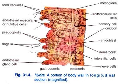
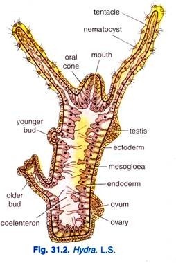
The body wall consists of two cellular layers, an outer epidermis derived from ectoderm and an inner gastro dermis derived from endoderm. In between the outer epidermis and inner gastro dermis, a thin non-cellular layer of jelly-like substance called mesogloea is present. Both the epidermis and gastro dermis are composed of different kinds of cells, hence, they are described separately.
2. Epidermis:
The epidermis is made up of small cubical cells and is covered with a delicate cuticle. It forms a thin layer, about one-third of the thickness of body wall. This layer contains several types of cells-epitheliomuscular, interstitial, gland, cnidoblast, sensory, nerve, and germ cells. The epidermis is protective, muscular and sensory in function.
ADVERTISEMENTS:
1. Epitheliomuscular cells:
The epitheliomuscular cells have both epithelial and muscular parts in the same cells.
The epitheliomuscular cells of the epidermis are cylindrical with their inner ends produced into two or more processes which have myonemes or un-striped muscle fibres, these fibres branch and branches anastomose. The ectodermal myonemes run parallel to the long axis of the body and tentacles, they form longitudinal muscles which bring about contraction of the body.
ADVERTISEMENTS:
The epitheliomuscular cell has a large nucleus, and along the border there is a row of granules which secrete the cuticle.
The epidermal cells of the basal disc are granular and they secrete mucus for attachment of Hydra; the basal epidermal cells can also form pseudopodia by which the animal glides on its attachment. Some granular epidermal cells of the basal disc secrete a gas to form a bubble by which the Hydra breaks from its attachment and is lifted up.
Ultra Structure of Epitheliomuscular Cell:
The epitheliomuscular cells are large possessing columnar or cuboidal shape and contain a centrally or basally situated nucleus. The nucleus is large and irregularly outlined and contains coarsely granular material and a large dense central nucleolus. In the cytoplasm is present both rough and smooth surfaced endoplasmic reticulum.
Many free ribosomes and numerous typical mitochondria are present in the ground substance of the cytoplasm. One to few Golgi apparatus lie oriented parallel to the long axis of the cell. Golgi apparatus is composed of parallel lamellae, vesicles and vacuoles.
Some vesicles of Golgi apparatus are filled with dense material, In the apex regions of the cells, elaborated from Golgi apparatus, are present large numbers of membrane bound mucous granules. The mucous granules are spherical about 0.5 to 1.0 µ in diameter and contain finely granular material.
The surface of the cell is covered by a membranous cuticle supposedly formed by liberations of mucous granules. Large intracellular spaces fill the cell. Large vacuoles are present a short distance away from Golgi apparatus. In most cells, one to several membrane bound, irregularly shaped masses are present in the apex region.
ADVERTISEMENTS:
The plasma membrane is a smooth structure with a few outward projections. Immediately above the plasma membrane is a thin layer of homogeneous material about 0.1 µ thick; this layer is covered by a thick feltwork (0.5 µ) of fine granular, fibrillar or filamentous material. The thin layer is absent at the points of contact between two adjacent cells, only the thick felt-work comes in contact.
The function of these layers appears to be primarily protective. A few small membrane bound vesicles are present immediately below or connected to plasma membrane. Microtubules, 200 A° in diameter, are also present below the plasma in a membrane in the apex of the cell showing selective uptake of materials.
Junctions of epitheliomuscular cells have a characteristic complex consisting of three parts. At the upper part, the plasma membranes are straight and a constant distant apart (120 A°). Below is an occluded zone where the plasma membranes are indistinct and the space between them narrows and filled with dense material.
Following the occluded zone, are regularly spaced transverse bars extending across the 120 A° space between the lateral plasma membranes. Below these bars, also called septate desmosomes, an intercellular space may be formed by a separation of adjacent cell membranes.
ADVERTISEMENTS:
The base region of the epitheliomuscular cell lies above the mesogloea and has many muscular processes running parallel to the long axis of the animal. The muscular processes are filled with two types of myofibrils. Myofibrils of about 50 A° in diameter are more numerous, within which are scattered the myofibrils of about 200 A° diameter.
The myofibrils terminate obliquely on the plasma membrane and become thickened at the point of terminations. Small microtubules (200 A° in diameter) lie parallel to the muscular processes and are supposed to carry water or ions and may, therefore, be involved in changes in the electrical potentials of muscular processes.
Regional variations have been observed in epitheliomuscular cells. In tentacles, the cells are large and contain numerous cnidoblasts and more apical mucous granules and more elaborate endoplasmic reticulum. In the peduncle region, the cells are small and cuboidal and contain few intracellular spaces having mucous granules and irregular masses in abundance.
The cells of the base, called glandulomuscular, have their entire interior filled with mucous granules which are elaborated by Golgi apparatus. The endoplasmic reticulum is well developed. The basal regions of the cells contain smaller mucous granules, while the apex has larger ones.
ADVERTISEMENTS:
Functions:
The epitheliomuscular cells perform the following functions:
1. They form a protective covering of the body;
2. They help in contraction, shortening and bending of body;
ADVERTISEMENTS:
3. They help in locomotion;
4. They help in attachment with the solid object, and
5. They help in respiration through mucous layer at the cell surface.
2. Interstitial Cells:
Lying in the spaces between the inner ends of cells of epidermis and between outer ends of cells of gastro dermis are interstitial cells (Fig. 31.6) lying in groups.
These are small, oval or round cells with a large nucleus. Interstitial cells form a growth zone just below the tentacles, from this zone all kinds of new cells arise which push out the old worn out cells, which are shed at the proximal and distal ends.
ADVERTISEMENTS:
Interstitial cells form nematocysts and germ cells, they can also form epitheliomuscular cells, they renew all cells of the animal once every 45 days (Brein, 1955), thus, they are totipotent.
Ultra structure of interstitial cell:
Each interstitial cell is small, round or oval, about 5 µ in diameter. The cytoplasm is filled with free ribosomes and contains few smooth membrane bounded vesicles and mitochondria. The central nucleus contains scattered granules; the nucleolus is smaller or absent.
Functions:
The interstitial cells perform the following functions:
ADVERTISEMENTS:
1. These cells are the main agent in rebuilding tissues during growth, budding and regeneration;
2. They form gonads during breeding season to give rise to germ cells;
3. They reform the worn out cells of gastro dermis and also differentiate to form new nematocysts to replace to older and worn out ones.
3. Gland Cells:
These are tall cells found chiefly on the pedal disc and around the mouth region. They produce a secretion by which the animal can attach itself and sometimes a gas bubble by which the animals can rise and fasten on to the surface of the water to float.
4. Cnidoblasts:
Cnidoblasts (Gr., knide = nettle; blastos = germ) are found throughout the epidermis but specially on the tentacles.
Some interstitial cells of the epidermis give rise to highly specialised cells called cnidoblasts. These are somewhat oval-shaped cells, contain the cell organoid, the nematocyst (Gr., nema = thread; kystis = bladder) or stinging cell. The nematocyst is made up of rounded capsule which encloses a coiled tube or thread that is continuous with the capsular wall to which it is attached.
Ultra Structure of Cnidoblast:
The cnidoblasts possess a thin cytoplasmic rim which surrounds the large centrally located nematocyst. The nucleus of cnidoblast is situated between the nematocyst and plasma membrane. The nucleus contains a small inconspicuous nucleolus.
A few rough surfaced lamellae and isolated smooth surfaced vesicles of endoplasmic reticulum are present in the cytoplasm. Free ribosomes are scattered throughout cytoplasm. Golgi apparatus is small and lies in the basal region. Mitochondria, lipid droplets, and mulitivesicular bodies are also present in the cytoplasm.
A bundle of small myofilaments is present in the basal region of the cnidoblast extending from the capsule of the nematocyst. The nemataocyst lies in the cnidoblast enclosed by a thick capsule, and consists of a thread or tube containing spine and stylets. The capsule is composed of collagen-like protein.
At the apex of nematocyst, the capsular material narrows and invaginates towards the centre of the nematocyst. In the space left by the invagination of the capsule, the nematocyst membrane is extended. The space is filled with fine granular material and is called the operculum of the nematocyst. The invaginated capsular wall encloses the stylets and spines of the nematocyst.
The apices of the three stylets lie close to each other but their bases are turned outwardly. Each stylet is a large rod overlying the spines. The spines are small and more than 50 in number. These are stacked in a pyramidal manner with the apex pointing upward.
The invaginating capsule wall exhibits a trifoliate outward extension from the stylets towards the apex. At the base of stylets and spines, the invaginated wall forms the outer wall of the thread. The thread lies coiled in the basal region of the nematocyst.
All the intra-capsular spaces are filled by a fine granular matrix. Situated at the apical surface of the cnidoblast and projecting above the surface is a pointed spike-like structure called cnidocil (Gr., knide = nettle; cilium = hair). The cnidocil is composed of a central core surrounded by large rods. The core contains smaller fibrils. From the capsule, 20-21 hollow rods extend upward converging around the core.
The core appears to be a modified cilium. At the tip of the cnidoblast, rods terminate in close proximity to the plasma membrane.
Numerous dense granules are present at the periphery both intra-and extracellular. At the terminal regions of the rods, 20-21 projections of plasma membrane extend outward to form the external portion of the cnidocil. The external flagellar material of the epidermis overlies the membranes of the external rods. Microtubules extend from the region of the cnidocil to surround the underlying nematocyst capsule.
Nematocyst:
A nematocyst is not a cell because it is chitinous and non-living. A clear space arises in the cnidoblast, the space grows and the cell secretes a double-walled chitinous capsule which has a lid or operculum. One end of the capsule forms a tube lying coiled in the capsule, the tube may have a basal swelling called a butt, and a long coiled thread which may be open or closed at the tip, inside the tube may be some spines.
This structure secreted by a cnidoblast is a nematocyst. In the nematocyst is a poisonous toxin made of mixture of proteins and phenols.
On the wall of the capsule are contractile fibrils running into the cnidoblast. In some nematocysts, the cytoplasm of the cnidoblast forms contractile muscle fibrils. Some nematocysts have a lasso or restraining thread attached to the base of the cnidoblast, the lasso prevents certain nematocyst from being thrown out of the body of an animal.
Nematocysts are produced only on the stomach, cnidoblasts containing developing nematocysts migrate through the body wall or into the enteron from where they are taken up by pseudopodia of endoderm cells and transferred to mesogloea through which they travel and penetrate outwards again through the body wall to reach their ultimate positions where development is completed; the cnidoblast gets fixed in the ectoderm with its base reaching the mesogloea, the cnidocil bores through the cuticle and projects outside.
Hydra has four kinds of nematocysts confined to the ectoderm:
1. Penetrants or stenoteles:
Penetrants or stenoteles have a large capsule, the butt is stout with their spiral rows of spines on its distal half, the lowest spine of each row is a large stylet; the thread had spirals of small spines and it is open at the tip. Stenoteles are weapons of defence and offence, their thread penetrates the body of the prey, they are also used for obtaining food.
2. Desmonemes or volvents:
Desmonemes or volvents have a small oval capsule, there is no butt, the thread is thick with no spines and it is closed at the tip, it lies in a single loop inside the capsule. On being discharged, the volvents are thrown out of the body and the thread coils around the bristles of the prey; they are used for obtaining food.
3. Small glutinants or atrichous isorhizas:
Small glutinants or atrichous isorhizas have an elongated capsule, butt is absent, thread is open at the tip, and it has no spines, they fix the tentacles to an object when the animal walks on its tentacles.
4. Large glutinants or holotrichous isorhizas:
Large glutinants or holotrichous isorhizas have an oval capsule, the butt is narrow and the thread is open at the tip, there are small spines on the butt and thread. Their function is doubtful but they stick to the surface of the prey.
Distribution of nematocysts:
Nematocysts are plentiful on the tentacles and body, but they are absent from the basal disc. All four kinds of nematocysts are found on the tentacles in abundance, hypostome has only holotrichous isorhizas, the body has mostly stenoteles and some holotrichous isorhizas.
Discharge of nematocysts:
Nematocysts are discharged but once, after discharge they are cast off, though volvents are thrown out being discharged, new nematocysts are found all the time. The method of discharge of nematocysts is not clear, but they are not under the control of the nervous system, hence, they are independent effectors, they are functional even in the bodies of some other animals.
An anaesthetized Hydra will discharge its nematocysts in the usual way when stimulated; even nematocysts removed from the body will shoot out their thread if an adequate stimulus is applied to them. Some animals swimming near a Hydra will cause the nematocysts to discharge, yet some other animals can walk on the body of Hydra without discharging the nematocysts.
Following explanations are offered for discharge of nematocysts:
(a) There are two factors responsible for discharge, first the presence of certain liquid-like chemicals in the water, second a mechanical contact of the cnidocil and cnidoblast by food animal or prey, if both chemical and tactile sitmuli are present then the nematocyst is discharged.
(b) The thread within the capsule is gelatinous, on suitable stimulation the operculum opens and water enters the capsule, then the thread is liquefied and forced out like a jet, but on exposure the jet of liquid solidifies and becomes the external thread of the nematocyst.
(c) There is a mechanism in the cnidoblast which possesses both receptor and effector parts which explode the nematocyst on stimulation of cnidoblast under the combined influence of mechanical and chemical stimuli received by the cnidocil and conducted to the cnidoblast.
In discharge of a nematocyst the operculum opens, water enters the capsule, the tube is turned inside out and shot out with a force, the eversion causes the spines to come to the outer surface of the tube.
The thread either sticks to the prey (glutinants), or coils around its bristles (volvents), or it penetrates its body (penetrants), and injects a powerful toxin which paralyses even such large animals as a water-flea or small worms.
Functions:
The cnidoblasts are supposed to be the organ of offence and defence of Hydra. They also help in the function of food-capture, locomotion and anchoring with the substratum.
5. Sensory Cells:
They are long narrow cells with a large nucleus and one projecting flagellum or sensory hair, their base may be produced into nodulated processes which join the nervous system. Sensory cells are found in both germinal layers, but they are more abundant in the ectoderm, they are sensory to touch, light, temperature changes and chemicals.
A sensory cell acts both as a receptor and as a sensory neuron, that is, it both receives and transmits impulses. The tentacles are devoid of gland cells and sensory cells, and their endoderm cells have no muscle processes.
Ultra structure of sensory cell:
A sensory cell (Fig. 31.12B) is situated perpendicular to the long axis close to the apices of epithelial cells. A modified cilium emerges apically from the cell. The plasma membrane of the apical surface of the cell is notched to form a collar and a single cilium extends from the base of the notch.
The cilium consists of nine peripheral and more than two central fibres; all the fibres merge with the basal body from which small rootlets spread out into the cytoplasm. Mitochondria and small vesicles are present in the apical cytoplasm and the microtubules extend into the apical collar. A Golgi apparatus lies above the nucleus. The basal end of the cell is either situated above a ganglion, cell or gives rise to a process.
6. Nerve Cells or Ganglion Cells:
The nerve cells or ganglion cells are small and elongated having one or more processes. They are situated at the base of the epitheliomuscular cells just above their muscular processes. They are rarely found in gastro dermis.
Ultra structure of nerve or ganglion cell:
The plasma membrane of nerve cells (Fig. 31.12C) is irregular with numerous crests and indentations. The nucleus is small, oval, and bounded by a nuclear membrane bearing pores. Nucleoli may or may not be present. Free ribosomes may be many or few. Smooth and rough surfaced endoplasmic reticulum is present but it is not a prominent feature.
Mitochondria may be scattered or clustered. Golgi apparatus is most prominent and two or three separate Golgi regions may be present. Golgi apparatus is composed of flattened stacks of membrane bounded lamellae and small vesicles and is usually situated between the nucleus and a longitudinal process or neurite extending from the nuclear region. Cytoplasm also contains a number of small and large vesicles.
The cells have highly developed microtubules which extend long distances in the neurites. The microtubules follow a straight course and are either fused with the pores in the nuclear membrane or curve to come close to a nuclear membrane. The neurites may be longer than 10.0 µ and contain ribosomes, small and large vesicles, mitochondria and microtubules. The neurite is lined by plasma membrane of the neuron.
7. Neurosecretory Cells:
The neurosecretory cells (Fig. 31.12D) are named on the basis of their membrane bound dense granules. They are deeply situated and contain a cilium that extends towards the surface. The cilium arises from the base of an indentation of the plasma membrane. Finger-like projections extend into the space produced by the indentation but the projections do not reach the base of the indentation.
The projections surround the cilium. Below the cilium, striated rootlets extend for a considerable distance into the cytoplasm. The neurosecretory cells are identical to nerve cells except for numerous membrane bound granules. These granules are 1000 to 1200 A° in diameter and present in the cytoplasm in close approximation to be within the dilated ends of Golgi apparatus lamellae.
Neurites of these cells also contain dense granules which at the end of neurites are enclosed within smooth surfaced vesicles.
Nerve cells of the base region of Hydra differ in structure. They contain less ribosomes and lack the microtubules. Golgi apparatus is usually present and the granules are small (200-300 A° in diameter) lie in the lamellae and vesicles of the Golgi apparatus. It is presumed that these cells arise from one or the other types of neurons because interstitial cells from which neurons differentiate are absent at the base.
8. Germ Cells:
Germ cells originate by the repeated divisions of the interstitial cells in certain restricted regions of the body of Hydra during the summer. These form the gonads which later differentiate either into testes or ovaries.
3. Gastrodermis:
The inner gastro dermis, a layer of cells lining the coelenteron has a plan similar to the epidermis. It is made up chiefly of large columnar epithelial cells with irregular flat bases. The free ends of the cells give a jagged and uneven contour to the coelenteron in cross section.
The gastro dermis forms about two-thirds of the body wall and is secretory, digestive, muscular and sensory. The cells of gastro dermis include nutritive muscular, interstitial and gland cells.
1. Nutritive muscular or digestive cells:
The epitheliomuscular cells of gastro dermis are long and club-shaped, their outer ends have two processes containing a myoneme which does not branch; these myonemes lie at right angles to the long axis of the body, they form circular muscle layer by which the animal contracts and slowly expands the body.
Some of them serve as sphincters to close the mouth and cavities of tentacles because the gastro dermal myonemes are best developed in the hypostome and in the bases of tentacles.
The cells are highly vacuolated and often filled with food vacuoles. The free end of the cell usually bears two flagella. Gastro dermal cells in the green hydra (Chlorohydra) bear green algae (Zoochlorella) which give the hydra their colour. Nutritive muscular cells may also secrete digestive enzymes into the coelenteron for the digestion of foods.
Ultra structure of Nutritive Muscular Cell:
The apical border of each nutritive muscular cell (Fig. 31.13) bears numerous slender microvili of varying lengths projecting into the digestive cavity. Mitochondria, glycogen granules, vesicles, oil droplets and vacuoles fill the apex of the cell. The endoplasmic reticulum is sparse, consisting of a few smooth and rough surfaced cisternae. Free ribosomes are abundant in the cytoplasm. The Golgi apparatus is small and lies close to the nucleus.
The nucleus is central or basal in position and contains a nucleolus. Numerous heterogeneous membrane bounded structures are characteristically present.
Large membrane bounded vacuoles are found in the cytoplasm, the vacuoles of the apical region may represent digestive vacuoles, the vacuoles of the basal region may represent residual bodies containing undigested food material. Generally, one central space but at times many small intracellular spaces are present.
A felt-work of fibrillar or filamentous material covers the plasma membrane and is applied directly to the outer layer of the unit membrane without an intervening space.
The felt work is thin at some places and thick at others. Pinocytotic invaginations are common at the bases of microvilli and membranous vesicles, and channels are present immediately below the plasma membrane. Adjacent cells have septate desmosomes.
Circularly oriented muscular processes containing myofilaments lie above mesogloea. Each cell bears a pair of flagella which are typical in structure, that is, each flagellum consists of nine peripheral and two central fibres enclosed in a sheath.
Digestive cells of tentacles are pyramidal in shape and contain a large intracellular space surrounded by a thin rim of cytoplasm in which lipid droplets and food vacuoles are found. Free microvilli and pinocytotic vesicles are present. The digestive cells of hypostome are irregular in shape, between the bases of these are found numerous gland cells.
The cells of peduncle are small and cuboidal containing a large intracellular space. They are like the digestive cells of the tentacles but do not possess as many lipid droplets. The digestive cells of the base are large, cuboidal and contain large intracellular spaces. They have few microvilli and pinocytotic vesicles, but small lipid droplets are numerous.
Towards the base, within the coelenteron, are present many cell fragments containing various cytoplasmic inclusions. According to one view, the aged cells are extruded through the basal pore, while the other view holds that by endogenous fragmentation digestive cells cast out fragments which are carried to different regions by flagellar currents.
The ultra structure of the digestive cells of different regions suggests that the digestive cells of stomach, budding and hypostome regions perform ingestion and digestion, those of peduncle and tentacles of storage; and those of base provide energy for mucous secretion since they contain large amounts of lipid droplets.
2. Interstitial cells:
There are a few of these small cells scattered among the bases of nutritive cells. They may transform into other types of cells when the need arises, i.e., totipotent in nature.
3. Gland cells:
Gland cells are often club-shaped, with the larger end facing the coelenteron. They are interspersed singly between the digestive cells. Most are club-shaped tapering to a narrow base which extends towards the mesogloea but do not reach it.
Gland cells are of two kinds, viz.,:
(i) Mucous gland cells are found in the mouth and hypostome, they secrete mucus which helps in swallowing solid food,
(ii) Enzymatic gland cells are found in the stomach where they secrete digestive enzymes. The gastro dermis of the stalk and tentacles is devoid of gland cells. Gland cells pour their secretions into the coelenteron for extracellular digestion. Gland cells are not under the control of the nervous system, they are independent effectors.
Ultra structure of gland cell:
In the gland cell (Fig. 31.14), the nucleus is basal containing a large nucleolus in the differentiating cell but none in a mature cell, which contains full complement of the secretory granules. The mitochondria contain closely packed cristae. The endoplasmic reticulum is composed of stacks of rough-surfaced lamellae and fill the basal region of the cell and occupy the narrow spaces between secretory granules.
The plasma membrane possesses few microvilli and flagella and is covered by a feltwork of fibrillar nature. One or more Golgi apparatus is found near the nucleus and is surrounded by the rough-surfaced endoplasmic reticulum. The lamellae of Golgi apparatus are sometimes dilated and filled with material similar to secretory granules.
The larger secretory granules are apparently formed by fusion of smaller Golgi vesicles and vacuoles and are more numerous in the apical part of the cell. When more than one type of granule is present within a single cell, each type is associated with a separate Golgi apparatus.
Although several types of membrane-bounded granules can be distinguishable and histochemically divided into mucus and enzyme secreting types, yet it appears that there may be one basic type of gland cell which is capable of secreting any type of granules.
Gland cells are most abundant in the hypostome, numerous in stomach and budding zone, rare in peduncle and virtually absent in tentacles and base. The cells develop from interstitial cells most frequently in growth region but very little is known about the replacement of all gland cells exhausted during secretion.
4. Mesogloea:
The mesogloea (Gr., meso = middle; glea = glue) lies between the epidermis and gastro dermis and is attached to both layers. It is gelatinous or jelly-like and has no fibres or cellular elements. It is a continuous layer which extends over both body and tentacles, thickest in the stalk portion and thinnest on the tentacles.
This arrangement allows the pedal region to withstand great mechanical strain and gives the tentacles more flexibility. The mesogloea supports and gives rigidity to the body, acting as a sort of elastic skeleton.
Ultra structure of mesogloea:
The mesogloea is an acellular layer, about 0.1 µ thick within which are embedded small filaments, about 100 A° thick. The small filaments show transverse striations and are either randomly dispersed or lie parallel or obliquely to the longitudinal axis. The filaments are not inserted on the plasma membrane. Small dense granules of glycogen are present in the mesogloea.
The mesogloea is continuous with the intercellular spaces between the epitheliomuscular and digestive cells. Except the small differentiating interstitial cells near the regenerative area, no other cells or neurites of nerve cells cross the mesogloea. Processes of both epitheliomuscular and digestive cells extend for various distances into the mesogloea, sometimes interlocking with one another.
5. Gastro vascular cavity:
The L.S. (Fig. 31.2) and T.S. (Fig. 31.3) of Hydra shows a central cavity in its body called coelenteron (= hollow gut) functionally referred to as gastro vascular cavity. Mouth opens in this cavity and there is no other exit in it. However, this cavity remains continuous in the tentacles and, therefore, the tentacles are hollow. The gastro vascular cavity is the site of digestion and circulation.


