ADVERTISEMENTS:
In this article we will discuss about Grasshopper:- 1. Habit, Habitat and External Features of Grasshopper 2. Internal Anatomy of Grasshopper 3. Digestive System 4. Circulatory System 5. Respiratory System 6. Excretory System 7. Nervous System 8. Sense Organs 9. Reproductive System 10. Economic Importance 11. Control.
Contents:
- Habit, Habitat and External Features of Grasshopper
- Internal Anatomy of Grasshopper
- Digestive System of Grasshopper
- Circulatory System of Grasshopper
- Respiratory System of Grasshopper
- Excretory System of Grasshopper
- Nervous System of Grasshopper
- Sense Organs of Grasshopper
- Reproductive System of Grasshopper
- Economic Importance of Grasshopper
- Control of Grasshopper
1. Habit, Habitat and External Features of Grasshopper:
Grasshoppers have worldwide distribution and are found where there are open grasslands and abundant leafy vegetation. They feed on leafy vegetation. They are essentially solitary and residential species often abundant as individuals, but which may occasionally migrate.
ADVERTISEMENTS:
The locusts are gregarious and migratory forms. Sometimes, the locusts increase in large numbers and travel long distances in swarms, attack and cause incalculable damage to the crops and vegetation.
External Features of Grasshopper:
(i) Shape and Size:
ADVERTISEMENTS:
The body of grasshopper is narrow, elongated, cylindrical and bilaterally symmetrical. It is relatively a large insect measuring up to 8 cm in length.
(ii) Colouration:
The usual body colour is yellowish and brownish with different markings and colour spots. The pigment in the chitin provides the protective colouration to the body matching the environment.
(iii) Exoskeleton:
The body is covered by an exoskeleton that protects the delicate systems within. This exoskeleton is the cuticle which consists of chitin and is divided into a linear row of segments. The exoskeleton is formed into hard plates or sclerites separated by soft cuticle that permits movement of the body segments and appendages.
The softer regions are known as sutures. Each segment is made up of separate pieces known as sclerites. Usually some of the sclerites of a typical segment cannot be distinguished and the sutures are, therefore, said to be obsolete or indistinct. The body wall consists of the cuticle beneath which is a layer of cells the hypodermis which secretes it and under this a basement membrane.
ADVERTISEMENTS:
(iv) Division of body of Grasshopper:
ADVERTISEMENTS:
In grasshoppers, the body is divided into three typical regions, viz., the head, thorax and abdomen.
(a) Head:
The head in grasshopper is more or less ventral, although it appears to be hypognathous while feeding. It is enclosed in a chitinous capsule and is attached to the body by means of a small neck having cervical sclerites. Six segments are fused together to form the head. The head is made up of a dorsal portion, the vertex; a region in front, the frons; and the sides, or genae.
Below the frons is the plate, clypeus. On each side of the head is a compound eye. Three simple eyes or ocelli are located in the region between compound eyes. A pair of slender antennae are also found on the head. On the ventral side of the head are the mouth parts.
Appendages of the head:
The head bears a pair of antennae, a pair of compound eyes, three simple eyes or ocelli and mouth parts.
(b) Antennae:
The antennae are filiform. Each antenna consists of a small piece called the scape, an undifferentiated pedicel and a sufficiently long flagellum composed of about twenty-five segments. Sensory bristles, probably olfactory in nature, are present on the surface of antennae.
ADVERTISEMENTS:
(c) Compound Eyes:
Two compound eyes are placed dorsolaterally on the first segment of the head. These are sessile as stalks are absent. Each compound eye is covered by a transparent part of the cuticle, the cornea which is divided into a large number of hexagonal facets. Each facet is the outer end of a unit known as an ommatidium. Such a structure gives mosaic vision.
(d) Ocelli:
Three simple eyes or ocelli are placed between the compound eyes. An ocellus consists of a group of visual cells the retinulae and a thick transparent lens which is the modification of cuticle.
(e) Mouth Parts:
The mouth parts of grasshopper are chewing or mandibulate type.
ADVERTISEMENTS:
The mouth parts include the labrum, mandibles, maxillae, labium and hypo pharynx. There is a labrum or upper lip attached to the ventral edge of the clypeus. Beneath this is a membranous tongue-like organ, the hypo pharynx. On either side is a single, hard jaw or mandible with a toothed surface fitting for grinding. Beneath the mandibles are a pair of maxillae.
Each maxilla consists of a basal cardo, central stipes, a long curved lacinia, a long rounded galea and a maxillary palp which arises from the palpifer. The labium or lower lip comprises a basal submentum, a central mentum, two movable flaps, the ligulae and a labial palp on either side.
(f) Thorax:
The thorax is separated from the head and abdomen by flexible joints.
It consists of three segments:
An anterior pro-thorax, a middle mesothorax and a posterior metathorax. Each of these segments bears a pair of legs, and the mesothorax and metathorax each bear a pair of wings. On either side of the mesothorax and metathorax is a spiracle, an opening into the respiratory system.
ADVERTISEMENTS:
A typical segment includes eleven sclerites. The dorsal tergum (called pronotum, mesonotum and metanotum in pro-thorax, mesothorax and metathorax respectively) consists of four sclerites in a row, an anterior prescutum followed by the scutum, scutellum and postscutellum. The lateral pleuron consists of three sclerites, the episternum, epimeron and parapteron. The single ventral sclerite is the sternum.
The pronotum of the pro-thorax is large and extends down on either side; its four sclerites are indicated by transverse grooves. The sternum bears a spine. In mesothorax the mesonotum is small but the sclerites of pleuron are distinct. The sclerites of metathorax resemble the mesothorax.
(g) Legs:
Each thoracic segment bears a pair of jointed legs.
Each leg consists of a linear series of five segments as follows the coxa articulates with the body, then come the small trochanter fused with the femur, the tibia and the tarsus. The tarsus of each leg consists of three visible segments, the one adjoining the tibia has three pads on the ventral surface and the terminal segment bears a pair of claws between which is a fleshy lobe, the pulvillus.
(h) Wings:
In grasshopper, each of the mesothorax and metathorax bears a pair of wings. The forewings are narrow and mostly parchment-like.
These may be coloured uniformly or with deepening shades towards the bases or may be spotted. This pair is also named as tegmina because in the position of rest it covers the abdomen and the hind pair of wings. The hind wings are broad and membranous and kept folded in fan-like manner having a number of longitudinal folds in alternating directions.
Each wing develops as a sac-like projection of the body covering and flattens to a thin double membrane that encloses tracheae, nerves and blood sinuses. The cuticle thickens along the sinuses to form strengthening nervures or veins. Although these veins vary in their patterns among the different species, they are constant in individuals of certain species, where they serve for classification.
(i) Abdomen:
The abdomen is elongated and tapers towards the posterior end, where the terminal segment is specialised for copulation or egg-laying. It consists of 11 segments. Each segment typically has a dorsal tergum and a ventral sternum, there being no pleura. The sternum of first segment of the abdomen is fused to the thorax and its tergum bears on either side the oval tympanic membrane which covers the auditory sac.
The terminal segments are modified in both the sexes for copulation and egg-laying. In the male the end of the abdomen is rounded, while in female it is pointed. In both the sexes, the terga of 9 and 10 segments are partly fused. In the male the tergum of 11 segment forms the supra-anal plate over the anus.
A small process called the cercus projects on each side behind 10 segment and sternum of 9 segment is long and bears the sub-genital plate which terminates dorsally in two short projections. The sub-genital plate covers the male genital apparatus.
In the female the sternum of 9 segment is elongated and the abdomen terminates in two pairs of lobes or valves with a smaller pair hidden between the larger valves. The ovipositor is made up of these three pairs of valves. Eight pairs of spiracles are present, one spiracle situated on either lower side of the segments from 2 to 9.
2. Internal Anatomy of Grasshopper:
The internal cavity of grasshopper is a haemocoel, i.e., contains blood and is not a true coelomic cavity. The systems of organs lie within the haemocoel.
Muscular System Grasshopper:
The muscles are of striated type, very soft and delicate but strong. The number of muscles is very large. They are segmentally arranged in the abdomen but not in the head and thorax. The most conspicuous muscles are those that move the mandibles, the wings, the metathoracic leg and the ovipositor.
3. Digestive System of Grasshopper:
Alimentary Canal of Grasshopper :
The alimentary canal of grasshopper consists of three principal regions, viz., foregut, midgut and hindgut.
The foregut or stomodaeum starts at the mouth surrounded by the mouth parts and opening into a very short muscular pharynx. The pharynx leads into a short, narrow, slender and tubular oesophagus which enlarges into a dilated conical sac-like and thin-walled structure, the crop extending up to the posterior end of the thorax.
The crop abruptly dilates to form a thick hard slightly conical structure, called proventriculus or gizzard. The proventriculus is thick-walled owing to the presence of a large powerful circular muscle which operates a number of hard chitinous plates bearing teeth. This complicated structure is an important masticatory apparatus and grinds the solid food.
A large sphincter muscle at the hind end of proventriculus forms the cardiac valve to control the passage into the midgut. A pair of small branched salivary glands are found attached to the ventral side of oesophagus and crop. The ducts of salivary glands open into the mouth cavity at the labium. The foregut is lined internally with the chitinous intima.
The midgut or mesenteron is the ventriculus or stomach. It is a very prominent nearly straight tube situated within four to five abdominal segments from the cardiac sphincter to the points of origin of Malpighian tubules. Its transparent membranous walls are not lined with cuticle as in the foregut.
A series of six double finger-shaped hepatic caeca or gastric caeca arise from its anterior end in two groups; the first group of thick broad pointed tubes is directed forwards, while the second group of slender ones points posteriorly. These open independently into the anterior end of midgut.
The pyloric sphincter located in the sixth abdominal segment marks the posterior end of mesenteron. A number of fine thread-like pale yellow Malpighian tubules take their origin from this area and may be seen floating about in the haemocoelomic cavity independently.
The hindgut or proctodaeum consists of an enlarged anterior portion, the ileum, a narrow middle portion, the colon and a slightly dilated but very thin-walled prominent rectum opening outside by an anus. The hindgut is lined internally by the chitinous intima.
Feeding and Digestion of Grasshopper:
The grasshoppers feed on the vegetable food. Food is held by the forelegs, labrum and labium, lubricated by the salivary secretion (which contains some enzymes) and chewed by the mandibles and maxillae. Chewed food is stored in the crop. It passes onwards into the gizzard little by little, where it is further pulverized, strained and passed on into the stomach.
The glands in the walls of stomach and hepatic caeca secrete a few enzymes which bring about digestion. The slightly alkaline or acidic secretions of midgut contain maltase, lipase, lactase, protease, trypsin and erepsin. The absorption of food material takes place in the midgut.
By the time the food material reaches the rectum the maximum nutritive material has been availed and the excess water is absorbed in the rectum. The undigested material or residue is transformed into slender faecal pellets to be ejected out through the anus.
4. Circulatory System of Grasshopper:
The circulatory system is an open one (lacunar), for there are no capillaries or veins. It is much reduced compared with many other arthropods. There is a single, slender, tubular and pulsatile heart lying mid-dorsally in the abdomen. It is suspended in a shallow pericardial cavity formed by a delicate transverse diaphragm which stretches across the concave inner surface of the tergites.
The heart has a number of side- openings named ostia which are provided with valves to permit the flow of blood in one direction only. A number of muscular strands, the alary muscles are divergently spread out fan-wise over the diaphragm to enlarge and reduce the cavity of pericardium by their contraction and relaxation.
Fibres of one muscle meet those of the corresponding muscle of the other side beneath the heart. In between the points of attachment of the alary muscles, there are spaces on either sides, through which the blood from haemocoelomic cavity passes into the pericardial sinus.
A wave of contraction over the diaphragm closes, the interspaces and pushes the valves of ostia to pump the blood into the heart. On pulsation of this structure the blood flows anteriorly into the head region through a long dorsal aorta and returns to the haemocoelomic cavity.
Anatomically the heart and aorta can be distinguished by the presence of segmental dilations of the tube, known as chambers of the heart. There are generally such seven chambers in grasshoppers. Ostia are, thus, crescentic openings in their lateral walls. Aorta is the thoracic part of the dorsal vessel and after passing through the thorax, enters the head.
Besides the dorsal diaphragm, there is a ventral diaphragm which forms a continuous sheet from the pro-thorax to the end of the body and encloses a perineural sinus below. The blood circulates throughout even into appendages and wing veins, although closed veins or capillaries are wanting.
The blood plasma contains colourless blood cells which act as phagocytes to remove foreign organisms. The blood serves mainly to transport food and waste material. Fat bodies consist of loosely aggregated masses of yellow cells completely enveloping the various organs and nerve cord, more or less acting as a sheath. They store the food for use under adverse conditions.
5. Respiratory System of Grasshopper (Locust):
The respiratory system consists of a network of ectodermal tubes, the tracheae that communicate with every part of the body. The tracheae consist of a single layer of cells and are lined with cuticle. The largest tracheal tubes possess spiral threads of chitin, the taenidia which prevent them from collapsing.
The spiracles on each side of the body lead by branches into a longitudinal trunk. The finest tracheae, the tracheoles are connected directly to the body tissues to deliver oxygen and carry away carbon dioxide. The small blind endings of the tracheoles, on the muscles and other organs, are filled with fluid. During activity of the muscle the concentration of substances in the body fluid around the tracheoles increases.
This causes diffusion of water from the tracheole into the surrounding area, thus, bringing oxygen into closer proximity to the site where it is being used as the air moves farther down into the blind tip of tracheole. After activity stops, the metabolic products that changed the osmotic pressure are disposed of and the water returns to the tracheole.
There are also several thin-walled air sacs in the abdomen which pump air in and out of the tracheal system by the alternate contraction and expansion of the abdomen. In the grasshopper the action of spiracles is so synchronized that the first four pairs of spiracles are open at inspiration and closed at expiration, while the other six pairs are closed at inspiration and open at expiration.
6. Excretory System of Grasshopper:
The excretory organs are the Malpighian tubules which are coiled about in the haemocoel and open into the anterior end of the hindgut. The Malpighian tubules have a wall of a single layer of cells with striated inner border. Their free ends are completely closed.
The metabolic waste materials from the blood are extracted by the cells of the Malpighian tubules, passed into the lumen of the tubules and discharged into the intestine for being finally ejected out through anus. Since the Malpighian tubules lie in the haemocoel, they remove uric acid, urea, urates, calcium carbonate and oxalate and salts.
7. Nervous System of Grasshopper:
The brain or supraoesophageal ganglion lies dorsally in the head above the oesophagus. It comprises three pairs of fused ganglia (protocerebrum, deutocerebrum and tritocerebrum) which give nerves to eyes, antennae and labrum.
The brain is joined by two stout circumoesophageal connectives to the sub-oesophageal ganglion, again formed by the fusion of three pairs of ganglia, viz., mandibular, maxillary and labial. It is situated above the mouth parts in the middle of the head, slightly inclined to posterior side. From this eight paired nerves are given off to mandibles, maxillae, labium, hypo pharynx, neck, head and salivary region.
From the sub-oesophageal ganglion extends posteriorly the ventral nerve cord made up of paired ganglia and longitudinal connectives. Each thoracic segment contains a pair of ganglia supplying nerves to the legs, wings and internal organs. There are only five pairs of abdominal ganglia which send nerves to various posterior organs.
There is also a visceral or sympathetic nervous system, composed of an oesophageal portion or stomatogastric nervous system with ganglia and nerves connecting to the brain and supplying to the anterior part of the gut and a ventral sympathetic system supplying nerves to hindgut and reproductive system. A fine pattern of peripheral nerves lies beneath the epidermis of the body wall.
8. Sense Organs of Grasshopper:
The sense organs of the grasshopper are adapted for receiving stimuli from the air and other environment in which it lives and to make adjustment to the external changes by the movement or other responses.
This is achieved by the development of special cells of the body wall forming particularly designed structures called sensilla to receive the external stimuli, which are transmitted to the central nervous system through a mechanism of nerve tracts controlling motor tissues. Sense organs are widely distributed over the body surface and appendages occurring even in the anterior and posterior portions of alimentary canal.
The following sense organs are met within grasshopper:
1. Tactile Organs:
They are in the form of setae, spines, hairs, cones and bristles, etc., scattered on the various parts of the body especially the antennae, mouth parts, legs, wings, genitalia, etc. Tactile organs are sensitive to touch.
2. Olfactory Organs:
Olfactory organs are sensitive to smell. The antennae are supplied with principal organs of smell.
3. Gustatory Organs:
Organs of taste occur in a form, similar to the olfactory organs, on the mouth parts specially palps, pharynx, antennae, and tarsi.
4. Visual Organs:
The grasshopper has a pair of large compound eyes and three ocelli. The compound eyes are concerned with vision and ocelli for light perception. An ocellus consists of a group of photoreceptor cells or retinulae, each ending in a nerve fibre which leads to the brain. The outer end of each photoreceptor forms a rhabdome.
The cuticle covering the group of photoreceptor cells forms a thick biconvex, transparent lens. The real function of ocelli is not clearly known. The compound eyes are similar to those of cockroach, prawn or crayfish in structure as well as function.
5. Auditory Organs:
It is supposed that grasshopper can hear because it creates particular sound with the stridulating apparatus. The pair of auditory organs are located on the sides of the tergite of the first abdominal segment. Each auditory organ consists of a tympanum or tympanic membrane stretched within an almost circular chitinous ring.
It is set into movement by sound vibrations in the air. This in turn affects a slender point beneath the membrane which is connected to sensory nerve fibres.
9. Reproductive System of Grasshopper:
The sexes are separate and the distinction between male and female grasshopper can be determined by the posterior ends of abdomen. In the male it is round; in the female it is pointed because of ovipositor.
Male Reproductive Organs:
The male reproductive organs (Fig. 74.14A) consist of two testes, two vasa deferentia, two seminal vesicles, single ejaculatory duct, single penis and a pair of accessory glands. Both the testes lie embedded in a mass of fat bodies above the intestine. Each testis is composed of a series of slender tubules or follicles in which the spermatozoa develop.
A convoluted tube called the vas deferens leads from each testis. Each vas deferens is dilated posteriorly into a sac-like structure called seminal vesicle. It narrows down posteriorly and meets a stout thick-walled prominent accessory gland of its own side.
The size and shape of seminal vesicle and accessory gland differ in different species. The two seminal vesicles coming from either side meet together forming a common median ejaculatory duct. This duct opens at the end of a large ventral male copulatory organ, the penis or aedeagus. The accessory glands apparently secrete a fluid that helps in the transfer of spermatozoa to the female during mating.
Female Reproductive Organs:
The female reproductive organs (Fig. 74.14 B) comprise a pair of ovaries, oviducts, accessory glands, a median vagina, and a spermatheca or seminal receptacle. Each ovary is composed of several ovarioles or ovarian tubules in which a number of ova are produced. Each ovariole is a tapering tube, the thickness of its walls increases posteriorly.
Since ova are shed into its lumen and descend posteriorly as they grow in size, this tube gives a beaded appearance owing to varying dimensions of ova in various stages of development. The terminals of ovarioles are intertwined. Posteriorly the ovarioles meet together to form a common duct, the oviduct.
Oviducts from either side meet together to form a median short vagina which is slightly thicker and muscular. It runs posteriorly and opens ventrally between the plates of ovipositor. A pair of prominent accessory glands meet the vagina independently.
A small sac, the spermatheca or seminal receptacle, joins the vagina by means of a small narrow duct. During copulation the sperms are received and stored in the seminal receptacle. They fertilise the eggs as they pass through the vaginal region.
Copulation:
The copulation occurs during late summer. In copulation, the male grasshopper clings to the back of female and inserts his penis into her vagina and transfers spermatozoa. The spermatozoa are stored in the seminal receptacle until the eggs are laid. Copulation may take place several times before the female starts to lay eggs.
Fertilisation:
The mature eggs, 3 to 5 mm long, pass down the oviduct. Each egg is enclosed by a delicate inner vitelline membrane and a brownish flexible shell or chorion that contains a minute pore or micropyle through which sperm enters during laying and fertilises the egg. The sperm nucleus unites with the nucleus of the mature egg and a blastoderm is formed around the periphery of the egg from which an embryo develops.
Oviposition:
Egg-laying begins a short interval after copulation and continues into the autumn. The female uses her ovipositor to form a short tunnel or hole in the ground in which eggs are deposited and surrounded by a sticky secretion that fastens them together as an egg-pod. The eggs are usually laid in lots of twenty and a single female may lay up to ten lots. The adults die some days after mating and egg-laying.
Development:
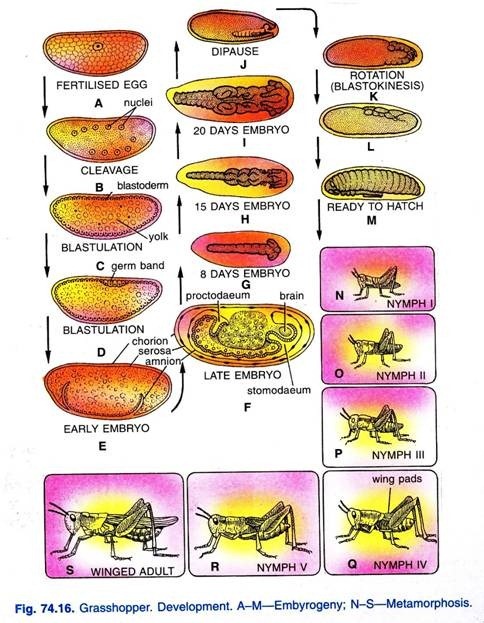
Embryonic development (Fig. 74.16) continues for about three weeks until the embryo is well formed. The development is then arrested and the embryo enters into a rest period, or dipause, to tide over the adverse conditions of cold and lack of food in winter. Growth begins again in the spring when the temperature is warmer.
The young grasshopper that hatches from the egg is called a nymph. It resembles its parent but has a large head compared with the rest of the body and it lacks wings and reproductive organs. It feeds upon vegetation and grows rapidly. As the young grasshopper grows and becomes too large for its inflexible chitinous exoskeleton, which is shed periodically.
The shedding of chitinous exoskeleton is a complex process called moulting. Wings gradually develop from wing-pads and after five moults the young grasshopper reaches the adult form. This type of development is called simple or gradual metamorphosis.
10. Economic Importance of Grasshopper:
(i) As Crop Pests:
Both nymphs and adults eat many kinds of vegetation, specially succulent types. They often migrate into new feeding grounds and may damage or ruin farm and garden plantings. Feeding is most active in the mid-morning hours of quite sunny days. When food is scarce, these insects will eat cotton or woolen fabrics, wood and disabled grasshoppers.
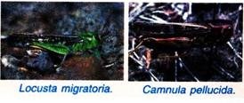
Grasshoppers also feed on grasses and, thus, render heavy damage to range and pasture fields. True locusts which are also grasshoppers migrating in long hordes, are of a different kind and cause heavy damage to our crop fields and other vegetation. Locusta migratoria, the migratory locust found in eastern hemisphere, has caused famines since Biblical times.
Melanoplus maxicans, the rocky mountain locust of North America give rise to a migratory phase called M. maxicans spretus which also cause great loss.
The grasshopper Camnula pellucida is a serious pest. During favourable conditions and lack of enemies, it develops and hatches in May or June. It is a migratory form and can fly long distances. The swarms of this grasshopper destroy the green vegetation and were called plague of grasshoppers by Egyptians.
(ii) As Food:
The grasshoppers are also of some use to man and other animals. They are used as good fish bait, either living or dead. They are sometimes used even for human food. They are still used as food in such countries specially in Japan, Mexico and Philippines. They are commonly eaten by North American Indians and primitive tribes in other parts of the world.
The Greeks ground the locusts by mortars and made flour of them and used the flour as food. The eggs, nymphs and adults of grasshoppers provide food for several predatory insects, spiders, frogs, reptiles, birds and mammals. In India also some people eat them as food either roasted or fried.
11. Control of Grasshoppers:
The grasshoppers are controlled by natural as well as artificial or chemical means. The grasshopper eggs are eaten by some beetles, bee flies, moles, skunks, and mice, the nymphs by robber flies and digger wasps and both nymphs and adults by large predatory insects and by frogs, reptiles, birds and mammals. Eggs of grasshoppers are also parasitized by certain insects.
Flesh flies (Sarcophaga) lay living maggots on adults, and tachinid flies deposit their eggs on grasshoppers in flight, the larvae of both burrow into their host and consume the fat tissues. Parasitized grasshoppers become logy and fail to reproduce or die.
The parasitic insects, thus, constitute a factor in grasshopper control. Both fungus and bacterial diseases also destroy numbers of grasshoppers at times. Eggs of grasshoppers are killed in the ground during winter, if the soil is exposed to the sun by ploughing. In olden days the control of grasshoppers was done by giving them food mixed with arsenic or some other stomach poison. But now insecticides are generally used.
Various insecticides are used in the form of sprays or dusts and poisoned baits that kill either by contact or when eaten. Some insecticides which are recently employed are aldrin, dieldrin, chloradane, heptachlor and toxaphane. Methoxychlor is also now used as insecticide for protecting fruits and vegetables and pasture fields, as it does not leave any residue which is harmful to man or domestic animals.


