ADVERTISEMENTS:
In this article we will discuss about:- 1. The Electron Micrograph of Mitochondria 2. The Electron Micrograph of Golgi Complex 3. The Electron Micrograph of Endoplasmic Reticulum 4. The Electron Micrograph of Lysosomes 5. The Electron Micrograph of Plastids 6. The Electron Micrograph of Nucleus.
Contents:
- The Electron Micrograph of Mitochondria
- The Electron Micrograph of Golgi Complex
- The Electron Micrograph of Endoplasmic Reticulum
- The Electron Micrograph of Lysosomes
- The Electron Micrograph of Plastids
- The Electron Micrograph of Nucleus
1. The Electron Micrograph of Mitochondria:
ADVERTISEMENTS:
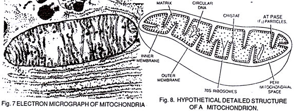
It is an electron micrograph of cell’s largest and most important organelle – the mitochondria and is characterized by the following features (Fig. 7 & 8):
(1) The name mitochondria was given by Benda (1898) and their ma n function was brought to light by Kingsbury (1912).
(2) Each mitochondria in section appears as sausage or cup or bowl shaped structure lined by double membranes. Theoretically, the membrane is similar n structure and chemical composition to plasma membrane.
ADVERTISEMENTS:
(3) Two membranes are separated by a 6-8 mm wide fluid filled space called peri-mitochondrial space.
(4) The inner membrane is projected into the central cavity as finger like outgrowths- the cristae.
(5) Numerous small, rounded & stalked particles – The oxysomes or F1 or ATPare are attached to the inner surface of inner membrane.
(6) The central cavity is filled with matrix which theoretically possesses circular DNA 55s ribosomes and respiratory enzymes.
(7) The main function of mitochondria is to synthesize chemical energy- ATP from glucose as substrate.
(8) From one molecule of glucose 38 ATP molecules (40%) are synthesized and the rest of the energy (60%) goes as heat.
2. The Electron Micrograph of Golgi Complex:
It is the electron micrograph of Golgi complex along with its line drawing and is characterized by the following features (Fig.9 & 10):
(1) It was discovered by Camillio Golgi (1898) and was named after his name.
(2) The Golgi complex, as is visible in electron microphotograph, is a stack (bundle) of hollow tubules, which in actual form are hollow flattened sacks arranged above each other. On either side certain large globular vesicles and smaller vacuoles are also visible.
(3) Each tubule or lamella is lined by membrane, which is theoretically similar to plasma membrane in structure and chemical composition.
ADVERTISEMENTS:
(4) The Golgi complex is more prominent and well developed in secretory cells and absent in RBC of mammals and prokaryotic cells.
(5) Its main function is to glycolise the proteins which are synthesized by ribosomes i.e., It converts these inert proteins into glycoprotein’s to act as hormones, enzymes and coenzymes.
(6) It also helps in the formation of lysosomes and acrosome of sperms.
3. The Electron Micrograph of Endoplasmic Reticulum:
ADVERTISEMENTS:
It is an electron micrograph of endoplasmic reticulum and is characterized by following features (Fig. 11 & 12):
 (1) It was discovered and named by Porter (1948).
(2) It is made up of large number of interconnected and branched tubules, long, flattened and sac-like cisternae and hollow approximately rounded vesicles present all over in the cytoplasm forming a continuous system.
ADVERTISEMENTS:
(3) Each tubule, cisternae or vesicle is made up of membrane, which is theoretically similar to plasma membrane in structure and chemical composition.
(4) Some cisternae and tubules bear small, dark, rounded and granular structures, ribosomes, along their surface. This endoplasmic reticulum is called rough or granular E.R. The endoplasmic reticulum without ribosomes is called smooth or agranular ER.
(5) The main function of rough endoplasmic reticulum is protein synthesis.
(6) The main functions of smooth endoplasmic reticulum are:
(a) Detoxification
(b) Synthesis of lipids & cholesterol
ADVERTISEMENTS:
(c) To mobilize Ca+++ and Mg++ ions and (1) Glycogenolysis.
(7) It is absent in R.B.C. of mammals and prokaryotic cells.
(8) Both types of reticulum provide mechanical support, transport with in the cell, conduction of nerve and electric impulses and formation of nuclear membrane at the time of cell division.
4. The Electron Micrograph of Lysosomes:
This is the electron micrograph of Lysosome, and is characterized by following features.
These are also called Suicide bags or Death bags of the cell (Fig. 13 &14):
(1) They were discovered by de Duve (1954).
(2) They are spherical or irregular membrane bound vesicles filled with digestive enzymes.
(3) The Lysosomes in a cell occur in three forms viz., primary lysosome, secondary lysosome and residual body.
(4) The primary lysosomes are nascent lysosomes which are in a dormant stage; the secondary lysosome are those which have fused with phagocytic vesicles and has released their enzyme contents into the vesicle. This is also called phagosome. The residual body is one which has completed its digestive function and is ready to be thrown out of the cell.
(5) They develop from Golgi complex.
(6) Besides digestion, their other function is autophagic digestion during extreme starvation or extreme toxicities.
They also promote:
(a) Aging
(b) Cancerous growth,
(c) Metamorphosis,
(d) Defence against disease, bacteria and viruses and
(e) Osteogenesis.
(7) These are absent in mammalian RBC, Prokaryotic cells and most plant cells.
5. The Electron Micrograph of Plastids:
This is an electron-micrograph of plastid or chloroplast, which is an integral component of all green plant leaves and is characterized by following features (Fig. 15 & 16):
 (1) They may be spheroidal, ovoid, stellate or collar shaped and differ in size and number in different cells.
(2) Each chloroplast is a sac-like structure, which is made up of double membranes separated from one another by periplastidial space.
(3) Two types of double membranous lamellae are embedded in the stroma or matrix filled cavity:
(a) Smaller flattened disc-shaped lamellae – The thylakoids, placed one above the other in a stack – the grana.
(b) Larger tubular lamellae between grana called lamellae or frets which connect adjacent granna.
(4) The Inner surface between the two membranes of a thylakoid bear countless granular chlorophyll particles the Ouantasomes.
(5) The plastids also have their own circular DNA 55s – Ribosomes and RNA
(6) The main function of chloroplast or plastid is to synthesize carbohydrate molecules from CO2 + H2O using light energy.
6. The Electron Micrograph of Nucleus:
This is an electron micrograph of nucleus. (Fig. 17 & 18):
(1) Nucleus was discovered by Brown (1831).
(2) It is a characteristic entity of almost all eukaryotic cells except mammalian RBCs.
(3) The nucleus is generally one but may also be two, four or many.
(4) Each nucleus is surrounded by double nuclear membranes perforated by numerous nuclear pores. Each nuclear membrane is just like unit membrane. Inside, there is present a large darkly stained nucleolus and a network of chromatin threads.
(5) The nucleolus is responsible for all the ribosomal RNA synthesis and chromatin (DNA) is responsible for controlling all the metabolic activities of cell as well as for all hereditary activities.
(6) The chromatin threads are made up of double helical DNA molecule which are the carrier of heredity units- the genes.


