ADVERTISEMENTS:
In this article we will discuss about the dissection of earthworm. Also learn about:- 1. The Alimentary System 2. Dissection of Nervous System 3. Dissection of Reproductive System.
Earthworms are delicate animals (Fig.2.1) and need careful handling to avoid damage to internal organs.
Killing:
ADVERTISEMENTS:
Wash the live specimens with water to get rid of mucus. Drop them in a petri dish containing 30% alcohol. Take out the specimens immediately after ceasation of movements and put in tap water in a large beaker.
Dissection:
Place the specimen on the fingers of your left hand. Insert the tip of one of the blades of a pair of fine scissors through the skin above the dorsal blood vessel at about 30th segment of the body. Hold the scissors almost in a horizontal position keeping the lower arm just below the body wall and cut the skin anteriorly for about 2 cm.
ADVERTISEMENTS:
Put the worm on the dissecting tray, keeping the dorsal surface upwards and fix it in a straight line on the wax with a few pins passing through the skin of the lateral sides and one at each of the anterior and the posterior end. Care should be taken not to damage the nerve ring at the anterior end and the anal region at the posterior end.
Starting from the initial incision cut the skin along the mid-dorsal line, proceeding anteriorly or posteriorly or both as required for dissection. Hold the skin with a pair of fine forceps and free it from septa with a fine needle. Care must be taken not to damage the gut or other organs. Pin down the loose flaps of the skin and proceed for dissection of organ systems.
The Alimentary System:
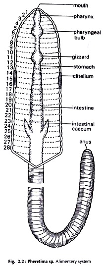
The alimentary canal is a straight tube (Fig. 2.2) running from the mouth to the anus. It is exposed when the skin is cut open. It runs along the whole length of the body. Remove the seminal vesicles in 10-12 segments to fully expose the gut.
Mouth:
A crescentic opening, ventral and posterior to the prostomium.
Buccal chamber:
A small chamber posterior to the mouth.
Pharynx:
ADVERTISEMENTS:
Pear-shaped, posterior to the buccal chamber.
Oesophagus:
The narrow, tubular portion between the pharynx and the gizzard.
Gizzard:
ADVERTISEMENTS:
A hard, muscular, almost round organ posterior to oesophagus.
Intestine:
The part of the alimentary canal from gizzard to the anus.
Intestinal caeca:
ADVERTISEMENTS:
Two conical outgrowths directed forward, one on each side of the intestine in the 26th segment.
Rectum:
The last portion of the gut in the posterior 23 to 25 segments.
Anus:
ADVERTISEMENTS:
The round opening of the rectum.
Dissection of Nervous System:
It is located on the ventral surface except the anterior end, which is oriented vertically.
Cut open the skin along the whole length of the body taking care that the nerve ring, which is dorsoventral in orientation, is not damaged. Separate the gut from the body wall. The anterior and posterior ends of the alimentary canal should be carefully detached from the body wall to which it is attached.
Cut the oesophagus and carefully pull out the anterior end of the gut from behind. The nerve ring is exposed. Remove the rest of the gut. The ventral nerve cord is clearly seen (Figs. 2.3 and 2.4).
Nerve ring:
ADVERTISEMENTS:
It is situated at the anterior end of the body and vertical in orientation. The pharynx runs through it.
a. Supraoesophageal or cerebral ganglia:
Two in number, fused to form the dorsal part of the ring or so-called brain.
b. Peripharyngeal connectives:
Two in number, constitute the lateral sides of the ring around the pharynx and connect the brain with the sub-pharyngeal ganglia.
ADVERTISEMENTS:
c. Sub-pharyngeal ganglia:
The two ganglia fused to form the ventral portion of the nerve ring.
Ventral nerve cord:
Situated ventrally, runs backward from the sub-pharyngeal ganglia to the posterior end of the body.
a. The nerve cord bears a ganglion in each segment.
b. Two pair of nerves arise from each ganglion.
Dissection of Reproductive System:
The earthworm is hermaphrodite, (Fig.2.5) i.e., both male and female reproductive organs are present in the same individual.
Cut open the skin and expose organs from about 30th segment to the anterior end of the worm. To detach the gut from the body wall starting from about 30th segment, cut the gut, hold the cut end with a pair of forceps in your left hand, lift it a little and remove the septa connecting the gut with the body wall, with a fine needle.
The ovaries are present in the 13th segment. Be careful from about 15th segment in removing the gut. Proceed up to the anterior end, cut the gut in the region of buccal chamber and remove it.
Male Reproductive Organs:
The organs are two pairs of testes, two pairs of testes sacs, two pairs of seminal vesicles, two pairs of vasa deferentia, one pair of prostate glands with ducts and two pairs of accessory glands.
Testes sacs:
Two pairs, one pair in each of the 10th and 11th segment, whitish or yellowish-white in colour.
Seminal vesicles:
Two pairs, whitish or yellowish-white in colour. One pair in each of the 11th and 12th segment.
Cut open the testes sacs to expose enclosed structures. The cavities of the two sacs of the same segment communicate with each other. Each testis sac communicates with the seminal vesicle behind it.
Testes:
ADVERTISEMENTS:
Two pairs. A testis is a minute white body close to the middle line of the testis sac, visible only with a powerful lens.
Vasa deferentia:
Two pairs, whitish, threadlike. A seminal funnel lies just beneath a testis. The ducts run backward and the two of each side enter the prostate gland of the side.
Prostate glands:
Two large, whitish, irregular bodies extending from 17th to 20th segment. The prostatic duct, one from each gland is muscular and horse shoe-shaped. The duct with two vasa deferentia of the side opens in the 18th segment.
Accessory glands:
Two pairs, globular in shape. One pair in each of the 17th and 19th segment.
Female Reproductive Organs:
The organs are a pair of ovaries, a pair of oviducts and four pairs of spermathecae. They are not connected with one another.
Ovaries:
Two minute white bodies attached to the posterior surface of the septum 12/13 segments, on either side of the nerve cord.
Oviducts:
Two very short, narrow tubes just beneath the ovaries with a funnel at the anterior end. Posteriorly they pierce the septum 13/14, converge to meet each other below the nerve cord and open to the exterior through a common aperture in the 14th segment.
Spermathecae:
Four pairs, located ventro laterally, one pair in each of the 6th, 7th, 8th and 9th segment. They open to the exterior by fine pores in the inter-segmental grooves 5/6, 6/7, 7/8 and 8/9.


