ADVERTISEMENTS:
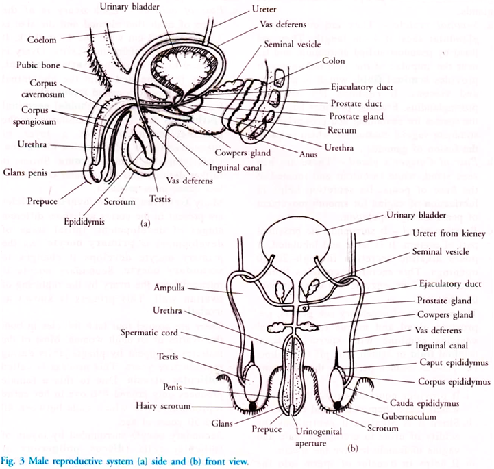
Male reproductive system consists of following parts:
a. Testes:
There is a pair of testis whose size is 4.5 cm x 2.5 cm x 3 cm. It is oval in shape and pink in colour. It is the primary sex organ in males. Testes is lodged in a thin walled skin pouch called scrotum or scrotal sac. Testes are extra abdominal. The reason behind this is that testicular temperature should be 2°C lower than the body temperature for normal spermatogenesis to occur. Rise in temperature kills spermatogenetic tissues.
ADVERTISEMENTS:
In case testes do not descend in the scrotum it causes infertility as the formation of sperms does not occur because of rise in temperature. When it is cold, testes shrink to bring it close to the body to keep it warm and in summers it is relaxed and thin. Scrotal sac is tilled with a tissue fluid called as hydrocoel. Testes is held in the scrotal sac by thick fibrous tissue called spermatic cord or gubernaculum.
There is a cavity between abdominal cavity and scrotal sac called as inguinal canal. When the testes descend in the sac they pull their nerves, blood vessels and conducting tubes after them. The connecting tissue along with the cremaster muscles form spermatic cord.
Any damage to inguinal tissue may cause budging out of intestine into scrotum. Such a condition is called inguinal hernia. Septum scorti divides the scrotum internally into two parts. Externally this division is marked by a scar, raphe.
Outer most cover of testes is called as tunica vaginali which is the visceral layer of peritonium. Under this there is a dense fibrous cover called as tunica albuginea. Under this coat there is a loose connective tissue and blood vessels which together form the tunica vasculosa. Internally tunica albuginea divides each testes into 200-300 lobules.
ADVERTISEMENTS:
Each of these lobes consists of 1—3 convoluted seminiferous tubules. Seminiferous tubules is a tubular structure which has both the ends terminating into short tubules called tubular recti. Tubular recti connects seminiferous tubules to rete testis. Rete testis is convoluted labyrinth of cuboidal epithelium.
Each testes consists of 1000 seminiferous tubules (Fig. 2). There are two kinds of cells found in seminiferous tubules. They are spermatogenic cells (germ cells) and Sertoli or supporting cells (nurse cells). Sertoli cells were discovered by Enrichno Sertoli, an Italian histologist. As the name signifies, germ cells form the spermatozoa by spermatogenesis and nurse cells provide nourishment to the developing sperm.
Between the seminiferous tubules, Leydig cells are found. These are polygonal in shape and secrete a male steroid hormone called testosterone. Testosterone controls the development of secondary sexual characters in males. Leydig cells were discovered by Franz von Leydig, a German anatomist.
b. Vasa Efferentia:
Rete testes gives rise to 10-20 ductules called as vasa efferentia or ductuli efferentes. Vasa efferentia enters the head of epididymis. They are lined by pseudo stratified epithelium which helps in sperm movement.
c. Epididymis:
It is a 6 metre long coiled tube found in the poster lateral side of each testes.
It is divided into three parts:
ADVERTISEMENTS:
i. Upper head or caput epididymis or Globus major — This part is wide and receives vasa efferentia.
ii. Corpus epididymis or Globus minor or body – It stores sperms for a short duration, which undergoes maturation. It lies in the lateral side of testes.
iii. Cauda epididymis or Globus minor or tail – Before entering the vas deferens the spermatozoa is stored here. This part lies on the caudal side of the testes and is wide.
d. Vas Deferentia:
ADVERTISEMENTS:
This is also called as seminal duct. It is around 30 cm long, narrow, muscular and tubular structure which starts from the tail of epididymis, passes through the inguinal canal, then over the urinary bladder and then joins the duct of seminal vesicle to form a 2 cm long ejaculatory duct. After passing through the prostate gland it joins the urethra. Before the sperms are transferred to urethra they are stored in spindle like ampulla of vasa deferens.
e. Penis:
It is the male genetalia. It is erectable, copulatory, cylindrical organ. It is made up of three erectable tissues. Two of the three are posterior and made of yellow fibrous ligament and is called as Corpora cavernosa. One is anterior, spongy and highly vascular Corpus spongiosum.
It surrounds the urethra. The tip of penis is highly sensitive and is called glans penis. There is a retractile fold of skin on glans penis and is known as fore skin or prepuce. The erection of penis is due to rush of arterial blood into sinuses of corpus spongiosum.
ADVERTISEMENTS:
Accessory Sex Glands of Males:
These are a pair of seminal vesicles, prostate gland and a pair of Cowper or bulbourethral glands.
a. Seminal Vesicle:
ADVERTISEMENTS:
They are convoluted, glandular sacs of 4 cm length. They are lined by pseudo stratified epithelium and lie near the ampulae of the vasa deferentia. It provides seminal fluid, which is alkaline and viscous. It contains fructose and prostaglandins. Fructose provides energy to the sperms for swimming and prostaglandins stimulate vaginal contraction which helps in the fusion of gametes.
b. Pair of Cowper’s Gland:
These are pea seed sized, white in colour and located at the base of penis. Its secretion helps in lubrication of vagina for smooth movement of penis during copulation.
c. Prostate Gland:
It surrounds the proximal part of urethra. It is large and lobulated. It pours alkaline secretion through 20-30 openings. This secretion contains lipids, bicarbonate ions, enzyme and small amount of citric acid.
The secretion of accessory sex glands, i.e. prostate gland and mucus from seminal vesicles combine with sperm to form seminal fluid or semen. The pH is alkaline, i.e. 7.3 – 7.5.
ADVERTISEMENTS:
Semen performs the following functions:
i. It provides nourishment to the sperms which keeps them viable and motile.
ii. Since it is alkaline it neutralises the acidity of urine in urethra of male and vagina of female to save the sperm.
iii. It helps in transfer of sperm into the vagina of female.
Hormonal Control:
The hormones responsible for the normal growth and functioning of seminiferous tubule and Leydig cells are, Follicle Stimulating Hormone (FSH) and Lutenizing Hormone (LH). These are secreted from anterior lobe of pituitary.


