ADVERTISEMENTS:
In this article we will discuss about the regulation of respiration in humans.
Oxygen requirement by the body differs depending on the activity. It is lowest at rest and increases during routine activity and further increases in muscular exercise. Similarly production of carbon dioxide also is dependent on the rate of metabolic activity in the body.
Respiratory system has the responsibility of meeting needs of the body by altering the rate and depth of respiration in order to keep the pO2 and pCO2 at normal levels.
ADVERTISEMENTS:
The regulation of respiration can be brought about by:
1. Neural mechanism.
2. Chemical influence.
3. Non-chemical influence.
ADVERTISEMENTS:
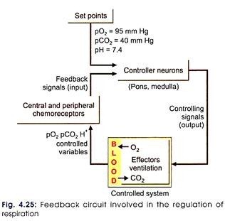
The chemical and non-chemical influence has to act through the neural mechanism only (Fig. 4.25).
I. Neural Mechanism of Respiration:
Centers are present in brainstem. The brainstem centers are required for rhythmic respiration whether during asleep or awake. The cerebral cortical center is required for voluntary alterations in respiration.
Brainstem centers are present in the reticular formation of pons and medulla oblongata.
In the pons, the centers present are:
i. Pneumotaxic
ii. Apneustic
In medulla oblongata, the centers present are:
ADVERTISEMENTS:
i. Inspiratory (dorsomedial group of neurons)
ii. Expiratory (ventrolateral group of neurons)
There is a lot of interconnection between the various centers. The interplay of the different centers is essential for a proper regulation of respiration. The medullary centers are termed as basic centers, whereas the pontine centers are called regulatory centers. The pontine centers act through the medullary centers and bring about smooth rhythmic respiration.
From the medullary centers, which are also spontaneously active, the impulses are sent to spinal cord through the reticulospinal pathway, which ends on the anterior horn cells in spinal cord. Both the phrenic (C3-C5) and intercostal nerve (T1-T11) take origin from spinal cord and influence the activity of diaphragm and intercostals muscles, respectively.
ADVERTISEMENTS:
So if there is a complete transverse section of spinal cord at the level of:
i. C2 segment person dies of respiratory paralysis.
ii. C6 person survives because the diaphragmatic respiration continues.
In a normal person, the inspiratory center (IC) appears to generate impulse on its own. During the course of the generation of impulse, it is presumed that the rate of impulse generation goes on increasing till it reaches a certain point and then there will be sudden cessation of impulse generation. Because of this, IC is known to act as a ramp generator.
ADVERTISEMENTS:
The impulses from the apneustic center have a regulatory influence on the inspiratory center. The apneustic center activity in turn is controlled by the impulses coming from the pneumotaxic center and through the vagus nerve from the stretch receptors of lungs.
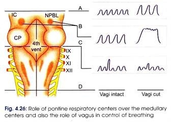
When the influence by the vagus and pneumotaxic center over the apneustic center is lost, there will be prolonged inspiration and a sudden expiration. This type of breathing is known as apneustic breathing (Fig. 4.26).
Sequence of events during normal regulation of respiration by neural mechanism:
ADVERTISEMENTS:
i. Onset and gradual increase in the number of impulses production in the inspiratory center because of the ramp generator.
ii. This leads to:
a. Impulses being sent from IC to spinal cord for stimulation of phrenic and intercostals nerves.
b. Reciprocal inhibition of expiratory center by IC.
c. Excitatory impulses from IC sent to pneumotaxic center through multisynaptic pathway.
iii. When inspiration is going on, there will be gradual inhibition of the apneustic center by the impulses coming from the pneumotaxic center and also from the afferent vagal fibers coming from the distended alveoli.
ADVERTISEMENTS:
iv. Apneustic center influence over the IC ceases completely. Hence the activity of inspiratory center stops and leads to no inhibition influence over the expiratory center (EC). No more impulses from the inspiratory center to motor neurons in the spinal cord.
v. The muscles of inspiration start relaxing. This starts the process of expiration which normally lasts for about 3 sec.
vi. After this, once again the activity in the IC starts, leading to the next respiratory cycle.
Location of the respiratory centers in CNS for rhythmic respiration can be experimentally studied from the following observations:
1. If transection is done above pons, the rhythmic respiration continues as usual.
2. If a mid-pontine section is done along with bilateral vagotomy, there will be a prolonged inspiration followed by a sudden short expiration (apneustic type of breathing).
ADVERTISEMENTS:
3. If transection is done between pons and medulla oblongata, though respiration continues on its own, it will be irregular. Sometimes it becomes shallow and sometimes it is deeper. This type of breathing is known as gasping.
4. If transection is done below medulla (at the beginning of the spinal cord), it leads to complete cessation of breathing.
So by the above studies, it can be concluded that the centers are present in brainstem. The pontine centers play role in smooth and rhythmic respiration.
Hering-Brueur reflex:
Inflation of alveoli brings about cessation of inspiration and expiration commences.
The details are as under:
i. Inflation of alveoli
ii. Leads of stimulation of stretch receptors present in the alveoli.
iii. Afferent impulses are carried by vagal fibers.
iv. Inhibit the activity of the respiratory center, cessation of inspiration.
v. Leads to relaxation of muscles of inspiration.
vi. Expiration commences.
This reflex is not very well seen in adults. The reflex probably helps to prevent over distension of the alveoli.
II. Chemical Influence on Respiration:
This is brought about by the chemoreceptors.
They are called:
i. Peripheral
ii. Central chemoreceptors.
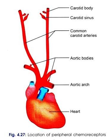
Peripheral Chemoreceptors (Fig. 4.27):
a. Carotid bodies which are present at the branching of internal carotid artery.
b. Aortic bodies are present in the arch of aorta.
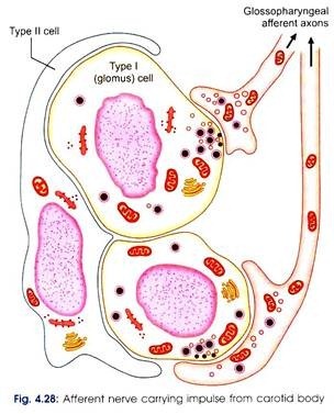
From the carotid bodies, the afferent impulses will be carried by the sinus nerve (Fig. 4.28) a branch of glossopharyngeal nerve and from the aortic bodies by the aortic nerve branch of vagus nerve.
The peripheral chemoreceptors respond to:
i. Decrease in pO2
ii. Increase in H+
iii. Increase of pCO2 of blood.
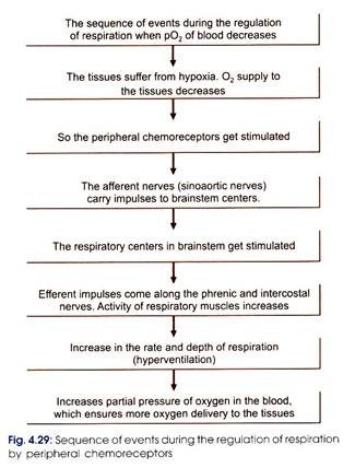
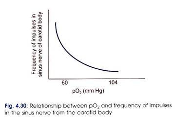
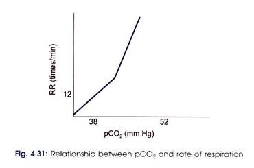
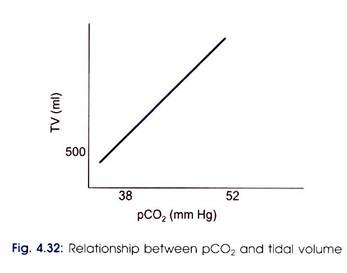
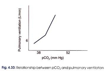
Details of the role of peripheral chemoreceptors in regulation of respiration are shown in Figs 4.29 to 4.33.
Central Chemoreceptors:
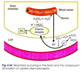
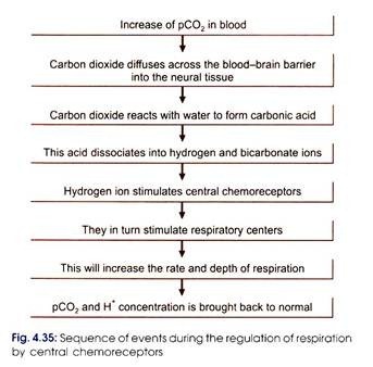
They are present in the brainstem near the respiratory centers. They are more sensitive to hydrogen ions, but the hydrogen ion of blood cannot stimulate them because the blood brain-barrier is impermeable for the hydrogen ion to diffuse through. Hence, the increase in partial pressure of carbon dioxide forms the stimulus (Fig. 4.34).
Decreased pO2, increased pCO2 together (asphyxia) will have an additive effect on chemoreceptors. Hence there will be maximum respiratory response in such a situation (Fig. 4.35). Asphyxia occurs in conditions, like drowning or strangulation.
III. Non-Chemical Influence on Respiration:
Non-chemical influence on respiratory centers pertains to impulses coming from:
i. Baroreceptors
ii. Muscle spindles of respiratory muscles to control depth of respiration.
iii. Pain receptors
iv. Intracranial tension
v. Irritant receptors stimulation in lungs while coughing.
vi. Higher parts of CNS
vii. Irritation of nasal mucosa (sneezing)
viii. Mechanoreceptors in pharynx (deglutition).
ix. Receptors of muscles and joints.
Depending on the location of the receptors influencing the respiratory centers, there will be appropriate alterations in the respiration.

