ADVERTISEMENTS:
In this article we will discuss about the anatomical structure of human tongue with the help of suitable diagrams.
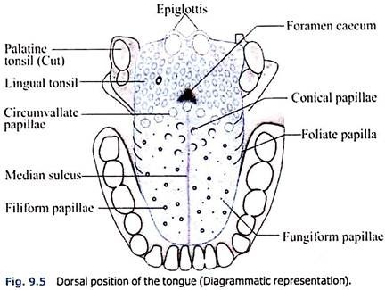
Tongue (Fig. 9.5) is made up of three elements; epithelium, muscles and glands.
The epithelium is stratified and non-cornified. Two types of special structures are seen on it; the papillae (Fig. 9.6) and the taste buds.
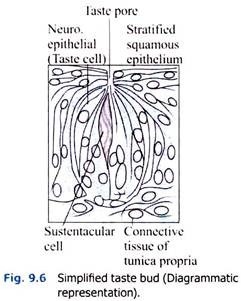
The taste buds (Fig. 9.7) are the sense organs of taste. These buds are lined by stratified squamous epithelium and are flask-like with a wide bottom. A taste pore pierces the short and narrow neck of each taste bud.
The taste bud possesses melon-shaped and frequent supporting (sustentacular) cells and also scanty, narrow and long neuro-epithelial (taste) cells to its outer ends. The first are spindle-shaped and their ends surround a small opening, the inner taste pore. The taste cells vary from 4 to 20 per taste bud.
ADVERTISEMENTS:
They have slender rod-shaped form with a nucleus in the middle, and on the free surface short taste hairs which project freely into the lumen of the pit. These cells are responsible for detection of taste which is to be dissolved in salvia for proper sensation.
The papillae are minute projection of the mucous membrane and are as follows:
i. Circumvallate Papillae:
Circumvallate papillae are large and can be easily seen with the naked eyes. They are only 10 -12 in number situated at the back of the tongue and arranged in the form of a ‘V’ with its limbs opening anteriorly. At the apex of the angle there is small invagination, foramen caecum. A circumvallate or vallate papilla (Fig. 9.7) consists
of a central-rounded elevation, surrounded by the non-cornified stratified squamous epithelium on lamina propria. External to this groove the mucous membrane is raised and is known as vallum.
ii. Fungiform Papillae:
Fungiform papillae having a flat-rounded head like fungus are covered by the non-keratinising squamous epithelium on the fibrous lamina propria, tip being broader than the base (Fig. 9.8). Circumvallate and fungiform papillae carry taste buds. The fungiform papillae are rich in blood vessels and hence have a marked red colour.
iii. Filiform:
ADVERTISEMENTS:
Filiform, also known as conical papillae due to presence of conical pointed cap with keratinising squamous epithelium on the lamina propria (Fig. 9.9). In man this cap consists of epithelial filaments.
iv. Conical (Conic) Papillae:
Conical (Conic) papillae, situated at the dorsum of the tongue, are scattered among the filiform papillae and similar to them, but they are shorter than the filiform.
ADVERTISEMENTS:
v. Foliate Papillae:
Foliate papillae, found on lateral margin of the posterior part, are arranged in several transverse folds. In man they are rudimentary. The filiform and conic papillae possess tactile sensitivity (perceive touch) and all the other papillae gustatory.
Muscles:
ADVERTISEMENTS:
The muscles of tongue are voluntary and consist of cross-striated muscular fibres.
Glands:
The glands are small and scattered.
They are of three types:
ADVERTISEMENTS:
1. Mucous glands.
2. Serous glands.
3. Lymph nodes (glands).
The glands of the last variety are very prominent at the posterior part of the tongue, and are collectively known as the lingual tonsil.
Nerve Supply:
The sensation of taste is carried by the chorda tympanic branch of the facial nerve (anterior two-thirds of tongue) and the glossopharyngeal nerve (posterior one-third). The general sensations of touch, pain, temperature, pressure, etc., are carried by the trigeminal nerve. The muscles are supplied by the hypoglossal nerve.
ADVERTISEMENTS:
Functions:
Tongue serves the following functions:
i. Mastication:
It helps in the act of chewing.
ii. Deglutition:
It helps in the act of swallowing.
ADVERTISEMENTS:
iii. Taste:
Subserves taste and general sensation.
iv. Speech:
Essential for speech.
v. Secretion:
Secretion of mucus and of serous fluids with which it keeps the mouth moist.
Salivary Glands:
There are three pairs of salivary glands parotid, sub-maxillary or submandibular, and sublingual. One of each pair remains on one side and opens into the oral cavity. The parotid gland opens upon the inner surface of the cheek opposite the second upper molar tooth, by a single duct called the duct of Stensen.
The sub-maxillary gland similarly opens by Wharton’s duct upon the floor of the mouth on the side of the frenulum of the tongue. The sublingual gland, on the other hand, opens by several fine ducts, upon the floor of the mouth by the side of the frenulum. These are called the ducts of Rivinus (Fig. 9.11).
Histology:
Salivary glands are typical racemose glands, consisting of lobules which are made up of alveoli and the interlobular septum. Each alveolus is enclosed by a basement membrane upon which the gland cells are arranged. These cells are wedge-shaped, with the thin end of the wedge directed towards the alveoli.
Between these cells and the basement membrane, there are a few scattered, flat cells with long cytoplasmic processes. Under electron microscope, myofibrils have been detected in them which lead to the postulation that these cells are contractile, the contraction of which compresses the alveoli and expels its contents into ducts. These cells are known as myoepithelial basket cells.
The gland cells may be of two types- serous and mucous, and accordingly the glands may be of two types. The parotid gland (Figs. 9.12 & 9.15) is composed entirely of serous cells. The sublingual gland (Figs. 9.14 & 9.17) is predominantly of the mucous type, whereas submandibular or sub-maxillary gland (Figs. 9.13. & 9.16) is mixed, but in man predominantly of the serous type. Each serous cell has a rounded nucleus towards the base of the cells.
There are also chromidial substances which are found to be arranged in parallel lines under the electron microscope. They are probably formed of granules containing ribonucleic acid. In the mixed glands, the serous cells remain compressed at one side of the alveolus in the form of a crescent being sandwiched between the basement membrane and the more numerous mucous cells, lining the alveolus.
These crescentic elements are called the demilunes or crescents of Giannuzzi. The serous cells towards the apices contain numerous fine granules called zymogen granules which are more opaque and appear darker in sections than the mucous cells. The mucous cells have flattened nuclei towards the base and have got less chromidial substance. These cells contain coarser and less numerous granules called mucinogen granules. In addition myoepithelial basket cells described above are present.
The secretion of the serous glands is thin, watery, poor in solid, but rich in enzymes, such as the starch-splitting enzyme, amylase, also known as ptyalin. The secretion of the mucous glands is thick and viscid containing much mucus.
The secretion of the glands is conveyed through various salivary ducts. The ducts also modify the composition of the saliva. The striated duct cells which are present in some of the ducts secrete calcium and iodide into saliva and absorb sodium and water from it. There are three types of ducts, e.g., excretory, striated and intercalated ducts composed of four different types of cells.
The intercalated ducts lead away from the alveoli
and are lined by cuboidal epithelium. The cells are practically filled by large nucleus, cytoplasm is scanty and few if any granules. The intralobular ducts are lined by columnar epithelium, which have peculiar rod-shaped appearance.
The cells have a centrally placed nucleus and there is a marked striation of the basal one-third of the cell. The interlobular or excretory ducts are lined by a two-layered epithelium, composed of columnar superficial layer and a flattened deep layer. At the termination the lining of the duct changes to a layered stratified squamous epithelium.
In addition to these three pairs, there are small accessory buccal glands scattered throughout the mucous membrane and containing cells chiefly of the mucous type. It has been observed that the histological structures of salivary glands are influenced by endocrine secretion of thyroid and sex hormones.


