ADVERTISEMENTS:
Cardiac function does not require intact innervations. Frog’s heart removed from the body, and in human beings who have transplanted heart, the heart continues to function. The perfusion of the isolated heart in the laboratory will also continue to beat at regular intervals.
In a frog’s heart when the chambers are separated, each of the chambers shows regular contractions on its own. The rate of contraction per minute varies. In the case of frog’s heart, the sinus venosus shows maximal rate of contractions when compared to any other chamber of heart.
This can also be proved by applying first and second Stannius ligature or effect of temperature on sinus venosus. It proves that sinus venosus is pacemaker of the frog’s heart.
ADVERTISEMENTS:
Mammalian Heart/Human Heart:
The SA node or sinoatrial node is the pacemaker. Automaticity and rhythmicity are the properties responsible for this. Action potentials from the various chambers (regions) of human heart have been shown in Fig. 3.4.
SA Node:
ADVERTISEMENTS:
i. It is about 8 mm long, 2 mm wide.
ii. Lies posteriorly in the groove at the junction between the superior vena cava and right atrium.
iii. Contains two types of cells namely small round cells (pacemaker cells)—P cells, and slender, elongated cells responsible for conducting impulses within the cells.
Action Potential from the Pacemaker Cells of SA Node:
i. The membrane potential of SA node is not stable. Hence there is nothing as resting membrane potential. Before the impulse generation from the SA node occurs, the membrane potential at SA node is about -60 mV.
ii. During the process of depolarization, the polarity of the interior gets reversed and it becomes positive. In the recording, the upstroke is called as depolarization and this part is less steep.
iii. After the depolarization, there will be reestablishment of the polarized state of the tissue. The down stroke (re-establishment of polarity) is known as repolarization and this phase is steeper. Figure 3.5 shows the action potential recording from ventricle, SA node and atrium.
Ionic Basis:
ADVERTISEMENTS:
Tetradotoxin, which blocks the sodium channels, has no influence on the action potential recorded from the pacemaker region. This suggests that the upstroke of the action potential is not due to sodium influx. Effect of stimulation of sympathetic and parasympathetic nerves on the activity of the pacemaker is shown in Fig. 3.6.
Adrenergic transmitter—increases all the currents whereas acetylcholine increases hyperpolarization by increasing K+ efflux, though acetylcholine regulates potassium channels.
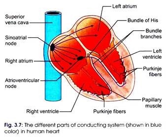
The different parts of conducting system in human heart are (Fig. 3.7):
i. SA node (sinoatrial node)
ii. Internodal fibers
iii. AV node (atrioventricular node)
iv. Bundle of His and its branches
ADVERTISEMENTS:
v. Purkinje fibers
The velocity of conduction of the impulse in the different parts of human heart is:
i. SA and AV nodes—0.05 m/sec
ii. Atrial and ventricular muscles—1.0 m/sec
ADVERTISEMENTS:
iii. Internodal fibers, Bundle of His and its branches— 1.0 m/sec
iv. Purkinje fibers—4.0 m/sec
AV Node:
It is 15 mm long, 10 mm wide and 3 mm thick. Located posteriorly on the right side of interatrial septum near the opening of coronary sinus.
Functions of AV node are:
1. Nodal delay
ADVERTISEMENTS:
2. Conduction of the impulse to bundle of His
3. When SA node fails to act as pacemaker, it can take up the function of pacemaker, but at a decreased rate of impulse production.
The nodal delay does not permit the impulse that has reached the AV node from SA node to get conducted immediately. The normal delay in the AV node is about 0.1 sec. This facilitates proper filling of the ventricular chambers and also prevents the ventricles from contracting simultaneously when atria are contracting.
Vagal stimulation, calcium channel blockers, decrease the velocity of conduction, whereas sympathetic stimulation increases the velocity of conduction at the nodes.
Ventricular Conduction:
Purkinje fibers:
ADVERTISEMENTS:
Purkinje fibers are 70-80 microns. Therefore, conduction velocity is as much as 4 m/sec. This facilitates simultaneous contraction of both right and left ventricles. Like ventricular muscle fibers, the Purkinje fibers also have a long absolute refractory period.
Right vagus nerve predominantly supplies the SA node whereas the left vagus nerve influences the activities of the AV node.
ADVERTISEMENTS:
When vagus nerve is stimulated, acetylcholine is liberated, and it inhibits the activity.
The last portion to be depolarized is the posterior basal part of the ventricles, pulmonary conus and upper most part of the septum.
Arrhythmia refers to irregularity in the beating of heart.
Heart rate varies depending on the phase of respiration. This is known as sinus arrhythmia. This occurs even in normal human beings and hence it is quite physiological one.
i. When the pacemaker is located in the SA node and heartbeats accordingly, it is known as sinus rhythm.
ii. If AV node acts as pacemaker, it is known as nodal rhythm.
iii. If ventricular muscle fibers act as pacemaker, it is known as idioventricular rhythm.
Ectopic beats occurs when regions other than the SA node produces the impulses.
In complete heart block, there will be delayed conduction or no conduction of impulse to ventricular muscle fibers from the SA node.
Heart blocks are:
1. First degree block wherein there will be delayed conduction of impulse from the SA node.
2. Second degree block in which some impulses from SA node only get conducted to the ventricles.
3. Third degree block is also known as complete heart block in which the impulses generated from the SA node will not get conducted to ventricular muscle fibers. The ventricle starts contracting on its own. There is complete dissociation between atria and ventricular contractions.
Vasovagal attack/syncope occurs due to intense stimulation of vagal fibers.
Sick sinus syndrome—ectopic foci producing 100- 150 impulses per min. Normal SA node activity is inhibited by the ectopic foci. Before the SA node starts producing impulses, there is delay during which cardiac output falls to almost zero. Hence the person loses consciousness.


