ADVERTISEMENTS:
In this article we will discuss about Cycas:- 1. External Features of Cycas 2. Anatomy of Different Parts of Cycas 3. Reproductive Structures.
External Features of Cycas:
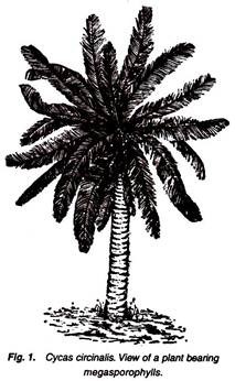
1. Sporophytic plant body attains a height of 8 to 15 feet or more and appears like a small palm.
2. The plant body is differentiated into roots, stem and leaves (Fig. 1).
3. Roots are of two types: normal roots and coralloid roots.
4. Normal roots grow deep into the soil and are well- branched and positively geotropic.
5. Coralloid roots (Fig. 2) are coral-like, dichotomously branched, fleshy, negatively geotropic and arise from the lateral branches of normal roots.
6. Coralloid roots are green in colour because of the presence of an algal zone. In this zone are present members of Myxophyceae, such as Nostoc and Anabaena and some bacteria.
ADVERTISEMENTS:
7. Due to the presence of endophytic algae, these roots become swollen, appear like a coral and hence named coralloid.
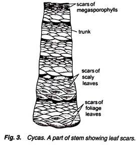
8. The stem is erect, stout and unbranched and remains covered with hard armours (Fig. 3) of woody leaves and sporophyll bases.
9. At the apex of stem is present a crown of leaves arranged spirally.
10. Leaves are of two types: foliage leaves and scaly leaves.
11. Foliage leaves are green, large, pinnately compound and may reach up to 3 to 5 feet or more in length.
12. A foliage leaf consists of a hard rachis having many pinnae or leaflets arranged in two rows on both the sides (Fig. 4).
13. Each leaflet is sub-sessile and lanceolate in shape having an acute apex. A midrib is present in each leaflet.
ADVERTISEMENTS:
14. The margin of leaflets is revoluted or curved downward in C. revoluta, while it is flat in C. circinalis and C. rumphii.
15. Leaves are circinately coiled when young (Fig. 5).
16. Scaly leaves are dry, brown-coloured, covered with many hair and present at the apex of stem.
ADVERTISEMENTS:
17. Sometimes, many “bulbils” arise in between the leaf bases of the stem. They are covered with scaly leaves at the base and germinate into new plants in favourable conditions (Fig. 6).
Anatomy of Different Parts of Cycas:
Cut thin transverse sections of different parts (young normal root, old normal root, coralloid root, young stem, old stem, rachis and leaflets of Cycas revoluta and C. circinalis), stain them separately in safranin- fast green combination, mount in glycerine and observe the anatomical details.
ADVERTISEMENTS:
T.S. Normal Root (Young):
1. Outermost layer of the root, which is circular in outline, is called epiblema. It consists of many tangentially elongated cells. From some cells arise the root hair (Fig. 7).
2. Inner to the epiblema is the parenchymatous cortex with many intercellular spaces. The cells are filled with starch.
ADVERTISEMENTS:
3. Tannin cells and sometimes mucilage canals are also present in cortex.
4. Inner to the cortex is a single-layered endodermis having casparian strips and multilayered pericycle.
5. Vascular bundles are radial, i.e., xylem and phloem are present on different radii. Xylem is exarch and generally diarch, but sometimes protoxylem strands range from 3 to 8 in number.
6. The exarch protoxylem contains spiral thickenings, while the metaxylem has scalariform thickenings.
7. The phloem consists of sieve tubes and phloem parenchyma and present alternate to the xylem groups.
8. Pith is generally absent.
ADVERTISEMENTS:
T.S. Normal Root (Old):
1. Epiblema is ruptured and lacks root hair.
2. Below the epiblema is present multilayered cork followed by cork cambium and many-layered parenchymatous cortex (Fig. 8).
3. Endodermis and pericycle are same as in young normal root.
4. Cambium cuts secondary phloem towards outer side and secondary xylem towards inner side. Vascular rays are also clear.
ADVERTISEMENTS:
5. Outside the secondary phloem may also present a layer of crushed primary phloem.
T.S. Coralloid Root:
It is similar in many aspects to the normal root expect a few following differences:
1. It is circular in outline and the outermost layer is epiblema. But at maturity cork as well as cork cambium develops. Root hairs are normally absent.
2. Cortex is parenchymatous and divisible into outer cortex and inner cortex having a middle algal zone (Figs. 9, 10).
Following algae and bacteria have been reported from algal zone:
(i) Noxtoc punctiforme,
(ii) Anabaena cycadeae,
(iii) Oscillatoria,
(iv) Members of Bacillariophyceae (diatoms),
(v) Bacteria, such as Pseudomonas and Azotobacter.
3. Details of endodermis, pericycle and vascular bundles are same as in normal root. Xylem is exarch and triarch.
4. Normally, the secondary growth is absent (Figs. 9, 10).
T.S. Stem (Young):
1. It is wavy or irregular in outline.
2. Outermost layer is the epidermis which consists of compactly arranged thick-walled cells. Due to the presence of persistent leaf bases and woody scales, g the epidermis is irregular and sometimes not very clear (Fig. 11).
3. Cortex is very large, parenchymatous and contains many girdle traces and mucilaginous ducts.
4. Each mucilaginous duct remains bounded by many epithelial cells or secretory cells.
5. Endodermis and pericycle are not very clear.
6. The vascular cylinder is ectophloic siphonostele and many vascular bundles are arranged in a ring. Each vascular bundle is conjoint, collateral, open and endarch.
7. Xylem consists of tracheitis and xylem parenchyma. There is no vessel.
8. Outside the xylem is the phloem which consists of sieve tubes and phloem parenchyma.
9. Companion cells are absent.
10. In the centre of the stem is present pith.
11. Many mucilage ducts are present in the pith.
T.S. Stem (Old):
1. Secondary growth is present.
2. A thick outer periderm followed by large parenchymatous cortex, having many leaf traces and mucilaginous ducts, are present in the old stem.
3. Vascular strands are present in the form of rings (Fig. 12). Medullary rays are also common.
4. The number of vascular rings is variable from 2 to 14 in different species, thus showing polyxylic condition.
5. Other details are similar to that of young stem.
T.S. Rachis:
1. It is rhomboidal, biconvex or roughly cylindrical in outline, if the section passes through the base, middle or apex of the rachis, respectively.
2. Two arms are present on rachis, one on each side. These are the bases of the leaflets, which arise from the rachis (Fig. 13).
3. The outermost layer consists of thick-walled epidermis which is heavily cuticularized.
4. The continuity of epidermis is broken by many sunken stomata present on upper as well as lower sides of rachis.
5. Below the epidermis is present chlorophyll-containing cells of chlorenchyma followed by thick- walled sclerenchymatous region (Fig. 14).
6. Sclerenchyma is four to six-layered.
7. Below the sclerenchyma is present a large region of ground tissue consisting of thin-walled parenchymatous cells. In this region are present many mucilaginous canals and vascular bundles.
8. Each mucilaginous canal is a double-layered structure consisting of an inner layer of epithelial cells surrounded by an outer layer.
9. Vascular bundles are arranged in omega (Ω) – shaped manner (Fig. 13).
10. Each vascular bundle is conjoint, collateral and open, and remains surrounded by a bundle sheath.
11. In vascular bundles, the xylem is present towards inner side consisting of tracheids and xylem parenchyma with no vessels. It is separated from the phloem by the cambium. The xylem is diploxylic, i.e., consisting of centripetal and centrifugal xylem (Fig. 14).
12. Phloem is present on outer side and consists of sieve tubes and phloem parenchyma with no companion cells.
The vascular bundles show different structures at different levels of rachis starting from the base to the apex with regard to their diploxylic nature as under:
(a) Vascular Bundles at the Base of Rachis:
1. Only the centrifugal xylem is well-developed (Fig. 15A).
2. Its protoxylem faces towards the centre showing endarch condition.
3. Centripetal xylem is not developed.
(b) Vascular Bundles in the Middle of Rachis:
1. Centripetal xylem as well as centrifugal xylem are present showing diploxylic condition (Fig. 15B).
2. Centripetal xylem is present just opposite to the protoxylem of the centrifugal xylem.
(c) Vascular Bundles at the Apex of Rachis:
1. Centripetal xylem is well-developed, triangular and exarch (Fig. 15C).
2. Centrifugal xylem is much reduced and present in the form of two patches lying one on each side of the protoxylem elements of centripetal xylem.
3. At the extreme tip, the centrifugal xylem is totally absent.
T.S. Leaflet of Cycas revolute:
1. It can be differentiated into a swollen midrib portion and two lateral wings.
2. The wings are curved downward or revoluted at the margins (Fig. 16).
3. Outermost layer consists of thick-walled epidermis surrounded by a layer of cuticle.
4. Upper epidermis is a continuous layer while the continuity of lower epidermis is broken by many sunken stomata.
5. Below the upper epidermis is present the sclerenchymatous hypodermis which is more cells thick in the midrib region.
6. Hypodermis is absent below the lower epidermis, except in the midrib region.
7. Mesophyll is differentiated into palisade and spongy parenchyma (Fig. 17).
8. Palisade is present in the form of a continuous layer below the sclerenchymatous hypodermis. Cells of the palisade are radially elongated and filled with chloroplast.
9. Spongy parenchyma is present only in the wings directly above the lower epidermis. The cells are oval, filled with chloroplast and are loosely arranged having many intercellular spaces, filled with air.
10. Few layers of transversely elongated cells are present in both the wings (blades) just in between the palisade and spongy parenchyma. This is secondary transfusion tissue.
11. Primary transfusion tissue present just on either side of the centrally located vascular bundle.
12. A single vascular bundle is present in the midrib portion of the leaflet.
13. The vascular bundle is conjoint, collateral and open. It is diploxylic. The triangular centripetal xylem is well-developed with endarch protoxylem. Centrifugal xylem is represented by two small groups on either side of protoxylem. Phloem is arc- shaped and remains separated from the cambium. Each vascular bundle is surrounded by a bundle sheath.
14. The portion of the midrib in between the palisade layer and lower hypodermal region is filled with parenchymatous cells, of which some cells contain calcium oxalate crystals.
T.S. Leaflet of Cycas circinalis:
It resembles very much with the Cycas revoluta, discussed above, except following differences:
1. The margins of wings are straight and not revoluted.
2. The upper side of the midrib is much ejected out.
3. Stomata, which are present on lower epidermis, are not much sunken.
4. Hypodermis in wings is present only at the corners.
5. Palisade is present only in the wings and not in the midrib region.
Structure of vascular bundle, spongy parenchyma and other details are similar to Cycas revoluta.
Reproductive Structures of Cycas:
1. Plants are strictly dioecious.
2. Male structures are in the form of a compact conical body called male strobilus or male cone (Fig. 18).
3. Female structures are not present in the form of compact cones but they are loosely arranged and called megasporophylls.
Male Cone:
1. It is very large (Fig. 18), conical or ovoid structure, reaching sometime up to 0.5 metre in length.
2. In the centre of each male cone is present a cone axis, which is clearly seen in L.S. (Fig. 19).
3. On the cone axis are attached many leafy structures at right angle. These are called microsporophylls.
4. At the base of the male cone are present many young leaves.
5. All the microsporophylls in the male cone are fertile, except a few at the base and a few at the apex.
Microsporophylls, Microsporangia and Microspores:
Separate a microsporophyll from the male cone and observe the shape and arrangement of microsporangia on its lower surface.
1. Microsporophylls are flat, leaf-like, woody and brown-coloured structures with narrow base and expanded upper portion.
2. Upper expanded portion becomes pointed and called apophysis.
3. Narrow base is attached to the cone axis with a short stalk.
Each microsporophyll has two surfaces: an adaxial or upper surface and an abaxial or lower surface.
4. On the adaxial surface is present a ridge-like projection in the middle and an apophysis at the apex (Fig. 20).
5. On the abaxial surface are present thousands of microsporangia in the middle region in groups of 3 to 5. Each such group is called a sorus (Fig. 21 A).
6. In between these groups are present many hair-like structures (Fig. 21B).
7. Each microsporangium is an oval or sac-like structure with a short stalk. It encloses many microspores or pollen grains.
8. Each pollen grain is a rounded, uninucleate structure, surrounded by an outer thick exine and inner thin (or thick on lateral sides) intine.
T.S. Microsporophyll:
Cut transverse section of microsporophyll, stain in safranin-fast green combination, mount in glycerine and study:
1. Many microsporangia (Fig. 22) are present on abaxial side.
2. Each shortly-stalked sporangium is surrounded by many layers with the innermost layer of tapetum. Many pollen grains are present in each sporangium.
3. Many mucilaginous canals and vascular bundles are present in the microsporophyll.
Female Reproductive Organs:
There is no true female cone or strobilus. Female reproductive organs are present in the form of megasporophylls.
Megasporophylls:
Observe the specimens of megasporophylls of different species and note the following features:
1. Like foliage leaves, megasporophylls are spirally arranged at the apex of stem, in very large number, and thus appear like a rosette.
2. They are loosely arranged in acropetal succession without showing any effect on apical meristem.
3. They are formed once in a year in the mature plant.
4. Each megasporophyll is considered as a modification of foliage leaf and reaches up to 20 cm or more in length.
5. Each megasporophyll is a flat body consisting of an upper dissected or pinnate leafy portion and a lower stalk. On stalk, the ovules are arranged in two rows.
6. Megasporophylls are covered by many yellow or brown-coloured hair.
7. The ovules are green when young but at maturity they are fleshy and bright orange or red-coloured structures.
8. The ovule of C. circinalis is the largest amongst the living gymnosperms, measuring about 6 cm in length.
External morphology of megasporophyll is different in different species of Cycas with regard to the number of ovules and the dissected nature of the upper portion.
(A) Megasporophyll of Cycas rumphii:
1. The pinnae are reduced in size (Fig. 23A).
2. The number of ovules is 4 to 6.
3. The base of the megasporophyll is covered by scaly leaves.
(B) Megasporophyll of Cycas revolute:
1. Upper part is much dissected and pinnate (Fig. 23B).
2. Tip of each pinna is generally acute.
3. The size of megasporophyll ranges between 15 to 20cm.
4. The number of ovules is 2 to 12.
(C) Megasporophyll of Cycas circinalis:
1. The upper part is not much dissected.
2. The margin of the upper part is serrate (Fig. 23C).
3. The ovule is largest amongst the living gymnosperms.
(D) Megasporophyll of Cycas Siamensis:
1. The leafy portion is much dissected.
2. Few upper pinnules unite to form a solid structure (Fig. 23D).
3. The number of ovules is only two.
L.S. Mature Ovule:
1. The ovule (Fig. 24) is orthotropous and unitegmic.
2. The single integument is very thick and covers the ovule from all the sides except at mouth-like opening called micropyle.
3. Single integument consists of following three layers (Fig. 24).
(a) Outer, green or orange, fleshy layer called sarcotesta;
(b) Middle, yellow, stony layer called sclerotesta and;
(c) Inner, fleshy layer.
4. Integument remains in close association with the nucellus.
5. The nucellus develops the nucellar beak in the micropylar region.
6. In the nucellar beak is present a hollow small cavity or chamber called pollen chamber.
7. In the centre of the ovule is present a female gametophyte, in which an archegonial chamber develops just below the pollen chamber.
8. Two archegonia are present in the female gametophyte near the archegonial chamber.
9. Ovule gets vascular supply by the vascular strand in the outer and inner fleshy layers. There is no vascular supply for middle stony layer.
L.S. Seed:
1. Ovule as a whole develops into the seed after fertilization.
2. It is very large, ovoid or globose in shape and attains a size of 2.5 to 5 cm.
3. Seed is red to orange-coloured structure.
4. Only one embryo is present in each mature seed.
5. Two cotyledons (Fig. 25) are present in the embryo.
6. Embryo remains surrounded by the endosperm.
7. Endosperm stores a considerable quantity of food material for the growth of embryo.
8. Seed remains covered by an outer thick fleshy layer, middle stony layer and the innermost papery layer.
Identification:
(a)(i) Sporophytic plant body is differentiated into roots, stem and leaves.
(ii) Ovules are naked.
(iii) Xylem lacks vessels.
(iv) Phloem lacks companion cells.
(v) Plant unisexual ………… Gymnosperms.
(b) (i) Leaves large, frond-like.
(ii) Wood manoxylic.
(iii) Dioecious plants with motile male gametes.
(iv) Seeds show radial symmetry………………… Cycadopsida.
(c) (i) Plants are not large trees.
ADVERTISEMENTS:
(ii) Stem unbranched and covered by leaf bases.
(iii) Plant is palm like.
(iv) Young leaves are circinately coiled.
(v) Mucilage canals are present both in pith as well as cortex.
(vi) Ovule is orthotropous and unitegmic…………… Cycadales.
(d)(i) Coralloid roots are present.
(ii) Leaves large, pinnately compound and circinately coiled when young.
(iii) Leaf-like megasporophylls …………………… Cycadaceae.
(e) (i) Roots are of two types-normal and coralloid.
(ii) Leaves are of two types-foliage and scaly.
(iii) Foliage leaves are circinately coiled, when young.
(iv) Male cones are very large, occur rarely and singly.
(v) Diploxylic vascular bundle in leaflets.
(vi) Leaf-like megasporophyll contains orthotropous and unitegmic ovules.
(vii) Embryo contains two cotyledons……………. Cycas.


