ADVERTISEMENTS:
Megasporogenesis and Development of the Female Gametophyte!
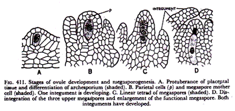
The ovule or the megasporangium develops as a small protuberance of the placental tissue. In the very young ovule a single hypodermal cell is differentiated as the archesporium (Fig. 411 A).
This archesporium cell may or may not cut off some parietal cells and then becomes the megaspore mother cell (Fig- 411B). The megaspore mother cell now undergoes meiosis or reduction division, and, usually, a linear row of four haploid megaspore cells (‘linear tetrad’) is formed (Fig. 411C).
ADVERTISEMENTS:
Meanwhile, two integuments develop from the base of the ovule. Of the linear tetrad of megaspores, usually the lower most one enlarges and becomes the functional megaspore while the three on top disintegrate (Fig. 411D).
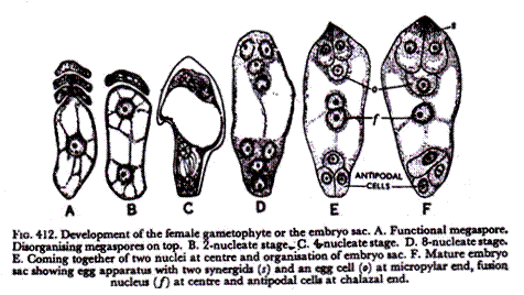
The functional megaspore now develops the female gametophyte or the embryo sac. In Angiosperms, the development of the female gametophyte is completely endosporous, i.e., within the megaspore. In a typical case, the nucleus of the embryo sac (Fig. 412A), which is the same as the functional megaspore (Fig. 411D), divides into two (Fig. 412B), then four (Fig. 412C) and finally, eight daughter nuclei (Fig. 412D) four of which are located at each pole.
Then, one nucleus from each pole moves to the centre of the embryo sac and fuses there forming the fusion or secondary nucleus (Fig. 412E). Finally, the embryo sac or the female gametophyte becomes organised (Fig. 412F).
ADVERTISEMENTS:
Three nuclei at the base form the antipodal cells. The secondary nucleus remains at the centre. On the top, three cells from the egg apparatus which consists of two flask-shaped synergids and a round egg cell (or ovum or oosphere) hanging between and below them.
The synergids usually are somewhat notched by an indentation and they show striations at the tip (‘filiform apparatus’). There is prominent vacuolation below the synergid nuclei and above the egg nucleus, showing accumulation of cytoplasm at different regions.
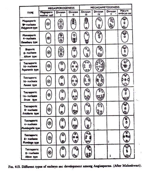
While the above method of gametophyte formation is usually described as the typical or normal type it has been found that many Angiosperms do not conform to this rule but show variations. The more intelligent students should be able to follow the different types of embryo sac development from the accompanying diagram (Fig. 413) prepared by Professor P. Maheshwari.
First, there is a great variation in megasporogenesis itself. Often, the usual four separate megaspore cells do not develop. If four megaspores develop the female gametophyte (embryo sac) is monosporic, being developed from the usual single megaspore. Here we have the so-called normal type which is the Polygonum type in the diagram being shown only by a few plants like Polygonum. In Oenothera, the development is monosporic but only 4 nuclei (the antipodals are eliminated) develop instead of 8 (Oenothera type).
In Allium, the megaspore mother cell divides only once giving rise to two haploid cells one of which develops a bisporic (as it contains the nuclei of two megaspores) embryo sac which is not normal although it develops the normal 8 nuclei (Allium type). In a number of cases no cell wall formation accompanies meiosis so that the normal separate megaspores are not formed. Here the embryo sac is tetrasporic as all the nuclei of the four megaspores are within the single cell wall.
Reduction division (meiosis) of course takes place but only separate nuclei, and not cells, are formed. There are many variations of the tetrasporic embryo sac as shown in the diagram. In Peperomia, 16 nuclei are formed inside the megaspore mother cell (Peperomia type) and the diagram shows how these are arranged. In Adoxa, the megaspore mother cell develops 8 nuclei simulating the normal (Polygonum) type although it is tetrasporic (Adoxa type).
There may be great variations in the appearance of the ultimate gametophyte, viz., the egg apparatus may show only one synergid (Peperomia), the fusion nucleus may involve only one (Oenothera type) or more than two (Peperomia, Penaea, Plumbagella and Fritillaria types) polar nuclei, the antipodals may vary from none in Oenothera type to a large number (Peperomia, Penaea and Drusa types; as many as 300 antipodal cells are reported in Sasa paniculata of Bambusae) and so on.


