ADVERTISEMENTS:
In this article we will discuss about the reproduction in phycomycetes. This will also help you to draw the structure and diagram of reproduction in phycomycetes.
Asexual Reproduction in Phycomycetes:
ADVERTISEMENTS:
The mode of reproduction in the Phycomycetes is asexual and sexual. Asexual reproduction is by the development of spores which may or may not be borne in a sporangium. In certain simpler forms the vegetative body itself behaves as a spore.
But in majority of cases after the mycelium has attained sufficient growth, apices of hyphae undergo certain changes to develop into sporangia with their contents being metamorphosed into spores.
These spores are single-celled and may be flagellate or non-flagellate. The flagellate spores or zoospores are of two kinds uniflagellate and biflagellate. The uniflagellate zoospores may have anteriorly or posteriorly placed whiplash type of flagellum.
Whereas, the biflagellate zoospores whose one flagellum is whiplash type and the other of tinsel type may be of two kinds:
ADVERTISEMENTS:
(i) Pear-shaped or pyriform with anteriorly placed flagella and are known as primary zoospores, and
(ii) Kidney- or bean-shaped having flagella inserted in the groove on the lateral side and are called secondary zoospores.
A zoospore usually swims for a while in water, then loses its flagellum or flagella as the case may be, then gives rise to new individual.
The biflagellate zoospores may behave in a different manner:
(i) A secondary zoospore on being liberated out from a vesicle of the zoosporangium swims around in water, after some time sheds its flagella, comes to rest, becomes rounded, encysts, and germinates by a germ tube which then develops into hypha of a new individual.
This type of zoospore is known as monoplanetic zoospore and the phenomenon is monoplanetism, exhibited by species of Pythium (Fig. 152).
(ii) A primary zoospore behaves initially like a monoplanetic zoospore and goes to rest by forming a rounded structure which instead of producing a new individual develops into a secondary zoospore.
The secondary zoospore so formed swims for some time and goes to rest by the loss of flagella, encysts, and eventually germinates by a germ tube which becomes the hypha of a new individual. Here, two types of zoospores alternate with two resting stages. Such a zoospore is called diplanetic zoospore and the phenomenon exhibited by it is diplanetism, found in Saprolegnia (Fig. 153).
(ii) A secondary zoospore after swimming goes to rest in a manner as stated above. After a period of rest, again produces a secondary zoospore, which again goes to rest and finally develops into a new individual. The process of formation of zoospore and resting condition may be repeated several times before a new individual is formed.
Such a process is known as repeated emergence, encountered in Dictyuchus. It is also known as polyplanetism (Fig. 154). In some terrestrial forms due to certain unfavourable atmospheric conditions, the zoospores fail to develop flagella and become non-motile and are known as aplanospores which behave like non-flagellate spores.
The non-flagellate spores are:
ADVERTISEMENTS:
(i) sporangiospores (some designate them as aplanospores) developed in a sporangium
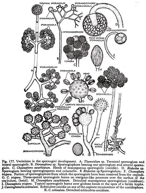
(ii) conidia. A sporangium contains large number of spores and may or may not have columella in it. Besides this, few-spored sporangia of much smaller size known as sporangiola (sing, sporangiolum) (Fig. 177) are also developed. A sporangiolum may be with or without columella and may be even monosporous.
The monosporous sporangiolum has evolutionary significance with regard to the origin of conidia from the sporangial condition. A cylindrical sporangiolum designated as merosporangium (pi. merosporangia) (Fig. 178E), represents as advanced evolutionary condition. Both sporangiospore and conidium germinate by germ tube which ultimately develops into new mycelium.
Members of the Entomophthorales reproduce asexually by conidia. Here each conidium is developed at the apex of the conidiophore. The conidiophore is again produced from hyphal body which is nothing but one of the segments of the segmented mycelium. But Thaxter showed that here conidium is aonespored sporangiolum which constitutes important evidence of the origin of conidium from a sporangiolum.
In certain fungi, for example, Phytophthora infestans, the behaviour of sporangia is very much controlled by conditions of moisture and temperature, depending on which a sporangium either produces zoospores or germinates directly by a germ tube behaving like a conidium.
Unsually a sporangium at an optimum temperature range of 18°C.-22°C. and at an optimum relative humidity of 100 per cent, produces zoospores. But at an optimum temperature of 24°C. and under less humid condition the sporangium germinates directly by a germ tube to form a new individual (Fig. 155).
ADVERTISEMENTS:
The sporangium thus behaves as a conidium. This type of sporangium with a dual function is called a conidiosporangium.
Besides the development of different types of spores and sporangia, in some Phycomycetes, the hyphae may directly produce thick-walled resting spores known as chalmydospores which can stand unfavourable condition and with the return of favourable condition the chlamydospores develop into hyphae of new individuals.
Sexual Reproduction in Phycomycetes:
There is a great diversity in the method of sexual reproduction and, very similar to the green algae, all gradations, from isogamous, anisogamous to oogamous types occur in the Phycomycetes. Again sexual reproduction may involve union of gametes which may or may not be well organized and may or may not be borne in gametangia.
Different methods of sexual reproduction are the following:
(a) Gametic Union:
ADVERTISEMENTS:
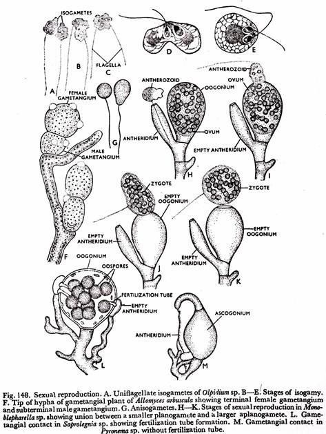
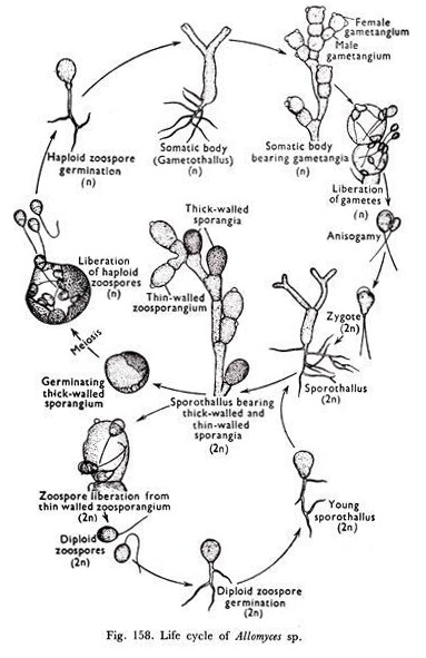
Union takes place between the two well-organized gametes. The fusing pair of gametes may be isogamous as in Olpidium (Fig. 148A to E); anisogamous, (i) with two free anisogametes found in Allomyces (Fig. 158) or (ii) between an antherozoid developed in an antheridium and an ovum which remains enclosed in an oogonium exhibited by Monoblepharella (Fig. 148H to K).
(b) Gametangial Contact:
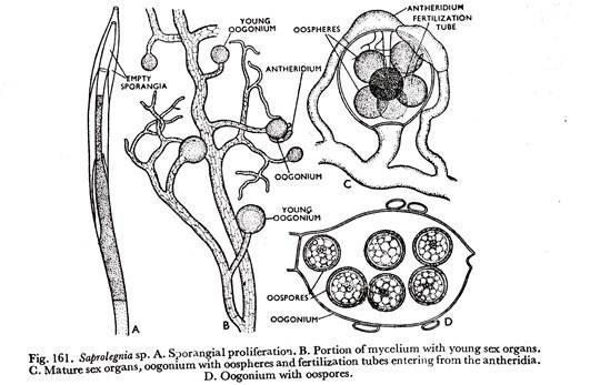
Two gametangia taking part in the process are distinguishable as antheridium and oogonium. Contact between their contents is established through a tube-like structure developed from the antheridium which pierces the wall of the oogonium, such a tube-like structure is known as the fertilisation tube.
The oogonium develops well-organized female gamete whose number may be more than one, as in the genus Saprolegnia (Fig. 161C) or just one, in the genus Pythium (Fig. 163N). But the antheridium does not bear well-organized male gametes. The male gametes are represented by nuclei embedded in a mass of cytoplasm.
ADVERTISEMENTS:
The nuclei and cytoplasm are discharged in the oogonium through the fertilization tube and come in contact with the female gamete (Fig. 163N).
The absence of the development of well-organized male gametes indicates the tendency of gradual degeneration of visible sexuality. The entities of the two gametangia are not lost at any stage of the sexual process. This type of sexual reproduction is encountered in the genera Saprolegnia, Pythium, Phytophthora, Albugo, and others.
(c) Gametangial Copulation:
Union between the two gametangia takes place in such a manner that they lose their entities during the process. The fusing pair of gametangia are multinucleate, nuclei being embedded in a mass of cytoplasm. The gametangia do not bear well-organized gametes. The gametes are nothing but a large number of nuclei embedded in a mass of cytoplasm. This again indicates a further degeneration of visible sexuality.
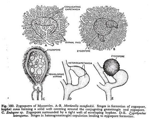
The fusing gametangia may be isogametangia, found in Mucor sp. (Fig. 149B to E) or heterogametangia, as in Zygorhynckus heterogamus (Fig. 180D & E). In the latter case of heterogametangial condition the two gametangia differ only in size but there is no difference in their contents and behaviour during the sexual process.
The gametangial copulation exhibited by species of Rhizophidium is different from that of Mucor sp. and Zygorhynchus sp. In Rhizophidium sp. the two gametangia are distinguishable as male and female from their external morphology and behaviour. The male gametangium empties its contents into the female gametangium through a pore or short fertilization tube.
The two gametangia originate from two equal-sized zoospores which behave as holocarpic thalli.


