ADVERTISEMENTS:
In this article we will discuss about Polyporus. After reading this article you will learn about: 1. Habit and Habitat of Polyporus 2. Vegetative Mycelium of Polyporus 3. Reproduction.
Contents:
- Habit and Habitat of Polyporus
- Vegetative Mycelium of Polyporus
- Reproduction in Polyporus
1. Habit and Habitat of Polyporus:
Polyporus (bracket fungus) is a large genus of family Polyporaceae. It is represented by about 50 cosmopolitan species. These species may grow as saprophytes on dead fallen tree trunks or as parasites on the roots, tree trunks.
ADVERTISEMENTS:
The species of Polyporus are known as wood rotters because they cause wood rot of various timber trees such as conifers, oak, apple, maple walnut, pear, Acacia etc. P. sulphureus, commonly called sulphur mushroom, causes wood rot of oak and other trees and has large sulphur yellow fruiting bodies P. betulinus causes the heart rot of birch and some other coniferous trees. P. squamosus causes heart rot of Ulmus.
The fruiting bodies of Polyporus are annual and appear in the form of brackets and slhelves (fig. 1) with stalk. These are fleshy when young but become hardened and leathery or corky at maturity. Young fruiting bodies of P. sulphureus are edible while mature fruiting bodies of P. lucida are used as decorative pieces in the houses due to its attractive brown colour.
2. Vegetative Mycelium of Polyporus:
It is of two types:
(a) Primary Mycelium:
ADVERTISEMENTS:
It originates by the germination of the uninucleate and haploid basidiospores which are produced from previous fruiting bodies. It consists of many white, slender, branched and septate hyphae. The cells are uninucleate i.e. monokaryotic It is short lived and soon becomes bi-nucleate (dikaryotic) by hyphal fusion occurring between cells of the hyphae of two apposite (compatible) strains.
(b) Secondary Mycelium:
It is subterranean, perennating, and originates by the fusion of two cells of the monokaryotic mycelium. The bi-nucleate or dikaryotic cells by divisions and by clamp connection form the secondary or dikaryotic mycelium.
It soon ramifies in the bark tissues and outer portions of the woody cylinder. The hyphae secrete enzymes which digest the lignified walls of the wood cells. Later it develops into the fruiting bodies called basidiocarps. Fresh crop of basidiocarp develops every year from this mycelium.
Basidiocarp:
The secondary mycelium collects in hyphal knots and soon forms a small button like strand in the bark of the wood. Later these hyphal strands grow in size, bursts through the bark on the surface of the tree and form the basidocarps or fruiting bodies. The basidiocarps may be sessile or stalked (having black or brown stalk measuring 2-6″ in height and 1/2″ diameter e.g. P. frondosus) bearing an umbrella shaped cap or pileus.
If stalk is present it may be lateral or central in position. The pilues portion develops as an expanded bracket or shelf like body with brown colour. Its upper surface is smooth, or ridged and undulating in concentric manner. The concentric rings are often alternately brown and white in colour especially in the peripheral regions of the pileus.
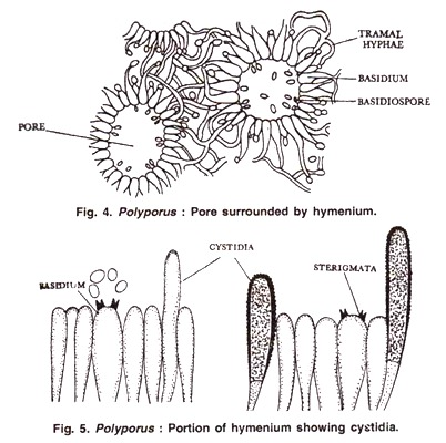
The lower surface of the pileus is smooth and flat, and does not bear any gills. The characteristic feature of Polyporus is that the pileus contains numerous small pores on under-surface (therefore named as polypore’s) which continue deep into the tissues as hollow tubular canals. These tubes are lined on inner surface by the fertile hymenium layer.
The vertical section of mature basidiocarp shows that it can be differentiated into four regions (Fig. 2):
1. Pileus Surface:
It is the upper surface of the basidiocarp. It may be smooth or incrusted.
2. Context:
ADVERTISEMENTS:
This region lies between the upper surface of pileus and the tube layer. This region is composed of anastomosing hyphae with large intercellular spaces between them.
The hyphae are of three types:
(a) Generative Hyphae:
Thin walled with dense cytoplasm, clamp connections are present.
ADVERTISEMENTS:
(b) Skeletal Hyphae:
Thick walled and un-branched.
(c) Binding Hyphae:
Thick walled but branched (Fig. 3).
Sometimes its upper region is soft and lower region is hard then it is called as duplex.
3. Tube Layer:
This region consists of vertically placed tubes attached to the lower surface of the context.
4. Pore Surface:
It is the lower surface of the mature basidiocarp. Tubes open at this surface.
On the basis of the hyphae present in the fruiting body, the basidocarps are differentiated into three types:
ADVERTISEMENTS:
(i) Trimitic:
All the three hyphae i.e. generative hyphae, binding hyphae & skeletal hyphae are present e.g. P. versicolor.
(ii) Dimitic:
Basidiocarps are made up of only two types of hyphae-generative hyphae and binding hyphae e.g. P. sulphureus.
ADVERTISEMENTS:
(iii) Monomitic:
Basidiocarps consist of only generative hyphae e.g., P adustus.
3. Reproduction in Polyporus:
Species of Polypore’s are heterothallic. Sex organs are absent. In tube layer of basidiocarp in between the tubes the tissue is made up of generative and skeletal hyphae. It is called dissepiment. From these hyphae arise short branches at right angles at the length of the tube.
These branches develop into fertile single celled calavate structures basidia and sterile structures paraphysis, cystidia and setae. These fertile and sterile structures form the inner lining of the pores and is known as hymenium (Fig. 4, 5).
Each basidium is club shaped bi-nucleate sterigmata. At maturity it projects slightly into the cavity of the pore. The two nuclei of the basidium fuse to form the synkaryon (karyogamy). The diploid nucleus undergoes meiosis to form four haploid nuclei. Four short sterigmata develop from each basidium from which small.
Oval and uninucleate basidiospores are abstracted. Large number of basidiospores (in billions) are formed in the pores. Through these pores the basidiospores are dispersed and after falling on suitable substratum each basidiospore germinates into a primary mycelium of plus or minus strain.


