ADVERTISEMENTS:
In this article we will discuss about Phytophthora. After reading this article you will learn about: 1. Habitat of Phytophthora 2. Symptoms of Phytophthora 3. Vegetative Structure 4. Reproduction 5. Some Common Diseases 6. Control.
Contents:
- Habitat of Phytophthora
- Symptoms of Phytophthora
- Vegetative Structure of Phytophthora
- Reproduction in Phytophthora
- Some Common Diseases Caused by Phytophthora
- Control of Phytophthora
1. Habitat of Phytophthora:
Phytopluhora (Greek phyton = plant; phthora = destruction) is represented by 48 species (Water house, 1973) which are cosmopolitan in distribution. Most of the species attack higher plants, mostly angiosperms and cause diseases of economic significance. Some species are facultative parasites and others as facultative saprophytes.
ADVERTISEMENTS:
One of the most common and well known species of Phytophthora is P. infestans, causing the disease called late blight of potato or Potato blight. Cool temperature (between 22-23°C) and excess of water favours the growth of this fungus. In India P. infestans has been reported from Nilgiri Hills and Darjeeling. It also occurs periodically in the plains particularly of northern India. However, in the Indo-Gangetic plain blight of Colocosia antiquorum (Vern. arvi) caused by P. colocasiae is quite common.
2. Symptoms of Phytophthora:
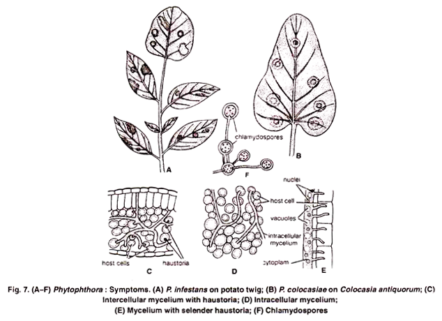
The disease appears as small black or purplish black areas at the margins and tips of the leaf (Fig. 7 A). These patches gradually enlarge and soon the entire crown may rot. Under favourable conditions all parts of the host undergo browning and rottening.
If the soil is very moist, tubers are also affected and may rot completely. In Colocasia anti-quorum large, circular, oval or irregular yellowish brown areas appear on leaves (Fig. 7 B). Drops of a yellow liquid oozes out from the leaf surface, and severe infection may result even in the rottening of corms.
3. Vegetative Structure of Phytophthora:
The mycelium is coenocytic, aseptate, hyaline and profusely branched (monopodial branching). The septa are formed at the time of reproduction or at maturity. The cell wall consists of glucan. Chitin is, however absent. Cytoplasm contains many nuclei, mitochondria, endoplasmic reticulum, ribosomes, dictyosomes, vacuoles and many oil globules. The mycelium is intracellular, and directly kills the invaded cells (Fig. 7 D).
ADVERTISEMENTS:
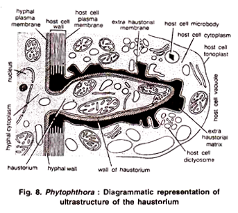
However, in some cases it is intercellular, present in the intercellular spaces of the host tissue. Some of the species develop haustoria to absorb their food material. In P. infestans the haustoria are selender and finger like (Fig. 7 E). Haustoria develop as lateral outgrowths from the intercellular hyphae.
Young haustorium invaginates the host cell. It remains surrounded by an extra-haustorial sheath, an extra haustorial haustorium membranes and the cytoplasm of the host cell. Cytoplasm of haustoria contains mitochondria, ribosomes, endoplasmic reticulum and nuclei. (Fig. 8.).
4. Reproduction of Phytophthora:
The fungus reproduces by Vegetative, asexual and rarely by sexual methods.
(i) Vegetative Reproduction:
Many species of Phytophthora (P. colocasiae and P. parasitica) reproduce by means by Chlamydospores. These vegetative reproductive bodies may be terminal or intercalary. They germinate by giving rise to 3-11 germ tubes which generally develop sporangia at their tips. (Fig. 7 F).
(ii) Asexual Reproduction:
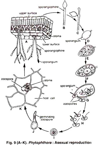
The asexual reproduction takes place by means of sporangia which are borne on aerial sporangiphores. Low Temperature (12-20°C) and high relative humidity (91-100%) favours the growth of sporangia. The sporangiophores arise directly from the internal mycelium and emerge out of the host singly or in clusters through stomata or by piercing through the epidermal wall (Fig. 9 A).
Each branch of sporangiophore bears, sporangium at its tip. With the growth of the hypha below, the sporangium is shifted to lateral position and another sporangium is formed at the tip (Fig. 9 B, C). The process may be repeated several times. Thus, the sporangiophore in Phytophthora is sympqdially branched (Fig. 9 A).
ADVERTISEMENTS:
The sporangia may vary in shape (i.e. lemoni form, ovoid or elliptical). It is hyaline to light yellow in colour, terminally papillate and has a basal plug. In P. colocasiae the sporangia are 38-60 µ long and 18-26 µ broad. The sporangia are deciduous (fall off) and are disseminated by water or are blown by the wind. At the place of detachment of sporangia, the sporangiophores bear nodular swellings which are typical for this fungus (fig. 9 C).
On falling upon a suitable host, the sporangia germinate. The germination of sporangium is governed by two main factors i.e., moisture and temperature. At high temperature (20-30°C), the sporangium germinates directly by a germ tube.
However, lower temperature (12°C) and presence of moisture favours indirect germination i.e., by zoospore formation. The sporangia are also susceptible to dessication. They loose their viability above 20°C temperature in 1-3 hours in dry air and 5-15 hours in moist air.
ADVERTISEMENTS:
Direct Germination:
ADVERTISEMENTS:
In the absence of moisture and high temperature (25 °C), sporangia germinate directly by germ tube and behave as conidia. The germ tube enters through a stomata and infects the leaf.
Indirect Germination:
In the presence of moisture and lower temperature (12°C) it behaves as zoosporangium and produces zoospores. The protoplasm of the sporangium is cut off into many uninucleate polyhedral pieces (Fig. 9 F, 10 in P. infestans and about 20 in P. colocasiae). Each polyhedral piece later rounds up and metamorphoses into zoospore (Fig. 9 G).
Zoospores are kidney shaped, biflagellate and possess flagella on lateral side. Of the two flagella one is of whiplash type and the other of tinsel type. The zoospores are liberated by the bursting of the sporangial wall. After swimming for some time they come to rest, encyst (Fig. 9 I) and germinate by a tube (Fig. 9 J).
ADVERTISEMENTS:
The germ tube adheres on the epidermis of the host and produces a flattened pressing organ i.e., appresorium, at its tip. From the appresorium a fine tubular, peg like outgrowth arises. It is the infection hypha. It peneterates the host tissue through stomata or epidermal cells (Fig. 9 K).
After penetration it develops into a profusely branched mycelium. The mycelium is intercellular and develops haustoria in the host cells. Under favourable conditions numerous sporangiophores emerge from the stomata and give rise to large number of sporangia. They are again disseminated by the wind and infect new plants. Thus, under favourable conditions the pathogen can reproduce several times by asexual method in one growing season.
(iii) Sexual Reproduction:
Clinton (1911) reported for the first time the sexual stages (oospore) in P. infestans. The sexual reproduction in Phytophthora is highly oogamous. The fungus is heterothallic i.e., requires two opposite strains, + and – for sexual reproduction. The male and female reproductive organs are called antheridia and oogonia, respectively.
Antheridium:
The antheridium is of following two types:
ADVERTISEMENTS:
(a) Amphigynous:
Attached to oogonium as a collar e.g., P. infestans
(b) Paragynous:
Attached laterally to the oogonium e.g. P. cactorum. The antheridium arises earlier than the oogonium showing a protandrous condition. It develops as a terminal, more or less club shaped structure on a short lateral hypha of one strain. In young stages, it is thin walled with non-vacuolar cytoplasm and possessing only one or two nuclei.
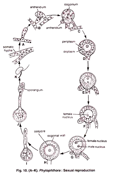
The mature antheridium is funnel shaped and forms a collar like structure at the base of the mature oogonium (Fig. 10 A-E). The two nuclei divide mitotically and forms 12 nuclei. All nuclei disintegrate except one in mature antheridium.
Oogonium:
ADVERTISEMENTS:
It is initiated laterally or below the antheridium on a hypha from other strain (Fig. 10 B). The young oogonium pierces the developing antheridium from below and swells above it into a pear shaped or spherical structure (Fig. 10 C). When young, it is multinucleate (up to 40 nuclei) and contains dense cytoplasm.
On maturity it becomes vacuolated and differentiated into an outer multinucleate periplasm and a central uninucleate ooplasm. (Fig. 10 D). The nucleus of the ooplasm divides mitotically and out of the two one survives and it functions as an egg or oosphere nucleus (Fig. 10 E, F).
Fertilization:
The oogonial wall bulges out at one point inside the antheridium and forms the receptive papilla. Later on the wall at the receptive spot dissolves and the antheridium pushes a short fertilization tube towards the oogonium. It penetrates the periplasm and passes into the ooplasm. It’s tip opens and liberates a male nucleus and some of the cytoplasm.
However, in P. himalayensis 2 to 3 papilla like outgrowths develops from the antheridium. One of these grows upwards and establishes a connection with the oogonium. The male nucleus passes into the oogonium through papilla and brings about fertilization (Fig 10 G). The oospore may also develop parthenogenetically in some cases.
Oospore:
During fertilization, first of all the plasmogamy takes place. The fertilized oospore secretes a wall and undergoes rest. Fusion of the two nuclei is very late and occurs even until after the oospore walls are laid down (Fig. 10 H). A mature oospore consists of an outer thick wall called exospore and an inner thin wall endospore. Exospore is made up of pectic substances and endospore is composed of cellulose and proteins.
Germination of Oospore:
It is of rare occurrence and observed in a few species like P. cactorum. P. palmivora etc. The fusion nucleus divides meiotically and later on successive divisions result in the formation of few or many nuclei in the oospore.
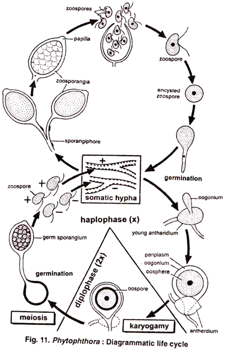
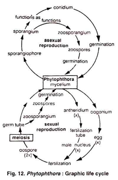
The exospore cracks and the endospore comes out in the form of a germ tube which develops a sporangium at the tip (Fig. 10 I, J). The contents of sporangium may divide to form zoospores (Fig. 10 K) or sometimes may directly develop into a mycelium (P. cactorum). Thus, it completes its life (Figs. 11, 12) cycle only within its host tissue. It has no saprophytic existence. It lives as dormant mycelium in the dead host remains lying in the soil.
5. Some Common Diseases Caused By Phytophthora:
6. Control of Phytophthora:
(1) Cultivation of resistant varieties.
(2) Spraying of fungicides like Bordeaux mixture.
(3) Destruction of the infected foliage by spraying with sulphuric acid; copper sulphate, Dithane and Fytolan.
(4) Seed tubers should be obtained from areas where the disease does not occur.
(5) Tubers should be covered with a thick layer of soil. It will avoid the chances of spores coming in contact with them.
(6) Good drainage system should be maintained.
