ADVERTISEMENTS:
In this article we will discuss about:- 1. Description of Mucor 2. Vegetative Structure of Mucor 3. Reproduction.
Description of Mucor:
The genus Mucor (L. muceo, be moldy) is represented by about 80 species, found throughout the world and about 17 species from India, commonly known as mold.
They grow mostly as saprophytes on decaying fruits and vegetables, in soil (Mucor strictus, M. flavus), on various food- stuff-like bread, jellies, jams, syrups. M. mucedo, is a coprophilous species (grows on dungs of herbivorous animals like cow etc.), known as black mold.
ADVERTISEMENTS:
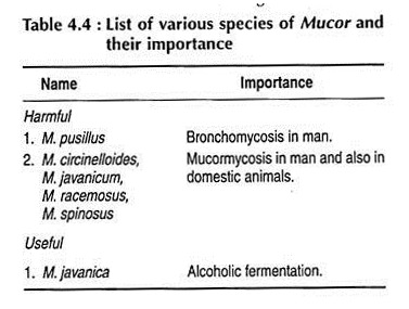
Species like M. mucedo and M. racemosus are well-known air contaminants. M. javanica is known to cause alcoholic fermentation.
List of various species of Mucor, harmful and useful to the mankind, are given below:
Vegetative Structure of Mucor:
ADVERTISEMENTS:
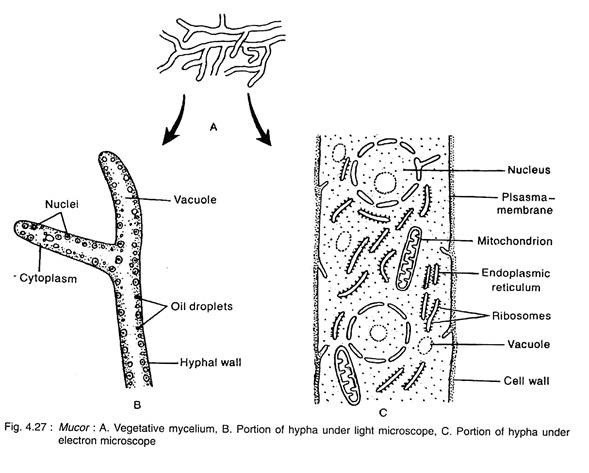
The vegetative plant body is eucarpic, consists of white cottony coenocytic much-branched mycelium. The mycelia ramify all over the substratum. The hyphae are usually prostrate, but some of them penetrate into the substratum and serve the function of both anchorage and absorption of nutrients (Fig. 4.27A).
The hyphal wall is microfibrillar, consist mainly of chitin-chitosan. In addition, other substances like other polysaccharides, lipids, purines, pyrimidines, protein, Ca and Mg are also present. Inner to the cell wall, cell membrane is present which covers the protoplast. The protoplast contains many nuclei, mitochondria, endoplasmic reticulum, ribosomes, oil droplets, small vacuoles and other substances (Fig. 4.27B, C).
Reproduction in Mucor:
Mucor reproduces by vegetative, asexual and sexual means.
1. Vegetative Reproduction:
ADVERTISEMENTS:
It takes place by fragmentation. Due to accidental breakage, the mycelium may break up into two or more units. Each unit is capable to grow as mother mycelium.
2. Asexual Reproduction:
It takes place by the formation of sporangiospore, oidia and chlamydospore (Fig. 4.28).
(a) Sporangiospore Formation:
ADVERTISEMENTS:
During favourable condition, the nonmotile spores known as sporangiospores or aplanospores are formed inside the sporangium.
The sporangiophores develop singly and scatteredly on the upper side of the superficial mycelium (Fig. 4.28B). The sporangiophore is generally unbranched, however it is branched in M. brunneus and M. racemosus.
After attaining a certain height, the nuclei and cytoplasm push more and more towards the apical side, consequently the apex of the aerial hyphae swells up (Fig. 4.28C, D). The swollen part enlarges and develops into large round sporangium.
With maturity, the protoplast inside the sporangium is differentiated into a thick dense layer of multi-nucleate cytoplasm towards the peripheral region just inside the sporangial wall, called sporoplasm and a vacuolated portion with a few nuclei towards the centre, called columellaplasm.
ADVERTISEMENTS:
A series of small vacuoles then appear between the sporoplasm and columellaplasm (Fig. 4.28E). These vacuoles become flattened and coalesce to form a continuous cleavage cavity (Fig. 4.28F).
This is followed by the formation of a septum towards the innerside of the cavity, which differentiates into inner columella and upper sporoplasm region. With further development, the septum becomes dome-shaped and pushes its way into the sporangium.
Protoplast of the sporoplasm then undergoes cleavage to produce many small multinucleate (2-10 nuclei) or rarely uninucleate segments. These segments transforms into globose non- motile sporangiospores (Fig. 2.28F, G).
Dehisence of sporangium takes place after maturation of spores. Minute needle-shaped crystals of calcium oxalate are formed on the external surface of sporangium wall. They imbibe water and make the wall soft. Consequently, the sporangiophore secretes water around the sporangium. The unused protoplast of the sporangium absorbs water and swells up, thereby it creates pressure.
ADVERTISEMENTS:
This pressure along with the pressure exerted by the bulging of the columella causes the soft wall of the sporangium to rupture. The dehisced sporangium thus shows a dome-shaped columella with attached spores at the top and remains of the sporangial wall as collar around its base (Fig. 4.28H). The spores are dispersed chiefly by insect and also by wind.
(b) Oidia:
Oidia are thin walled bid-like structures formed by mycelium grew in a medium rich in sugar. After detachment, the oidia increase by budding like yeast. This stage is called torula stage. Later, they develop to mycelia.
(c) Chlamydospore:
During unfavourable condition, thick-walled, nutrition rich, intercalary mycelium segments are developed by septation of mycelium which are termed as chlamydospores. They get separated from each other when the connecting mycelium dries up. In favourable condition, the chlamydospore germinates and gives rise to a new mycelium.
3. Sexual Reproduction:
ADVERTISEMENTS:
Sexual reproduction takes place during unfavourable condition by means of gametangial copulation. The gametangia look alike and by conjugation, they give rise to zygospore. Most of the species of Mucor are heterothallic (M.-mucedo, M. hiemalis), but few species (M. tenuis, M. genereosis) are homothallic (Fig. 4.28).
In heterothallic species, zygospores are produced by the union of two gametangia developed from mycelia of compatible strains; whereas, in homothallic species, the uniting gametangia develop from mycelia that derived on germination of a single spore.
When two mycelia of compatible strains come close to each other, the mycelia produce small outgrowth, called progametangia (Fig. 4.28L, M). The apical region of the two progametangia come in close contact. Nuclei and cytoplasm of each progametangium push more and more towards the apical region and its tip swells up with dense protoplasm.
The rear region becomes vacuolated. A septum is laid down, separating the apical region, which is called gametangium; and the basal region is called suspensor (Fig. 4.28N). The undifferentiated multi-nucleate protoplast of the gametangium is called aplanogamete or coenogamete.
After maturation of gametangia, the common wall at the point of their contact dissolves and the protoplast of both the gametangia unite to form zygospore (Fig. 4.280, P). The nuclei of opposite gametangia fuse together to form diploid (2n) nuclei, unpaired nuclei gradually degenerate.
ADVERTISEMENTS:
The diploid nuclei undergo meiosis before resting stage of zygospore. In heterothallic species normally 50% of the nuclei are of “+” strain and the other 50% of “-” strain.
Accordingly, the spores are of + or – type, but in M. mucedo (a heterothallic species) the spores are of either + or – type. All the nuclei except one degenerate and the surviving nucleus undergo repeated mitosis to produce large number of nuclei of the same strain.
The young zygospore enlarges and probably secretes five layered (two in exospore and three in endospore) thick wall, of which the outer one is black and warty. The zygospore then undergoes a period of rest.
After resting period, the zygospore germinates and, on germination, the innermost layer comes out after cracking the outer walls and produces a promycelium. The content of the zygospore migrates into the tip of the promycelium which swells up and differentiates into a lower stalk like germsporangiophore and the upper spherical germsporangium (Fig. 4.28Q).
The haploid nuclei of germsporangium form haploid spores called sporangiospores inside the germsporangium. These spores are also known as meiospores (Fig. 4.28R). Each meiospore, after liberation, germinates like sporangiospore and forms a new mycelium like the mother thallus (Fig. 4.28S).
Sometimes failure of gametangial copulation results in parthenogenic development of zygospore by any one gametangium called azygospore or parthenospore. It is, however, haploid in nature and its nucleus does not undergo meiosis before spore formation.


