ADVERTISEMENTS:
In this article we will discuss about Aspergillus. After reading this article you will learn about: 1. Habit and Habitat of Aspergillus 2. Vegetative Structure of Aspergillus 3. Reproduction 4. Economic Importance.
Contents:
- Habit and Habitat of Aspergillus
- Vegetative Structure of Aspergillus
- Reproduction in Aspergillus
- Economic Importance of Aspergillus
1. Habit and Habitat of Aspergillus:
Aspergillus (commonly known as black mold) is represented by about 100 species (Raper and Fennell, 1965) which are widely distributed from Arctic to tropical regions. Species of Aspergillus grow as contaminant in the laboratory cultures as their spores (conidia) are present in the air. A large number of species have been found in the soil.
ADVERTISEMENTS:
Majority of the species are saprophytic and grow on decomposing organic substances such as fruits, vegetables, jams, cheese, wood, leather etc. A few species are parasitic and cause diseases of animals and human beings.
Aspergillus can be grown easily by keeping a piece of cheese or bread in a warm moist chamber. It appears in the form of greenish, smoky patches along with, Mucor, Rhizopus and Penicillium. In India, it is represented by about 33 species.
2. Vegetative Structure of Aspergillus:
The plant body is mycelial. The mycelium consists of slender, tubular, pale coloured, extensively branched, thin walled hyphae. Some hyphae ramify superficially upon the substratum while some penetrate into the substratum to absorb the food material.
Each cell is multinucleate and is filled with granular cytoplasm, mitochondria, endoplasmic reticulum, ribosomes and vacuoles. The cross walls between the cells have a simple pore through which the cytoplasm of the adjacent cells remain continuous. Reserve food material is in the form of oil globules.
3. Reproduction of Aspergillus:
Aspergillus reproduces by vegetative, asexual and sexual methods (Fig. 14, 15).
(i) Vegetative Reproduction:
It takes place by the following methods:
(a) Fragmentation:
The vegetative mycelium breaks up into small pieces (fragments) and each fragment grows independently into a new thallus under favourable conditions.
(b) Sclerotia:
Some species e.g., A niger, A. terreus produce sclerotia. It is more a means of keeping the fungus alive than of propagation.
(ii) Asexual Reproduction:
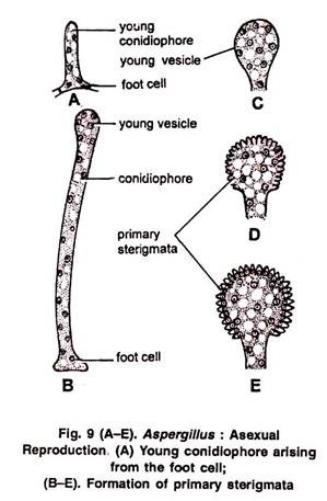
Asexual reproduction takes place by the hyphae called conidiophores. Some cells of the of fungus) and are known as foot cells (Fig. 9 A). Each foot cell produces a special erect branch as an outgrowth. It is the young conidiophore.
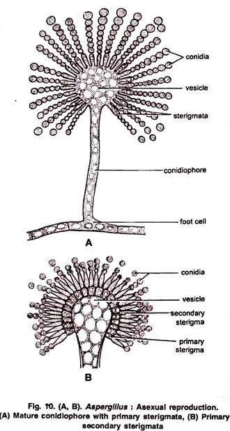
The tip of the conidiophore swells up into on elliptical or globular multinucleate head called vesicle. It forms many radially arranged tubular outgrowths called sterigmata or phialides (Fig. 9 B-E). In some species primary sterigmata (uniseriate) bear secondary sterigmata. (bi-seriate) (Fig. 10 A, B).
Formation of Conidia:
ADVERTISEMENTS:
Conidia (sing. = conidium), arise exogenously from the sterigmata or phialides (therefore, conidia are also called phialospores or phialoconidia) by abstraction method. They are arranged in basipetal succession (i.e., the youngest conidium is at its base and the oldest at the tip) (Fig. 10 A).
The sterigmata elongate at the tip to form a tube. The conidia are formed inside this. The sterigmata are uninucleate. At the time of formation of conidia the single nucleus of the phialide divide mitotically into two daughter nuclei. One of the daughter nuclei passes into the tube (Fig. 11 A-C). It is the first conidium. As the first conidium is formed the upper broken wall of the phialide serves as a cap around it (Fig. 11 D).
ADVERTISEMENTS:
The second conidium is formed by phialide just below the first (Fig. 11 E). The cytoplasm of both the conidia is confluent through a narrow cellular link called isthmus (Fig. 11 F, G). The continuity of the cytoplasm is stopped by the formation of the inner conidial wall. The isthmus becomes empty and now it is called connective.
Structure and Germination of Conidia:
Conidia are small, globose, unicellular, uninucleate (Fig. 12 A) or multinucleate, black, brown or yellow green in colour. They have two layered wall. Outer wall layer is thick spiny, pigmented and known as epispore, whereas the inner one is thin, delicate and is called endospore. Conidia are dispersed by wind. They germinate on suitable substratum by giving out a germ tube. The germ tube becomes septate, branched and forms a mycelium.
(iii) Sexual Reproduction:
The sexual reproduction is of rare occurrence. Majority of the species of Aspergillus are homothallic. However, a few species are heterothallic e.g., A. heterothallicus. It takes place by the formation of male and female sex organs. Male sex organ is known as antheridium and the male branch is called Pollinodium. Female sex organ is called ascogonium and female branch is called as archicarp.
Ascogonium:
Archicarp develops on the mycelium in the form of septate, loosely coiled structure.
ADVERTISEMENTS:
The young archicarp can be differentiated into three parts (Fig. 13 A-D):
(i) The basal multicellular, multinucleate stalk.
(ii) Middle unicellular, multinucleate ascogonium (gametangium).
(iii) Apical unicellular, multinucleate receptive organ called trichogyne.
At first archicarp is loosely coiled but later on the coil approaches nearer and nearer and finally touch each other to form a cork screw like structure (Fig. 13 A, E).
Antheridium:
ADVERTISEMENTS:
Pollinodium grows up beside the archicarp on the same or adjacent hyphae (Fig. 13 B). It gets spirally coiled around the archicarp and arches over the apex of ascogonium.
It can be differentiated into two parts:
(i) Upper part, slightly broader, unicellular, multinucleate and behaves as antheridium.
(ii) Lower unicellular and multinucleate part called stalk.
Fertilization:
The tip of the archegonium arches over the trichogyne and fuses with it. The wall at the point of contact dissolves, thus making a continuous passage. It is plasmogamy. The contents of the antheridium pass into the ascogonium. The pairing of male and female nuclei takes place in ascogonium (Fig. 13 E-G). In A. herbariorum antheridium is very well developed and pairing takes place in ascogonium.
ADVERTISEMENTS:
However, in some species the antheridium is very well developed but the male contents do not pass and fuse with the contents of the ascogonium (e.g.,A.repens). In still some other species the antheridium may be completely absent (e.g., A. flavus, A. fisheri). In such cases the pairing takes place between ascogonial nuclei. It shows the degeneration of sex organs in Ascomycetes.
Development of Ascocarp:
Whether the antheridium is functional or not, the ascogonium in all cases develops into a fruiting body called ascocarp (Fig. 13 G). After the pairing of the nuclei, the ascogonium becomes septate. Each segment consists of one male and one female nucleus (dikaryon). From these dikaryotic segments arise ascogenous hyphae. Each ascogenous hypha is multicellular with a pair of nuclei and produces asci by crozier formation.
Crozier Formation:
The terminal bi-nucleate cell of the ascogenous hyphae elongate (Fig. 13 H) and then been itself to form a hook like structure known as crozier (Fig. 13 I). Both nuclei divide in such a way thin spindle apparatus are oriented parallely in vertical direction.
Now the separation takes place in crozier and it is differentiated into three cells (Fig. 13 I, J):
(i) The terminal or ultimate uninucleate cell.
(ii) Sub-terminal or penultimate bi-nucleate (‘+’ and nuclei) cell, occurs at the curve position.
(iii) A basal uninucleate, anti-penultimate cell.
The penultimate bi-nucleate cell acts as ascus mother cell. The nuclei in these cells fuse to form diploid nucleus (karyogamy). The diploid nucleus first divides meiotically and forms four haploid nuclei. Each haploid nucleus divides meitotically and thus 8 haploid nuclei are formed (Fig. 13 K-N).
Each nucleus later on gets surrounded by cytoplasm and develops a wall. Thus, 8 haploid ascospores are formed in each ascus. The cytoplasm left over in each ascus is known as epiplasm. The asci may be globose or pear shaped.
As the asci develop, a large number of sterile hyphae grow around them and form a protective covering called peridium. The entire structure is known as ascocarp. It encloses many asci. It is spherical and has no opening. Such an ascocarp is known as cleistothecium (Fig. 13 O).
Structure of Ascospore:
As the asci mature, the ascospores are set free by dissolution of the wall of the asci in the ascocarp. They are liberated only after the decay of the ascocarp wall. Each ascospore is pulley wheel shaped, unicellular, uninucleate and attains a diameter of approximately 5 µm.
Spore wall is differentiated into two layers, the outer thick, sculpturous epispore and inner thin endospore (Fig. 13 P). After falling on a suitable substratum each ascospore germinates to give rise to a germ tube which develops into a new haploid mycelium (Fig. 13 Q).
Economic Importance of Aspergillus:
The species of genus Aspergillus are of great importance to us in many ways, since they are both useful and harmful:
A. Useful Activities:
(i) Antibiotics:
Some species of Aspergillus produce antifungal and antibacterial antibiotics, such as:
(ii) Bioassay:
A. niger is used to treat the soil for the tracing out of elements like copper. It can detect copper even in traces. A. virens is used to detect arsenic in agricultural soils.
(iii) Organic acids:
Many species of Aspergillus are used to produce organic acids, such as:
Citric acid: A. niger
Gluconic acid: A. fumaricus, A. niger
Gallic acid: A. gallomyces, A. niger
Itaconic acid: A. itaconicus, A. terreus
Kojic acid: A. oryzae, A. flavus, A. tamari.
(iv) Vitamins:
Many species of Aspergillus e.g., A. gossypii are good sources of vitamins such as Riboflavin (vitamin B1).
(v) Enzymes:
Enzyme amylase is produced by A. niger and A. oryzae in culture.
(vi) Alcoholic Beverages:
A. oryzae is used for fermenting rice to produce ‘sake’ wine and in making ‘miso’ and ‘soja’ sauces.
(vii) Destruction of wasted organic products. Majority of the species of Aspergillus are saprophytic. These species remove litter and wastes.
B. Harmful Activities:
(i) Spoilage of Food and Other Articles.
Aspergillus is one of the most frequent contaminants of food. Saprophytic species of Aspergillus grow commonly on food stuffs, leather, paper, fiber etc. The most common species are A. niger, A. flavus etc.
A. repens: causes ‘button formation’ in canned condensed milk.
A. glaucus: spoils bread and makes it ‘musty’.
ADVERTISEMENTS:
A. repens, A. flavus, A. fumigatus: pasteurized milk.
A. oryzae: spoils cocoa, butter.
A. niger, A. calavatus, A. glaucus: spoils meat.
A cartdidus. A. fumigatus, A. terreus : deterioration of rubber.
(ii) Diseases of Human Beings.
Many species of Aspergillus e.g., A. flavus, A. niger. A. fumigatus, parasitize man. They cause a number of diseases grouped under the name Aspergilloses (sing. Aspergillosis).
Aspergillosis is a lung disease and appears to be similar to tuberculosis. The conidia of Aspergillus remain in the air and causes allergy to human beings. They also infect the human ear and cause otomycosis disease.
(iii) Diseases of Animals:
In chickens species of Aspergillus cause ‘Brooder’ pneumonia of lungs.
(iv) Diseases of plants:
A. niger causes fruit rot of pomegranate, mango, wood apple, date etc. crown rot disease in ground nut. ball rot in cotton etc.
(v) Contaminant of Culture Media:
Aspergillus niger, popularly known as the ‘black mould’, is considered as a ‘weed of laboratory’ as it often contaminates the bacteriological and mycological cultures.
(vi) Toxic substances:
A. flavus produces a carcinogenic fungal toxian aflatoxin.


