ADVERTISEMENTS:
In this essay we will discuss about the digestive system in humans. After reading this essay you will learn about:- 1. Organs of Digestive System 2. Accessory Glands for Digestion of Foods.
Essay # 1. Organs of Digestive System:
Digestion means simplification of complex foods. It is the process of breaking various foodstuff into simple products. The complex foods like carbohydrates, proteins and fats are converted into glucose, amino acids and fatly acids respectively by the action of digestive enzymes. These simple substances enter into the blood circulation after absorption and then they are utilized by the body.
Digestive system consists of two main organs:
ADVERTISEMENTS:
(1) Alimentary Canal
(2) Digestive Glands
1. Alimentary Canal:
This is also known as digestive tract or gastrointestinal tract. It is a long tube of varying diameter which begins at the mouth and ends at the anus. The length of this tube is about 8-9 meters. It opens at both the ends. The alimentary canal starts at the mouth into which cavity, the glands of the mouth pour the juice. As it passes backwards, it spreads into a funnel shaped cavity called-pharynx.
The tube then narrows into a soft muscular tube about ten inches in long, called the food pipe or gullet. This passes down the neck into the chest. It then opens into the stomach by piercing the diaphragm. The stomach is a large bag lying a little to the left under the diaphragm. It has two openings, one where the food pipe ends and the other where the intestines begin. The alimentary canal narrows again and passes into the small intestine which is about twenty two feet in length.
ADVERTISEMENTS:
The first ten inches of the small intestine is called as Duodenum which forms a ‘C’ shaped loop. The rest of the small intestine is like a coiling tube, whose ends opens into a wide but comparatively short tube known as large intestine. It is about six feet long. The last part of the Large Intestine is known as Anus.
2. Digestive Glands:
Various digestive glands help in the digestion of foods.
These are:
(1) Salivary glands in the mouth,
(2) Gastric glands in the stomach
(3) Pancreas,
(4) Liver,
(5) Intestinal glands in small intestine.
All these digestive glands secrete digestive juices containing different enzymes which digest carbohydrate, protein and fatly foods.
ADVERTISEMENTS:
Digestive juices:
Five digestive juices are secreted from digestive glands of the body. The enzymes present in these juices help in the digestion of different types of foods.
These juices are:
1. Salivary juice from salivary glands in mouth.
ADVERTISEMENTS:
2. Gastric juice from Gastric glands in the stomach.
3. Pancreatic juice from Pancreas.
4. Intestinal juice from Small Intestine.
5. Bile juice from Liver.
Why so many digestive juices are necessary for digestion of food?
There are three reasons for the presence of so many digestive juices:
1. One digestive juice cannot digest three types of foods i.e. proteins, fats, and carbohydrates up to their completion.
2. One digestive juice cannot digest one particular type of food up to its completion, because food cannot remain in one place for a longer period of time.
ADVERTISEMENTS:
3. The medium of action of enzymes present in different digestive juices are different. Some act on acidic medium and some on alkaline medium.
Digestion in Different Parts of Alimentary Canal:
The alimentary canal consists of the following organs in which foods are digested:
1. Mouth
2. Oesophagus
3. Stomach
ADVERTISEMENTS:
4. Duodenum
5. Small Intestine
6. Large intestine
1. Mouth:
The mouth cavity is the front spread out end of the food pipe. The sides of the cavity are formed by the cheeks, the roof by the palate, and the floor by the tongue. When closed, it is bound in-front by the upper and the lower sets of teeth meeting in the middle. The opening at the back of the mouth is known as throat on each side of which there is a mass of tissue called tonsils. In the outside of the mouth cavity there is a slit like opening which is bounded by two soft movable lips.
ADVERTISEMENTS:
Teeth:
During our life time two sets of teeth are developed:
1. Temporary teeth or milk teeth:
These are twenty in number, five in each half of the jaw. They appear at about the age of six months. They are usually smaller and delicate.
2. Permanent teeth:
These are thirty two in number sixteen in the upper and sixteen in the lower jaw. They appear gradually and push out all the temporary teeth. By the age of 14th, twenty eight permanent teeth are erupting. The last four teeth called as wisdom teeth appear after a person is twenty one years of age.
Functions:
The teeth are essential for efficient mastication. They cut, crush and grind the food.
Tongue:
The tongue is a muscular organ which is present on the floor of the oral cavity. The anterior part of the tongue is small which can move easily into the inner part of the mouth.
Functions:
1. It mixes the food well with the salivary juice by moving it into different direction.
2. The taste buds present on the tongue detect the nature of the food. Sweet taste can be felt by the taste buds present in the anterior tip of the tongue and salty taste at a small area behind this tip. Buds at the posterior part of the tongue feel bitter taste and sour taste can be easily known by the taste buds situated in the middle and side area of the tongue.
Salivary Glands:
There are three pairs of salivary glands inside the mouth which secrete a fluid called saliva.
These are:
(1) Parotid Gland
(2) Submandibular Gland and
(3) Sublingual Gland.
Perotid Glands:
A pair of perotid gland is situated in the cheeks, in front of each ear inside the mouth cavity. They are the largest of the salivary glands. Each gland is attached with a long duct known as Stenson’s duct by which salivary juice is carried to the mouth.
Submandibular Glands:
This is also known as Sub-maxillary gland. A pair of submandibular gland is situated on each side beneath the jaw bone. They are the next largest glands, and are about the size of a walnut. Their secretion is carried through a duct called as Wharton’s duct.
Sublingual Glands:
It is the smallest pair of salivary glands. It is present below the tongue. The secretion of these glands is carried through the Duct of Rivinus into the mouth cavity. The salivary glands secrete about 800 to 1500 ml of saliva perday. Saliva is a watery alkaline fluid. It contains 90% water, a thick lubricant called mucin, a small amount of calcium salts, maltose, urease and lipase. A starch splitting enzyme known as ptyalin and a bacteriolytic enzyme called as lysozyme are present in saliva.
Functions:
The action of saliva is both physical and chemical.
1. The enzyme ptyalin present in saliva acts on starchy food and converts them to dextrin and maltose.
Starch + Ptyalin → Dextrin
Dextrins + Ptyalin → Maltose
2. Saliva constantly moistens the mouth and tongue.
3. It lubricates and moistens the food, so that the food can be easily swallowed.
4. It cleanses the tongue and makes speech easier.
5. Saliva dissolves the food particles, which stimulate the taste buds. It makes food delicious.
6. The antibacterial enzyme “lysozyme” present in salive kills the bacteria. Salivary juice has antiseptic property.
Secretion of saliva is controlled by the parasympathetic nervous system.
Digestion in Mouth:
As the food enters into the mouth, even the smell, the sight or the thought of food, the salivary glands are stimulated and saliva is secreted. This is poured into the mouth. The teeth chew the food and breaks into small pieces while the tongue thoroughly mixes the saliva with the food.
The enzyme ptyalin in the saliva acts on starch and converts it first to dextrins and finally to maltose. Therefore, it is important to chew the food well to get the starch changed into maltose. When some cooked rice or a piece of bread is chewed well in mouth, it becomes slightly sweet in taste, because the starch present in them has been changed into sugar.
Then the chewed food is gathered into a ball or bolus and set down the pharynx into the gullet or food pipe. The wind pipe (passage for the air) is situated in front of the gullet. The food is prevented from going into the wind pipe by a lid like organ called as epiglottis.
It is a cartilaginous muscular flap which prevents the entry of food into the windpipe during swallowing and the food enters into the food pipe or gullet. If a particle of solid food or water enters into the wind pipe, it is expelled by violent coughing.
2. The Oesophagus or Gullet:
It is a collapsible muscular tube of 9 to 10 inches in length, which extends from pharynx to the cardiac orifice of the stomach. It passes downwards through the neck, the thorax and the abdominal cavity.
The oesophagus is composed of four layers of tissue:
1. An outer covering of connective tissues.
2. A muscular coat composed of two layers of muscle fibers.
3. A sub-mucous layer containing blood vessels, nerves and mucous glands.
4. An inner mucous membrane layer.
Functions:
The food passes through the oesophagus by peristaltic action. It passes down the food pipe by the action of its muscular wall which contract above it and push it along. Food does not simply fall down the gullet, but each part of the gullet contracts after the part next above and so squeezes the food along till it passes through the opening and enters the stomach.
3. Stomach:
The stomach is a dilated part of the alimentary canal. It is a bag like expansion of the food pipe with its broad end to the left and narrow end to the right. It is situated between the oesophagus and the beginning of the small intestine. It lies close to the diaphragm and slightly towards the left of the abdominal cavity. The size of the stomach varies according to the age and sex.
Structure:
The stomach consists of an upper part, the fundus. The main part of the stomach is called the Body and this narrows at its lower end to become the Pyloric Antrum.
The walls of the stomach consist of four layers of tissues:
1. An outer covering of serous membrane.
2. A muscular layer consisting of longitudinal, circular and oblique muscle fibers.
3. A sub mucous layer.
4. A lining mucous membrane layer.
Gastric Glands:
In the mucous membrane there are a large number of minute blind tubes which are called as gastric glands. These are tiny holes which form the mouth of these gastric glands. Connective tissue is present between the tubules which contain blood vessels and lymphatic’s.
When the food enters the stomach, the blood vessels dilate and bring extra blood to the stomach. Then the gastric glands secrete gastric juice. The gastric juice is poured into the cavity of the stomach and mixed with the food. Besides the gastric glands, the walls of the stomach also contain other glands like pyloric glands, cardiac glands, and mucous secreting cells which secrete mucous, pepsinogen, hormone gastrin etc.
Gastric juice is a colourless, acid liquid containing water, minimum quantity of salt, 0.2 – 0.4 percent hydrochloric Acid (HCL) and enzymes like Renin, Pepsin and Gastric Lipase. The stimulation of the secretion of gastric juice is partly nervous and partly chemical.
Digestion in Stomach:
After the food enters the stomach for some fifteen to twenty minutes, the action of saliva continues that is conversion of starchy food to maltose. The gastric juice by that time is secreted in sufficient amount and mixed with the food completely. When the food of the stomach becomes acidic by hydrochloric acid, the action of ptyalin of saliva stops, because it can act only upon alkaline medium. Then the action of the digestive enzymes present in gastric juice start, as they become active on acidic medium.
The functions of gastric juice are as follows:
1. The enzyme Renin coagulates milk and converts milk protein into casein.
2. The enzyme pepsin in the presence of hydrochloric acid acts upon protein food and the casein, and converts them into more soluble substances called peptone.
3. Gastric Lipase (small amount is secreted) helps in splitting the fat molecules.
4. Hydrochloric acid acidifies all foods. It has an antiseptic and disinfectant property which kills germs and bacteria’s from the food.
5. The stomach acts as the reservoir of food for a short time.
6. An anti-anaemic factor is formed in the stomach.
7. Stomach also secretes a protein compound called intrinsic factor which is necessary to facilitate the absorption of vitamin from alimentary canal.
8. Water, alcohol and some drugs are absorbed inside the stomach.
9. Besides digestion, the important functions of stomach are to protect the small intestine from injury especially very hot and very cold foods, chemical irritants, chilies, alcohol etc.
10. It also helps in softening hard particles of food which have escaped chewing in the mouth.
The gastric juice has no action upon the carbohydrate food. The time taken for the gastric digestion is about three to four hours. The acid mixture of gastric juice and partly digested food is called “chyme”. Some of the sugar and peptone is absorbed into the blood vessels of the stomach. This ‘chyme’ then passes into the Duodenum, the first part of small Intestine.
Small Intestine:
The small intestine is a coiled tube extending from the pylorus of the stomach and leads into the large intestine. It is approximately 5 to 6 meters (16-20 feet) long tube. It lies in the umbilical region of the abdomen and is surrounded by the large intestine. There are four coats of small intestine like the stomach. Small-intestine is consisting of three parts which are continuous with each other.
There are:
(1) Duodenum
(2) Jejunum
(3) Ileum.
4. Duodenum:
The duodenum is the first part of the small intestine. It is approximately 25 – 30 cm (10 – 20 inches) long and is curved like English letter ‘C’. The shape is like a horse-shoe. Inside the curve, the head of the pancreas is encircled. It is fixed to the posterior abdominal wall by peritoneum. The bile duct from the liver and the pancreatic duct from the pancreas unite to open together into the lower portion and from the back side of the duodenum. This place of entrance is known as “Ampulla of Vater” which is situated 10 cm (4 inches) from the pylorus.
Digestion in Duodenum:
The bile duct brings bile juice and the pancreatic duct, pancreatic juice. These two fluids mix with the chyme and carry on the process of digestion a stage further.
Pancreatic Juice:
The pancreatic juice is an alkaline colourless liquid. It acts upon alkaline medium. It contains three enzymes.
(1) Amylopsin or Amylase
(2) Trypsin,
(3) Lipase which digest carbohydrates, protein and fats respectively.
Amylopsin acts upon uncooked as well as cooked carbohydrate foods and converts them into disaccharides. Trypsin is produced by the enzyme trypsinogen. It acts upon undigested protein foods and peptones and converts them into polypeptides. Lipase acts upon fatty food and converts them into fatty acid and glycerol. The fat splitting action of lipase is greatly facilitated by the presence of bile juice. Fat digestion is completed by lipase in the duodenum.
Bile Juice:
Bile juice is carried by the bile duct from the liver. Though it has no direct action on digestion of food, but it aids the pancreatic juice in its action for digestion of fat.
The functions of bile juice are as follows:
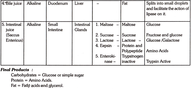
1. Bile juice helps in splitting the fat into small particles and thus helps the action of lipase for its digestion.
2. As bile juice is alkaline, it helps to convert the acidic food of the stomach to alkaline medium in duodenum, so that the enzymes of the pancreatic juice can easily act upon all types of foods.
3. Bile juice has important antiseptic properties and kills the germ and bacteria’s from the food in the intestine.
4. It also promotes the absorption of the products of digestion.
5. Bile prevents formation of gall stone.
6. It gives colour to the stool.
5. Small Intestine (Jejunum):
It constitutes the upper two fifth of the remaining small intestine. It is the middle portion of the small intestine. Intestinal juice is secreted from simple tubular glands which are situated in the mucous membrane layer of the small intestine.
Intestinal Juice:
The digestive process is completed by the work of intestinal juice secreted form the intestinal glands. It completes the digestion of carbohydrates and proteins. About 1800 ml of intestinal juice is secreted per day. This is also known as Saccus Entericus. The enzymes present in this ferment act upon alkaline medium.
Saccus Entericus contains enzymes like:
(1) Invertase /sucrase,
(2) Lactase,
(3) Maltase,
(4) Enterokinase,
(5) Erepsin.
1. Invertase:
It acts on cane sugar and converts them into simple sugar or glucose.
2. Lactase:
This enzyme splits lactose into glucose and galactose. This galactose is again converted into glucose in the liver at time of necessity.
3. Maltase:
It converts maltase into dextrin.
4. Enterokinase:
This enzyme activates the proteolytic enzyme of pancreatic juice. It activates trypsinogen to trypsin which helps in the digestion of protein foods.
5. Erepsin:
The enzyme completes the digestion of protein foods. It converts peptone polypetide and other undigested protein foods into amino acids. In this part of small Intestine the digestion of all the three types of foods are completed. Carbohydrate foods are simplified into glucose, protein into amino acids and fat into fatty acids and glycerol.
Ileum:
When the process of digestion is completed, they are carried to the last part of the small intestine i.e. Ileum. The digested food reaches this place in about four hours. Ileum constitutes the last three fifth of small intestine. It is relatively thinner than jejunum. From this place the absorption of simplified and digested food starts.
By the action of various digestive juices, saliva, gastric, juice, pancreatic juice and intestinal juice, different food materials are simplified and ready for absorption. Proteins have been broken down to peptone by gastric juice, polypeptide by pancreatic juice and finally to Amino acids by intestinal juice.
Carbohydrates have been converted into maltose by salivary juice, other disaccharides by pancreatic juice and lastly mono-saccharides of glucose or simple sugar by Intestinal juice. Fat is simplified into fatly acids and glycerol by the action of gastric lipase and pancreatic lipase.
Absorption of Digested Food:
Absorption of digested food take place in the epithelial surface of the villi in the Ilium part of small intestine. The mucous membrane of the small intestine is line by a number of projections like substances known as villi. These help in absorption by increasing the surface area. The absorption of digested food takes place entirely in the small intestine through two channels, the capillary blood vessels and the lymphatic’s of the villi on the inner surface of the small intestine.
A villus contains a lacteal, blood vessels, epithelium and muscular tissue, which are connected together by lymphoid tissue. Carbohydrates in the form of glucose are directly picked up by the blood vessels from the villi. Proteins are absorbed by the blood vessels from the villi in the form of amino acids. Fats, in the form of fatly acids and glycerol are absorbed into the lacteals.
Small amount of emulsified fats from water soluble compounds with bile, that are readily absorbed. Fat soluble vitamins get absorbed along with fats. Mineral salts and small amount of water pass into the blood vessels along with sugar and amino acids. Amino acids, glucose and fatly acid are passed from the small intestine to the liver for oxidation and metabolic process. The excess amount of glucose is converted to glycogen and is stored in the liver for future use.
After absorption, intestinal contents are slowly propelled along the Alimentary canal. The onward movement of the food is affected by means of waves of contraction of the intestines called peristalsis. This peristaltic activity is called “Segmentation”.
6. Large Intestine:
The large intestine or colon is about 1-5 meters or 5 feet long. It is wider than small intestine. The colon begins as a dilated punch the “Caecum” to which the “Vermiform Appendix” is attached. Most of the digested food having been absorbed in small intestine, a semifluid mass is left to pass into large intestine.
The large intestine absorbs the substances which are not absorbed in small intestine as well as a large amount of water. The contents of the large intestine thus become firmer. The materials ultimately thrown out are called faeces which consist of undigested and un-absorbable portions of food. These faeces are decomposed and putrefied by a type of bacteria Bacillus coli and give rise to bad odour, which are passed out through the Anus.
Functions of Large Intestine:
1. Water, minerals, salts and some drugs are absorbed into the blood capillaries.
2. Mucin is secreted which helps in lubricating the faeces and facilitates their passage.
3. Excess of calcium, iron drugs of heavy metals are excreted from the walls of the large intestine and mix with the faeces.
Movements of Gastro Intestinal Tract:
Movements of Gastro intentional Tract is due to the presence of muscular tissue in the alimentary canal. Different parts of the alimentary canal have different kinds of movement.
Movements in the tract are essential for three reasons:
1. To push the food particles forward so that different digestive juices can be mixed properly with the food in different parts of alimentary canal.
2. To ensure blood circulation through the walls of the tract so that the secretion of juices can become easier and the absorption work may proceed rapidly.
3. Movements are essential for the process of Defecation.
Moreover, for the proper functioning of the Gastro intentional tract, movements are essential. There are many types of movements like peristalsis, antiperistalsis, mass peristalsis, segmentation, pendular, tonus rhythm etc.
All these movements can be grouped under two headings:
1. Translatory movement in which the food are propelled forward. It includes peristalsis, antiperistalsis and mass peristalisis. Generally this movement is present in every part of Gastrointestinal tract.
2. Stationary movements are localized and do not move the foods forward. It includes segmentation, pendular and tonus rhythm etc.
Food enters into the stomach through the oesophagus by the peristaltic movement of the circular muscles. By the contraction and relaxation of the circular muscles attached to the food pipe or oesophagus, food propel forward to the stomach. Inside the stomach the gastric juice is mixed with the food by the rippling waves of peristalisis called tonus or mixing waves.
The action of these waves moves the food forward. The strong waves of peristalsis are able to force the chyme through the pylorus of, the stomach into the Duodenum. Duodenum is the place where many peculiarities of movements are seen. It possess peristaltic and segmentation movements throughout the duct.
Mainly there are two types of movements in human small-Intestine:
1. Rhythmic Segmentation or Ludwigs Pendulum:
In rhythmic segmentation there is alternate contraction and expansion of the intestinal wall by which the intestinal juice can be mixed thoroughly with the “chyme”. It also brings the chyme into the contact with mucosa for absorption.
2. Peristalisis:
Propulsive peristaltic movement push the chyme along the length of the small intestine until it reaches the ileocaecal valve which opens to allow the contents of the ileum to enter into the large intestine. In the large intestine, there are two types of movements like stationary and translatory peristaltic.
Movements of the large intestine are generally very slow. It consists of a mixture of segmentation movements and mass peristalsis which transfers the faecal material into the colon. The movement helps to empty of the contents of proximal colon into more distal portion and finally into the rectum for defaecation. One of the greatest functions of large intestine is its capacity to move.
Carbohydrate Digestion:
Carbohydrates are composed of carbon, Hydrogen and oxygen. They are classified into Mono-saccharides disaccharides and polysaccharides. The Carbohydrate foods which arc taken in the diet are rich in sucrose, lactose, starch, dextrin and cellulose. Most of them are disaccharides and polysaccharides. So they require simplification. They are converted into simple sugar or glucose which is a monosaccharide by the action of digestive enzymes for their utilization in the body.
Digestion in Mouth:
Inside the mouth, the digestion of Carbohydrate begins. Salivary juice secreted from salivary Glands in the mouth act upon Carbohydrate foods. The enzyme ptyaline of saliva hydrolyzes starch into maltose and dextrins. Inside the mouth 3 – 15% of starch is digested.
Starch→ Ptyalin → Maltose and Dextrin.
Digestion in Stomach:
Ptyalin present in saliva continue to act on starch for 15 minutes inside the stomach because Hydrochloric acid of gastric juice converts the alkaline food into acidic medium by that time. The enzymes present in the gastric juice cannot act upon Carbohydrate food for digestion.
Digestion in Duodenum:
Pancreatic juice is carried through pancreatic duct from pancreas to the duodenum. The enzyme Amylopsin or pancreatic amylase acts upon starch and dextrins and converts them to maltose.
Starch → Amylase→ Dextrins
Dextrins → Amylase→ Maltose
Digestion in Small Intestine:
The intestinal juice or Saccus Entericus contains the enzymes like maltase, sucrase or Invertase, Lactase etc. All these enzymes act upon starch and other Disaccharides and convert them into simple mono-saccharides or glucose which is easily absorbed by the body. Carbohydrate digestion is completed in the small intestine.
Maltose → Maltase→ Glucose
Sucrose→ Sucrase→ Glucose and Fructose
Lactose → Lactase → Glucose and Galactose.
Carbohydrates are changed into simple sugar or glucose by the digestive enzymes. In the form of monosaccharides they are directly picked up by the blood from the villi. Simple sugars or glucose thus absorbed may be used for energy purposes after oxidation and metabolic process. The excess amount of glucose is converted to glycogen and is stored in the liver for future use.
Protein Digestion:
Proteins are the complex nitrogenous organic compounds. The smaller units of protein are called amino acids which are composed of elements like carbon, hydrogen, oxygen and nitrogen. Protein digestion starts in the stomach.
Digestion in Stomach:
Some of the starchy foods are digested by the enzyme ptyaline present in saliva inside the mouth. Then it enters into the stomach which is a bag like expansion of Alimentary canal through the food -pipe or gullet. The gastric juice secreted from gastric glands contain Hydrochloric acid and three enzymes. Renin, Pepsin and gastric lipase.
After the conversion of food into acidic medium by hydrochloric acid, the work of Renin and pepsin starts. Renin acts upon milk protein and converts them into casein. Pepsin acts upon casein and other protein foods and converts them into peptones.
Milk protein → Renin→ Casein.
Protein → Pepsin → Proteoses + Peptones.
Digestion in Duodenum:
The food when comes out of the stomach is called chyme or chyle. It enters into the ‘C shaped loop Duodenum. Protein foods are digested by the pancreatic juice which is carried from pancreas to Duodenum. The enzymes which digest protein foods in pancreatic juice is Trypsin. It acts upon peptone and other undigested protein foods and converts them into polypeptides.
Peptones + Proteoses→Trypsin → Poly Peptides.
Digestion in Small Intestine:
The semi digested food passes from the duodenum into the Jejunum part of the small intestine. Intestinal glands secrete a ferment saccus Entericus. The enzyme Erepsin present in this ferment helps in the digestion of protein foods. Erepsin acts upon poly-peptides and coverts them into Amino acids. Here the digestion of protein foods is completed.
Polypeptides → Erepsin → Aminoacids.
Simplified proteins are absorbed by the blood vessels from the villi as amino acids and carried to the liver via the portal circulation. Peptones may also be absorbed from villi. In the liver Amino acids are oxidised and after metabolism Ammonia is separated. This is converted to urea and excreted through urine. The essential substance of protein i.e. carbon, hydrogen, oxygen and nitrogen are utilized by the body.
Fat Digestion:
Fats or Lipids are complex foods which are mostly utilized for the supply of energy to the body. Different forms of Lipids are taken in the diet.
They are:
(1) Neutral fat,
(2) Phospholipids,
(3) Cholesteroids,
(4) Free Cholesterol,
(5) Fatly acids and glycerol.
Cholesterol and fatly acids do not require any digestion but other fats are digested by the digestive enzymes.
Digestion in Stomach:
Digestion of fat starts from the stomach. The enzyme gastric lipase present in gastric juice breaks the neutral fat into one molecule of glycerol and 3 molecules of fatly acids. Only splitting process begins in the stomach by gastric lipase. It acts best upon emulsified fat like egg yolk, milk etc.
Digestion in Duodenum:
Fat digestion is mainly carried out in the duodenum by the enzyme Lipase of pancreatic juice. Pancreatic lipase acts in a slightly alkaline medium. Bile juice helps in making the food alkaline and facilitates fat digestion. Some other enzymes of the pancreatic juice acts on other lipids for their digestion. Pancreatic lipase converts the fatly food into fatly acid and glycerol.
Fat → Lipase → Fatlyacids and Glycerol.
Under normal conditions fat digestion is completed by pancreatic juice in the Duodenum.
Digestion in Small Intestine:
Intestinal Lipase has of little importance. Little amount of fat is digested by intestinal lipase if some are accidentally escaped from digestion by pancreatic Lipase. Some emulsified fats and phospholipids are digested by intestinal lipase into fatly acids and glycerol.
Emulsified fat → Intestinal Lipase → Fatly acids and glycerol
Phospholipids→ Intestinal Lipeise →Phosphoric Acid, Fatly acid, Glycerol.
Fat absorption differs from that of sugar and amino acids. It takes place in the lacteals in the villi. Fatly acids get phosphorylated and these get transported across the lacteals as phospholipids. Small amount of emulsified fats, form water soluble compounds with bile that are readily absorbed fat soluble vitamins.
Essay # 2. Accessory Glands for Digestion of Foods:
The liver and pancreas are the accessory glands of digestion.
1. Liver:
Liver is the largest gland of the body. It is situated in the uppermost part of the abdominal cavity on the right side just below the diaphragm. Its weight is between 1.0 to 2.5 kg. It is heavier in males than in females. Liver is a soft, solid and chocolate coloured body.
Structure:
The liver is divided into two main lobes right and left. The right lobe is larger than the left. The under surface of the right lobe is further sub divided into quadrate and caudate lobes.
The liver has four surfaces like:
(1) Superior
(2) Inferior
(3) Anterior
(4) Posterior surfaces.
Gall bladder is situated in the ventral side of the liver.
The liver has a double blood supply by means of:
(1) Hepatic artery
(2) Portal vein.
(1) Hepatic artery:
The hepatic artery arises from the aorta and supplies ⅕ th the blood to the liver. This blood has an oxygen saturation of 95 -100%.
(2) Portal Vein:
It is formed from the splenic vein and supplies ⅘ th of the blood to the liver. This blood has an oxygen saturation of only 70%. The portal vein brings the nutrients absorbed by the small intestine.
The bile juice opens into bile duct which is carried to the duodenum and helps in the digestive process.
Functions:
The liver is the most active and versatile gland of our body. Along with the digestive function liver performs many other functions of the body.
The functions are:
a. Secretion of Bile juice:
The secretion of bile is an exocrine function of the gland, liver. Bile juice is produced by the liver cell which is passed to the gall bladder for storage and to assist the digestive process.
b. Glycogenesis:
Liver cells produce glycogen from glucose by the action of an enzyme. This is stored for future use. This conversion of glucose into glycogen is called glycogenesis.
c. Detoxification:
Sometimes some poisonous and toxic substances enter the body through diet and drugs. The liver is able to destroy or modify the toxic substances into harmless materials in the body. There by protects the body.
d. Metabolism of Fat:
Fats are converted into fatly acids and glycerol by the enzyme lipase in the presence of bile juice. After absorption they come to the liver for metabolism and is used by the tissues of the body.
e. Glycogenolysis:
When blood glucose level is reduced in the body or the requirement of glucose is increased for some reasons, the glycogen stored in the liver is converted to glucose and is utilized by the body. This conversion of glycogen to glucose is known as glycogenolysis.
f. Gluconeogenesis:
Liver also converts amino acids, fatly acids and glycrol to glucose when carbohydrate is deficient for a longer period.
g. Deamination of Amino Acids:
After digestion of protein, amino acids arc absorbed through villi into blood capillaries and carried to the liver where these are oxidized. Deamination process also take place in the liver cells. Deamination is the process of removal of nitrogen from amino acids. Ammonia is separated and combines with carbon dioxide to form urea which is excreted in urine by kidney.
h. Urea Synthesis:
Synthesis of urea from ammonia is an important function of the liver.
i. Storage of Vitamins and Minerals:
The fat soluble vitamins A, D and K are stored in the liver. Vitamin B12 also stored to prevent deficiency up to four months. The liver can synthesize vitamin A from carotene present in fruits and vegetables. The liver also stores iron in the form of a protein compound called as “Ferritin” which is derived from the hemoglobin after the destruction of R.B.C. This reserve of iron is again used to form haemoglobin in new R.B.C.
j. Maintenance of Normal Content of Blood:
Liver is concerned with the normal content of Blood:
1. It forms red-cells in fetal life.
2. It plays an important role in the destruction of R.B.C.
3. It stores haematin required for the maturation of new red cells.
4. It manufactures 90 to 95% plasma proteins, albumin, globulin and fibrinogen.
5. It removes bilirubin from the blood.
k. Production of Blood Clotting Factors:
Liver cells synthesize proteins like pro-thrombin and fibrinogen which help in clotting of blood. During hammer-age, the liver secretes an anti-coagulant called heparin which prevents coagulation of blood inside blood vessels.
l. Maintenance of Body Temperature:
The liver helps to maintain the temperature of the body through various metabolic activities.
Gall Bladder:
The Gall bladder is a pear-shaped muscular bag like organ. It is attached to the under surface of the right lobe of the liver. Its length is about 8 – 10 cm (3 – 4 inches) with a capacity of about 60 ml.
Structure:
The Gall bladder is divided into three parts, fundus, body and neck. It consists of three coats.
1. An outer peritoneal coat:
It binds the gall bladder in position on the under surface of the liver.
2. A middle un-striped muscular tissue coat:
The gall bladder is able to empty its contents into the bile duct by the contraction of this coat.
3. An inner mucous membranes coat:
It is composed of epithelial cells which secrete mucin and rapidly absorbs water and electrolytes but not bile salts or pigments. Thus bile becomes concentrated. The cystic duct passes from the neck of the gall bladder and joins the hepatic duct which forms the common bile duct, so that bile juice can be easily carried to the duodenum. The length of the cystic duct is about 4cm (1 ½ inches).
Function:
1. The gall bladder acts as the reservoir of bile juice.
2. It reabsorbs water and electrolyte content of bile and thus concentrate the bile about 10 times.
3. It absorbs inorganic salts from bile to some extent and reduces the alkalinity of liver bile.
4. It excretes cholesterol to some degree.
5. It secretes mucus, which is the main source of mucin in bile.
6. It helps in equalization of pressure within the biliary duct system.
Composition of Bile:
Bile is a product of secretion as well as the excretion of the liver. It is an alkaline fluid secreted by the liver cells. It is produced in the liver in a dilute form and is then concentrated by the gall bladder to yellowish-green fluid. The taste of bile is bitter. The amount secreted daily is from 500 to 1000 ml, on the average about 700 ml. The secretion is continuous. The rate of secretion is increased during digestion of fats. Bile is composed of water, bilesalts, bile pigments, cholesterol and mucin.
Bile Salts:
Bile salts are sodium glycocholate and sodium-taurocholate. These salts are synthesized by the liver. They have digestive function. Bile salts assist in the action of pancreatic enzyme lipase for the digestion of fat. It also helps in the absorption of digested fat like fatly acids and glycerol in the villi. Bile salts do not appear in the faeces as they are reabsorbed from the small intestine and returned to the liver.
Bile Pigments:
Bile pigments are formed in the reticula endothelial system of spleen and bone marrow. When the haemoglobin of the worn-out Red Blood cells are broken down, bile pigments are derived from the unused “haem”. These pigments are conveyed to the bile to the small intestine. The two chief bile pigments are bilirubin and biliverdin.
If there is an obstruction to the excretion of bile, the bile pigments accumulate in the blood in which the skin and mucous membranes becomes yellow colour. This condition is known as jaundice. Bile pigments have no digestive action. They are excreted from the body.
Cholesterol:
This is an excretory product. Its amount in bile varies with its level in blood.
Functions of Bile:
Bile is very essential for life. Though it has no direct digestive function it helps in the digestion of food.
Bile has following function:
a. Digestion:
Bile is essential for the complete digestion of fats by splitting them and activating the enzyme lipase.
b. Absorption:
Bile helps in the absorption of various substances like fat, iron, calcium, fat soluble vitamins like ADEK and pro-vitamin carotene.
c. Excretion:
Bile excretes the unused and toxic substances of the body. These are metals like copper, zinc, mercury, toxins and bacteria’s, bile pigments, cholesterol etc.
d. Laxative Action:
Bile salts stimulate peristalsis.
e. Maintenance of pH:
As an important source of alkali it neutralizes the hydrochloric acid and maintains a suitable pH of the duodenal contents.
f. Action as Buffer:
Mucin of bile acts as a buffer and a lubricant.
g. Antiseptic Function:
Bile has antiseptic properties. It kills the germs and bacteria’s from the food in the intestine.
2. Pancreas:
Pancreas is a soft, irregularly shaped compound gland. The length of this gland is about 23 cm (7 inches). It extends from the duodenum to the spleen. It is situated behind the stomach and parallel to it. Pancreas is inside the ‘C shaped curve of the Duodenum. The colour of the pancreas is greyish-pink.
Structure:
The pancreas consists of three parts.
1. The Head:
It is the broadest part which is situated to the right of the abdominal cavity and in the curve of the Duodenum.
2. The Body:
It is the main part of the gland, it lies behind the stomach and in front of first lumbar vertebra.
3. The Tail:
This part is the narrow part of the pancreas. This is situated to the left part touching the spleen.
The Pancreas consists of a number of lobules. Each lobule contains masses of secretory cells arranged in a grape like formation. Small ducts from the lobules are united together to form larger duct which is called as pancreatic duct. This duct is extended from left to right in the center of the gland and through the head of the pancreas enters the Duodenum at the Ampulla of Vater.
Another group of cells known as Islets of Langerhans are present mostly in the tail portion of the pancreas. There are about 1-2 millions of islets in the pancreas. The islets contain three major types of cells, the Alpha, Beta and Delta cells.
The Islets secrete two hormones:
(1) Insulin.
(2) Glucagon which are directly poured into the blood.
Functions of the Pancreas:
The pancreas is described as a dual organ which has two functions:
(1) Exocrine
(2) Endocrine Functions.
1. Exocrine Functions:
This is the digestive function of the pancreas. The Acinar cells of the pancreas secetes pancreatic juice which is carried by the pancreatic duct to the duodenum through Ampulla of vater. 1200 ml of pancreatic juice is secreted daily.
Pancreatic juice contains three enzymes:
1. Amylase:
Acts upon carbohydrate foods and converts the starch, glycogen and other carbohydrates into disaccharides.
2. Trypsin:
Acts upon protein foods and converts them into polypeptide.
3. Lipase:
Acts upon fatly-food and simplifies them to fatly-acids and glycerol in the presence of bile juice.
2. Endocrine Functions:
“Islets of Langerhans” is known as the endocrine tissues of the pancreas. Due to this function pancreas is also known as Endocrine gland. This ductless gland secretes two hormones which are directly poured into the blood.
These hormones are:
1. Insulin:
It is important for the metabolism of carbohydrate and maintaining blood sugar level. Diabetes is caused due to the deficient secretion of insulin.
2. Glucagon:
This hormone is secreted from the alpha cells of the pancreas which increases the blood sugar level. Pancreas is an important gland in the body as it performs both the endocrine and exocrine functions.


