ADVERTISEMENTS:
In this article we will discuss about:- 1. Meaning of Placenta 2. Types of Placenta 3. Organogenesis 4. Functions.
Meaning of Placenta:
The embryo, specially in eutherian mammals, becomes implanted to the uterine wall. The process of implantation involves tissue interaction and establishment of connection between the uterine wall and the extraembryonic membranes. The region of attachment between the embryonic tissue and the uterine wall is called the placenta and the process involved in implantation is called the placentation.
The placenta is usually defined as an apposition or fusion- between uterine and embryonic tissues for physiological exchange of materials. Human placenta is a round flattened mass from which the name placenta is derived. The name placenta has been derived from the Greek word meaning a flat cake.
Types of Placenta:
ADVERTISEMENTS:
The Placenta is divided into several types:
A. Depending on the Involvement of Embryonic Tissue:
(i) Yolk-Sac Placenta:
When the midgut extension of the splanchnopleure enclosing the yolk fuses with the extraembryonic somatopleure to make embryonic contact with the uterine wall. Examples: Mustelus.
ADVERTISEMENTS:
(ii) Chorio-Allantok Placenta:
The allantoic evagination of the hindgut unites with the extraembryonic somatopleure to make contact with the uterine tissue. Examples: Eutherian mammals and a lizard, Chalcides.
B. Depending on the Distribution of Villi:
(i) Diffused Placenta:
The villi are numerous and distributed uniformly over the whole of chorion. Examples: Ungulates, Cetacea.
(ii) Cotyledonary placenta:
The villi become aggregated in special regions to form small tufts. Examples: Ruminants.
(iii) Zonary Placenta:
The villi are confined to an annular zone on the chorion. Examples: Carnivora (Pinnipedia).
ADVERTISEMENTS:
(iv) Discoidal Placenta:
The villi become restricted to a discoidal area as seen iruro- dents and insectivores. In apes and man, the placenta is of metadiscoidal type.
C. Based on the Relationship of Villi with the Uterine Wall:
(i) Deciduate Placenta:
ADVERTISEMENTS:
The villi become intimately connected with the mucous membrane of the uterine wall which comes out with the embryo at the time of birth.
(ii) Indeciduate or Adeciduate Placenta:
The villi are loosely united with the uterine walls which separate from the uterus at birth.
D. Based on the Degree of Involvement of Foetal and Maternal Tissues:
ADVERTISEMENTS:
(i) Epitheliochortal Placenta:
The epithelium of uterus remains in simple apposition with the chorion of the embryo. Examples: Pig and Horse.
(ii) Syndesmochorial Placenta:
The epithelium of the uterus disappears and the chorion comes in direct contact either with the glandular epithelium or endometrium of the uterus. Example: Sheep.
ADVERTISEMENTS:
(iii) Vasochorial or Endotheliochorial Placenta:
Both the glandular epithelium and the. endometrium disappear and the chorion comes in close contact with the endothelium of the uterine capillaries. Examples: Dogs and Cats.
(iv) Haemochorial Placenta:
The glandular epithelium, endometrium and endothelium of the capillaries disappear and the chorion is bathed with circulating maternal blood. Example: Man.
(v) Haemoendothelial Placenta:
Like that of haemochorial type of placenta, the glandular epithelium, endometrium and the endothelium of the maternal blood capillaries disappear. With the disappearance of these maternal structures, the tropoblastic epithelium (outer layer of the blastocyst) of the foetus also disappears, as a result the foetal endothelium separates the maternal and foetal circulating blood stream. Examples: Many rodents.
Organogenesis of Human Placenta:
ADVERTISEMENTS:
In human females, implantation of the developing embryo occurs in the early luteal phase when the endometrium of the uterus remains in optimum condition. The developing egg reaches the uterus in blastocyst condition with a greatly enlarged blastocoelic space.
The placental organogenesis is described under two broad aspects:
a. Previllous period (6th-13th day).
b. Villous period (14th day to term).
Pre Villous Period:
The implantation of human embryo takes place about 6th to 9th days after fertilization (Figs. 5.44, 5.45).
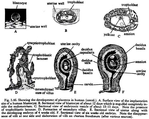
The end of the blastocyst containing the developing germinal disc attaches itself to the uterine wall (Fig. 5.46A). The uterine epithelium is eroded at the region of contact. The trophoblast tissue increases in thickness in this contact area due to the division of epithelial cells of the trophoblast layer.
Prelacunar Stage:
The implanted embryo, consisting of a bilaminar disc, is protruded into a cavity (lecithocoel). This cavity is enclosed by the trophoblast. Profound cytological changes occur in the trophoblast layer. The inner trophoblast cells remain cellular and are designated as the cytotrophoblast while the outer cells fuse together to form a syncytium called the syncytiotrophoblast.
The syncytiotrophoblast serves as the invading tissue of the embryo into the uterine wall. The growth of this syncytiotrophoblast is caused by differentiation of the cytotrophoblast and by amitotic division of the syncytial nuclei.
Lacunar stage (10th to 13th days of Placental development):
As the syncytiotrophoblast invades and increases in quantity, irregular spaces are produced in the syncytiotrophoblast. These spaces are called trophoblastic lacunae. (Fig. 5.46B).
Villous Period:
During the period between 14 and 18 days, the trophoblastic lacunae merge with one another to form large cavities bordered by syncytiotrophoblast. Such a cavity is named as the intervillous space and the primary villi are formed through the proliferation of the cytotrophoblastic elements into the syncytial trabeculae.
The primary villi, when first formed, lack mesodermal core. The mesoderm of the somatopleure invade into them to form the secondary villi (Fig. 5.46D).
With the formation of the blood islands and appearance of blood vessels, the secondary villi transform into the definitive tertiary villi. At this time, some endometrial tissue including blood vessels near to the invading chorionic vesicle break down to produce liquefied areas called the embryotroph. The liquefied
material from the embryotroph is assimilated by the syncytiotrophoblast for the growth of the embryo. This particular type of. nutrition is called the histotrophic nutrition.
From the physiological point of view the foetal villi developing from chorionic plate can be divided into three categories:
(i) Chorionic villi, through which physiological exchange of materials taking place between the foetus and the mother
(ii) Anchoring villi for mechanical anchorage of foetus and
(iii) Free villi.
The developing chorionic vesicle grows and invades the endometrium of the uterus. The uterine mucosa extends over the invading vesicle. The endometrial tissue covering the chorionic vesicle is called the decidua capsularis while the endometrial portion which is not concerned with the closure of such vesicle is called the decidua parietalis or decidua vera.
The endometrial portion lying between the musculature of the uterine wall and the invading villi is called decidua basalts.
Although the chorionic villi are formed over the entire chorionic vesicle, only the villi in relation to decidua basalis are retained and those grown to decidua parietalis are reabsorbed to form a smooth area named as the chorion leave. The villi within decidua basalis become greatly enlarged to serve the main role of physiological interchange (Fig. 5.46E).
This area of chorionic vesicle with the villi is the chorion frondosum which together with the tissue of decidua basalis forms the actual placenta. Thus the placenta is composed of decidua basalis (maternal placenta) and chorion frondosum (foetal placenta) (Fig. 5.46F).
The villi, at the early phase of development, consist of blood capillaries within mesodermal core covered over by cytotrophoblast and syncytiotrophoblast on the outer side. As development goes on the blood capillaries grow enormously and the cytotrophoblast layer is extremely reduced to few scattered cells below syncytiotrophoblast.
The villi are aggregated into groups called the cotyledons which are separated by incomplete placental septa. The villi in each cotyledon remain surrounded by a pool of maternal blood and by this way a hemochorial type of placenta is established.
Organogenesis of Placenta in Rabbit:
In rabbit, as in other eutherian mammals, placentation involves an interaction and attachment between the uterine wall and the extraembryonic membranes (chorion). The initial contact of the embryonic tissues with the uterine wall forms an epitheliochorial relationship in rabbit.
Subsequently in course of development, it changes into a hemochorial condition in which the endothelial tissue of the uterine blood vessels is destroyed by the erosive action of the foetal tissue. As a consequence, the chorionic epithelium of the embryonic part of the placenta comes in direct contact with the maternal blood.
During the later stages of pregnancy, even the chorionic epithelium also disappears thus leaving the endothelial lining of the foetal blood vessels in contact with the maternal blood. This type of placental relationship is called the hemoendothelial type (Fig. 5.47) and is regarded to be the most intimate grade of placental contact in animal kingdom.
Depending upon the distribution of villi, the placenta in rabbit is categorized as the discoidal type. At the onset, the chorion becomes uniformly covered with villi, but at late stage the villi remain only on one side. The villi continue to develop on the side which turned away from the restricted lumen of the uterus, while the villi on the other parts of villi become atrophied.
Events in Placentation in Rabbit:
The egg of rabbit, after fertilization, comes to lie in one of the crypts of the uterine wall (Fig. 5.48). As soon as the fertilization is completed the corona radiata (covering of the egg) breaks down to form a frothy substance (histotrophy which surrounds the entire fertilized egg.
The histotroph, around the egg, provides nutrition for further growth and development. The egg starts development and grows vigorously at the expense of the histotroph and soon completes the gastrulation stage to form the three primary germinal layers (viz., ectoderm, mesoderm and endoderm).
With the differentiation of the primary germinal layers in the embryo, remarkable physiological changes occur in the uterine wall of the mother. The region of the uterine wall where the embryo touches, continues to bleed due to some special enzymatic action.
The ‘maternal site of implantation’ becomes subsequently thick, spongy and vascularised. The superficial contact is made more intimate by the development of minute finger-like projections called the villi.
ADVERTISEMENTS:
The villi develop as outgrowths from the extraembryonic region of the foetus and penetrate into depressions in the wall of the uterus. These outgrowths (villi) are initially formed by the trophoblast but the connective tissue and blood vessels enter into the outgrowths later on. They are designated as the ‘chorionic villi’ and the blood vessels are actually the ramifications from allantoic blood vessels.
The region of the uterine wall which has passed the preparatory stage (vascularised) is called the trophospongia. The trophoblast and trophospongia come to establish intimate physiological relationship during placentation.
The villi of the trophoblast penetrate into trophospongia and ramify extensively deep into the maternal tissues. During this action, the villi are constantly bathed by the maternal blood vessels because of excessive vascular state of trophospongia.
After coming into close physiological unition, the trophoblast and trophospongia form a nutritional bridge where the maternal blood will bring nutrient materials for the developing embryo throughout the entire period of gestation. In rabbit, the epithelium of the chorion may disappear in some regions during the later stages of gestation—thus permitting an exposure of foetal blood vessels to the maternal blood.
The placenta is represented jointly by the trophoblast (chorion with its villi) and the trophospongia (uterine wall). At the time of parturition the chorionic villi are simply drawn out from the depressions in the uterine wall and the foetal tissues are separated without causing damage to the uterine wall and without causing any bleeding.
Functions of Placenta:
The functions of the placenta are many- fold.
It serves:
(i) Adhesion or anchorage of the developing embryo with the uterine wall.
(ii) Nutritional role—the food materials from the maternal blood circulation reach the blood stream of the embryo for supplying nutrition.
(iii) Excretory role—the waste products from embryonic circulation are eliminated to the maternal blood stream.
(iv) Respiratory role—it serves as the external respiratory surface for the developing embryo.
(v) Elaborate endocrine functions: Two ovarian hormones—estrogen and progesterone together with chorionic follicle- stimulating and luteinizing hormones are functionally elaborated by the placenta.
(vi) Protective role: Acts usually as barrier against the transportation of microbes into the embryo but the viruses and antigen do pass through the placenta.
(vii) Storage function: Glycogen, fats and some inorganic salts are stored in the placenta.
(viii) Source of nourishment for mother: Female of many mammals takes the placenta and after-birth tissues which provide nourishment.


