ADVERTISEMENTS:
Introduction to Recombinant DNA Technology:
Recombinant DNA technology has revolutionised life sciences, opening new vistas for research in molecular biology. It allows genetic manipulation using techniques for synthesizing, amplifying and purifying individual genes from any type of cell.
Genomics has emerged as an extension of recombinant DNA technology for high resolution mapping and characterization of whole genomes and gene products on a large scale, referred to as global analysis.
These rapidly advancing disciplines promise new insights into sequence data, organisation, expression, and regulation of the genetic material. Genetic engineering is a natural fallout of these techniques, exploring application in medical, biotechnological industry and agricultural fields.
ADVERTISEMENTS:
The success of recombinant DNA technology is based on some key discoveries: restriction enzymes that can cut and join DNA fragments (molecular scissors) for manipulations in a test tube; use of plasmids and bacteriophage DNA as vectors (vehicles) for foreign DNA that can replicate into identical copies and produce clones containing recombinant DNA; introduction of Southern blotting technique; and development of polymerase chain reaction for amplification of a specific sequence.
Recombinant DNA molecules can be purified and investigated for understanding gene structure and sequence, and can be inserted into another genome.
Recombinant DNA molecules are constructed artificially by incorporating DNA from two different sources into a single recombinant molecule. The process is distinct from recombination which is a natural process in sexually reproducing organisms, whereby a single individual gets a combination of genes from two parental organisms.
Natural recombination involves the coming together of similar nucleotide sequences in chromosomal DNA, breakage and exchange of corresponding segments and rejoining. This type of recombination, notably, produces new arrangements of alleles and usually occurs in closely related species. It does not take place in unrelated organisms due to natural barriers.
Restriction Endonucleases:
ADVERTISEMENTS:
Restriction endonucleases are a class of enzymes that can recognise and bind to specific DNA sequences of four to eight nucleotides, then cleave the sugar-phosphate backbone of each of the two strands at the site of binding. All restriction enzymes cut DNA between the 3′ carbon and the phosphate moiety of the phosphodiester backbone.
Therefore, fragments produced by restriction enzyme digestion have 5′ phosphates and 3′ hydroxyls. Most restriction enzymes are present in bacteria, only one has been found in the green alga Chlorella. In bacteria, restriction enzymes protect the bacterium against foreign DNA such as that in viruses, by cutting up the invading viral DNA. Thus they restrict entry of foreign DNA into the bacterial cell.
The bacterium modifies its own restriction sites by methylation, so that its own DNA is protected from the restriction enzyme it makes. Three scientists who discovered restriction enzymes and their applications namely, Adams, Nathan and Smith were awarded Nobel Prize in 1978.
More than 400 different restriction enzymes have been isolated. The restriction enzyme EcoRI (from E. coli) recognises the following six nucleotide base pair sequence in DNA of any organism: 5′-GAATTC-3′ and 3′-CTTAAG-5′.
This type of a nucleotide sequence is said to be symmetric, called a palindrome because both strands have the same nucleotide sequence in antiparallel orientation. Several different restriction enzymes recognise specific palindromes. Many restriction enzymes including EcoRI cut the sequence producing staggered ends. EcoRI cuts between G and A (Fig. 23.1).
The staggered cuts give rise to a pair of identical five nucleotide long single-stranded “sticky ends”. The ends consist of an overhanging piece of single-stranded DNA that are said to be sticky because they can base pair by hydrogen bonding to a complementary sequence. Sticky ends produced by restriction enzymes are desirable in recombinant DNA technology.
If two different DNA molecules are cut with the same restriction enzyme, the fragments of each will have the same sticky ends that are complementary, enabling them to hybridise with each other under appropriate conditions. Some restriction enzymes such as small cut the target sequence in the middle producing blunt ends which lack sticky ends.
These can also be used for making recombinant DNA molecules with the help of enzymes that join blunt ends, or other special enzymes that synthesise short single-stranded sticky ends on the exposed 3′ strand of the blunt end.
ADVERTISEMENTS:
Digestion of DNA by a restriction enzyme yields fragments of different lengths. For example, bacteriophage λ DNA cut with restriction enzyme EcoRI produces six fragments ranging in size from 3.6 to 21.2 kilo bases in length (one kilo-base or kb = 1,000 base pairs). These fragments can be separated according to size by gel electrophoresis (Figs. 23.2, 3) or by other methods.
The fragments together provide a map of the EcoRI sites in phage λ DNA. If multiple different restriction endonucleases are used, the locations of their cleavage sites can be used to generate detailed restriction maps of the DNA molecule.
Gel Electrophoresis for Separation of DNA Fragments:
Purification of DNA:
ADVERTISEMENTS:
The cells are homogenized and nuclei isolated for extraction of DNA. To lyse nuclei and release DNA, a negatively charged detergent sodium dodecyl sulphate (SDS) is used. The next step involves purification of DNA from contaminants such as RNA and proteins. To remove proteins, the mixture is shaken with phenol or chloroform.
Phenol makes the proteins insoluble and precipitates them out of solution. Because phenol and buffered saline are immiscible, they form two separate phases. Centrifugation gives DNA or RNA in the upper aqueous phase, while protein precipitate forms the boundary between the two phases.
The aqueous phase containing the nucleic acid is removed from the tube and shaken with phenol, followed by centrifugation. The process is repeated until no further protein can be removed from solution. Cold ethanol is then layered on top of the aqueous DNA solution, and the DNA is taken out with a glass rod at the interface between the ethanol and saline. RNA forms a precipitate that settles at the bottom of the vessel.
To make the DNA preparation free from RNA, the DNA is treated with ribonuclease. The ribonuclease is destroyed by treatment with protease, and the protease is removed by using phenol. Purified DNA is then re-precipitated with ethanol.
ADVERTISEMENTS:
Gel Electrophoresis:
Electrophoresis depends on the ability of charged molecules to migrate in an electric field. The small DNA fragments of a few hundred nucleotides or less, produced by a restriction endonuclease, can be separated by polyacrylamide gel electrophoresis (PAGE). The larger DNA fragments ranging in size from a few hundred base pairs to about 20 kb can be separated on agarose gels.
The mechanism for separation of DNA fragments involves migration of DNA molecules through the pores of the matrical gel. Agarose consists of a complex network of polymerised molecules, the pore size of which is determined by the composition of the buffer and concentration of agar used.
Visualisation by fluorescence microscopy has revealed that during electrophoresis DNA molecules display stretching in the direction of applied field and then contract into dense balls. The size of the DNA ball must be smaller than pore size in the gel to pass through. If the volume of the DNA ball exceeds that of pore size in gel, then the DNA molecule can only pass through by a serpentine motion resembling that of a snake.
ADVERTISEMENTS:
DNA molecules up to 20 kb can migrate through pores in gel by this mechanism. Studies have shown that both fragment length and molecular weight determine rate of migration of DNA molecules in a gel. For accurate size determination of a DNA molecule, marker DNA samples of known size are electrophoresed in the same gel.
The procedure for polyacrylamide gel electrophoresis is as follows. A mixture of linear DNA fragments of different sizes are driven by the current through the gel composed of organic molecules of acrylamide. Cross linking of acrylamide molecules forms a molecular sieve in a thin slab of the polyacrylamide gel between two glass plates. The gel slab is suspended between two compartments containing buffer in which the positive and negative electrodes are immersed (Fig. 23.2).
The sample containing linear DNA molecules (fragments) is placed in wells along the top of the gel, near the cathode of the electric field. Voltage applied between the buffer compartments allows current to flow across the slab. Because of its negatively charged phosphate groups, DNA migrates towards the positive electrode (anode) at speeds inversely dependent on their size. That is, the smaller fragments migrate most rapidly.
All DNA molecules regardless of their length, have a similar charge density, that is, the number of negative charges per unit of mass, and therefore, all have equivalent potential for migration in the electric field. The greater the molecular mass of the DNA fragment, the more slowly it moves through the gel. Hence, the smaller the fragment, the farther it migrates in the gel.
Therefore, if there are distinct size classes in the mixture of DNA fragments, these classes will form distinct bands on the gel. The fragments of different sizes thus get separated from each other (Fig. 23.3). The bands can be visualised by staining the DNA with ethidium bromide.
The migration of the DNA fragments can be compared with a set of control size standards that were loaded in the same gel, to determine the exact size of each fragment in the mixture. Moreover, if the bands are well separated, a single band can be cut from the gel and the DNA sample can be extracted and purified.
Larger DNA fragments that do not pass through the pores of polyacrylamide, are fractionated on agarose gels which have greater porosity. Agarose is a sea weed polysaccharide, it is dissolved in hot buffer and poured into the mould and a comb placed in the molten agarose. Lowering the temperature solidifies agarose as a gel. The comb is removed producing wells in the gel. DNA samples are loaded in the wells.
Gels having 0.3% concentration of agarose are used to separate larger DNA fragments. After running the gel, DNA fragments can be identified by using a labeled probe, for a specific fragment. Alternatively, the gel can be stained with a solution of ethidium bromide which intercalates into the bases in the double helix. The DNA bands can be viewed under ultraviolet light using a trans-illuminator.
Pulse-Field Gel Electrophoresis:
The separation of DNA molecules greater than 20 kb, up to 10 Mb can be accomplished by use of pulse-field gel electrophoresis (PFGE). In PFGE, the orientation of the electric field with respect to the gel is changed in a regular manner, which causes the DNA to periodically alter its direction of migration on the gel.
Each time there is a change in the electric-field orientation, the axis of the DNA must realign before it starts migration in the new direction. The difference between the direction of migration of DNA induced by successive electric fields determines the angle through which DNA must turn in order to change its direction of migration. To make the sample run in straight lines, improved methods for alternating the electric field have been devised. PFGE is used extensively in molecular biology laboratories.
ADVERTISEMENTS:
Southern Blotting:
When genomic DNA is subjected to restriction enzyme digestion it results in a very large number of fragments. A stained gel containing numerous fragments separated by electrophoresis shows a continuous smear of DNA instead of distinct bands. The technique of Southern blotting developed by E. M. Southern allows a single DNA fragment or a specific gene to be identified in this mixture.
This is a widely used technique that is based on labelling of DNA and hybridisation on membranes. It is referred to as a blotting technique because it involves transfer of nucleic acids from gels to a solid support of a membrane and immobilisation of DNA on to the membrane. Detection of the DNA fragment is done by using a complementary strand of DNA or RNA that is called probe (something that detects is a probe) which hybridizes with the DNA fragment.
The transfer of DNA from gel to membrane is accomplished by the flow of buffer through the wick, gel, membrane, and above onto adsorbent paper layers. The gel is overlaid on a filter paper wick (3 to 4 sheets) which dips into the buffer contained in a vessel (Fig. 23.4). The hybridisation membrane is placed above the gel.
A stack of paper towels comes above the membrane. The adsorbent paper towels serve to draw the buffer through the gel by capillary action. The flow of buffer carries the DNA molecules out of the gel on to the membrane (nitrocellulose or nylon membrane). Nylon membranes are considered superior in having greater binding capacity for nucleic acids and being stronger than membranes consisting of nitrocellulose.
Large DNA fragments require longer time to transfer out of gel than shorter fragments. To overcome such time differences, the electrophoresed DNA on the gel is pretreated for depurination, which also denatures the fragments into single strands, making them accessible for hybridisation with the probe.
After transfer from the gel, the DNA fragments are attached (permanently fixed) to the membrane by, heating at 80°C in the oven or by cross-linking using UV radiation. The DNA fragments become fixed or imprinted to the membrane at the same locations as in the gel (replica).
In other words, the spatial arrangement of DNA in the gel is preserved in the sheet. Following fixation, the membrane is incubated in a solution containing labelled single-stranded DNA or RNA probe that is complementary to the blot transferred DNA sequence to be detected. Under appropriate conditions, the labelled nucleic acid probe hybridizes with the DNA on the membrane.
After hybridisation the membrane is washed to remove unbound probes, so that the only labelled molecules left are those that hybridised with target DNA. The membrane is then placed in contact with X-ray film for autoradiography that will reveal the position of the desired DNA fragment.
By using size-calibration controls, the size of any fragment from the mixture of fragments can be determined. Variations of Southern blotting are called Dot and Slot blots. The sample is blotted directly on the nylon or nitrocellulose sheet without prior separation on the gel.
Northern Blotting:
This technique is similar to the Southern blotting technique and is used for identifying a specific RNA molecule from a mixture of RNAs separated on a gel. The separated RNA molecules on a gel are blotted on to a membrane and probed in the same way as DNA is blotted and probed in Southern blotting. A cloned gene having complementary sequence to the RNA being searched, can be used as a probe.
Stated briefly, it is a combination of the recent remarkable techniques that are finding widespread application. Generation of restriction fragments, gel electrophoresis, blotting techniques, cloning, together with nucleic acid hybridisation allow a specific DNA sequence to be analysed in a whole genome.
To identify an individual gene, the DNA from the organism is purified and cleaved by restriction enzymes. The restriction fragments are separated by gel electrophoresis. The fragments are denatured to get single strands, then labelled with a radioactive probe.
The probe consisting of single-stranded DNA or RNA must have a sequence that can base pair with the gene of interest. The probe hybridises with its complementary sequence under appropriate conditions of temperature, salt and pH. Exposure to X-ray film indicates the binding of the probe through the presence of the radioactive signal.
Recombinant DNA Molecules:
The generation of recombinant DNA molecules is a process of inserting a gene of interest or donor DNA into a DNA molecule from a different source that acts as a vector or vehicle for the donor DNA. The first step is to obtain donor DNA, that is, isolate the gene of interest. Genomic DNA from the donor organism is isolated, purified and cut with a restriction endonuclease that makes staggered cuts with sticky ends in the donor DNA.
The sticky ends consist of overhanging or cohesive single-stranded tails. Thus donor DNA will be cut into a set of restriction fragments according to the locations of the restriction sites. The vector DNA, which could be a bacterial plasmid or genome of bacteriophage λ (described later), is also cut with the same restriction enzyme that was used for donor DNA.
The fragments thus produced would have the same complementary sticky or cohesive ends, enabling them to hybridise with sticky ends in the donor DNA fragments. The hybridised molecules produced do not have complete covalently joined sugar phosphate backbones. To seal the backbones, the enzyme DNA ligase is used which makes phosphodiester bonds and links the two covalently to form a recombinant DNA molecule (Fig. 23.5).
Other Sources of Donor DNA:
Besides genomic DNA from a donor organism, it is possible to use RNA sequences as donors. The first step is to synthesise a DNA copy of the RNA using the enzyme reverse transcriptase. The complementary DNA or cDNA obtained can be used as donor DNA by incorporating it into vector DNA and making a recombinant DNA molecule.
In studies that require characterisation of the gene transcript, that is mRNA, a cDNA is prepared by using mRNA as a template for reverse transcriptase. This is particularly useful because mRNA is short-lived and techniques for isolating individual mRNA molecules are not available. cDNAs can be used to learn about the variety of mRNAs in a cell, and the number of copies of different mRNAs present in a cell.
If the sequence to be inserted for making a recombinant DNA molecule cannot be isolated from natural genomic DNA, nor as cDNA, then chemically synthesised DNA can be used. Chemical synthesis of oligonucleotides, that is, DNA fragments 15 to 100 nucleotides in length can be developed by highly automated techniques.
Vectors for Generating Recombinant DNA:
A vector must be a small molecule capable of independent replication in a living host cell; must have convenient restriction sites that can be used for insertion of the DNA to be cloned; must permit easy identification and recovery of the recombinant molecule. Vectors are also referred to as cloning vehicles or replicons. The basic vector systems use bacterial plasmid or bacteriophage λ DNA as vectors.
Plasmids are small circular DNA molecules that replicate independently of the bacterial chromosome. Plasmid molecules are partitioned accurately to daughter cells. Most plasmids exist as double-stranded DNA molecules.
If both strands of the DNA are intact circles, the molecule is described as a covalently closed circle DNA (CCC DNA), if only one strand is intact, then open circle DNA (OC DNA). Not all plasmids are circular, some exist as linear molecules such as those in Streptomyces and Borrelia.
Plasmid DNA is 2 to 4 kb in length and has a sequence which is origin of replication, that signals the host cell DNA polymerase to replicate the DNA molecule. Plasmids have the advantage of carrying genes for resistance to antibiotics, so bacteria carrying antibiotic-resistance phenotype (plasmids) can be selected.
Suppose we want to insert gene X in human genome into a plasmid vector. Fragments of human DNA are prepared by cutting with a restriction enzyme, for example EcoRI, and the same restriction enzyme is used to cut plasmid DNA (Fig. 23.3).
The sticky ends will have complementary single-stranded cohesive ends that would hybridise with each other. Among the large number of fragments produced from human DNA, only a very small fraction would have gene X.
To isolate the fragment with gene X, plasmid and human DNA restriction digests are incubated together, along with DNA ligase. During incubation, the sticky ends of the two types of DNA become hydrogen-bonded to each other, their broken ends sealed by ligase to form circular recombinant DNA molecules.
Now there would be a large number of different recombinant DNA molecules, each containing a bacterial plasmid with a human DNA fragment. To isolate those plasmids that have human gene X, the process of DNA cloning has to be done.
Bacteriophage λ vectors can carry foreign DNA inserts as large as 15 kb. There are two basic types of phage λ vectors, insertional vectors and replacement vectors. The wild type phage λ DNA contains several target sites for most of the commonly used restriction endonucleases, hence is not suitable as a vector.
Derivatives of the wild type phage have, therefore, been produced that either have a single target site at which foreign DNA can be inserted (insertional vectors), or have a pair of sites defining a fragment that can be removed and replaced by foreign DNA (replacement vectors).
Since phage DNA can accommodate only about 5% more than its normal complement of DNA, vectors are constructed with deletions to increase space within the genome. The shortest λ DNA molecules generally used are 25% deleted.
Foreign DNA is first ligated to phage DNA (Fig. 23.6). The recombinant molecules are then introduced into E. coli cells. DNA replication produces numerous phage progeny containing the foreign DNA fragment. This fragment can be isolated from the rest of phage DNA by restriction endonuclease digestion and gel electrophoresis.
Phagemids are plasmid vectors that carry the origin of replication from the genome of a single-stranded filamentous bacteriophage such as M13 or f1. Phagemids combine the best features of plasmids and single-stranded bacteriophage vectors.
They have two separate modes of replication: as a double-stranded DNA plasmid, and as a template to produce single-stranded copies of one of the phagemid strands. A phagemid can therefore, be used in the same way as a plasmid vector, or it can be used to produce filamentous bacteriophage particles that contain single-stranded copies of cloned segments of DNA.
Some studies require large fragments of foreign DNA to be inserted for which phage λ vectors are not suitable. Artificially prepared cosmid and yeast artificial chromosome (YAC) vectors are useful for this purpose. Foreign DNA up to 45 kb in length can be accommodated in cosmid vectors. Cosmid vectors can exist as plasmids but they also contain the complementary overhanging single-stranded ends of phage λ.
The presence of bacteriophage λ sequences in cosmid vectors permit packaging of the recombinant DNA into phage particles. The λ phage then introduces these large sized recombinant DNA molecules into recipient E. coli cells. Cosmids also contain origins of replication and genes for drug-resistance, so that they can replicate as plasmids in bacterial cells (Fig. 23.7).
However, because cosmid vectors lack the essential phage sequences necessary to form progeny phage particles, the recombinant DNA molecule depends on the plasmid sequences in the cosmid. Once inside the recipient E. coli cell, the recombinant cosmids form circular molecules that replicate extra-chromosomally in the same manner as plasmids.
The YAC vectors can accommodate still larger fragments, from a hundred to a thousand kilo-bases in length, that is one million base pairs. As the name implies, YACs are artificial versions of normal yeast chromosomes and replicate as chromosomes in yeast cells.
They contain all the elements of a yeast chromosome that are required for replication during S phase, one or more origins of replication, and segregation to daughter cells during mitosis, telomeres at the ends of the chromosome, as well as centromere to which spindle fibres can attach during chromosome separation.
In addition to these elements, YACs are constructed to contain the following: a gene whose encoded product allows those cells containing the YAC to be selected from those that lack the element; the DNA fragment to be cloned. Yeasts can take up DNA from the medium, allowing YACs to be introduced into host yeast cells.
For introducing such large-sized DNA fragments (100 to 1000 kb in length), it is necessary to use restriction enzymes that recognise particularly long nucleotide sequences, 7 to 8 nucleotides containing CG di-nucleotides.
For example, the restriction enzyme NotI recognises the 8 nucleotide sequence GCGGCCGC, which cleaves mammalian DNA into fragments approximately one million base pairs long. These fragments can then be incorporated into YACs and cloned within host yeast cells. YAC technology has been used extensively in the Human Genome Project.
Bacterial artificial chromosomes (BACs) are based on the F factor of E. coli, and are among the most widely used vectors for very large DNA fragments of up to 300 kb. BAC vectors are 6 to 8 kb in length and include genes for essential functions such as replication (genes repE, and oriS), genes for regulating copy number (parA and parB), and genes for resistance to the antibiotic chloramphenicol.
Genomic DNA Libraries:
Collections of DNA fragments obtained by DNA cloning from the entire genome of an organism constitute a DNA library. Basically, the genomic library is a collection of bacteria, typically E. coli, each carrying a fragment of DNA from the genome. In one approach for making a DNA library, all the DNA from an organism is cut into fragments with a restriction enzyme.
Each segment is inserted into a different copy of the vector, thereby creating a collection of recombinant DNA molecules, which collectively represent the entire genome. These are then used to transform separate recipient bacterial cells, where they are amplified. The resulting collection of recombinant DNA-bearing bacteria is called a genomic library.
If the cloning vector used could accommodate an average insert size of 10 kb, and if the entire genome size is 100,000 kb, then we can expect 10,000 independent recombinant clones to represent the library of the whole genome.
In another approach, genomic DNA is cleaved by one or two restriction enzymes that recognize very short nucleotide sequences such as Sau3A (recognises GATC) and HaeIII (recognises GGCC). The enzymes are used in low concentration so that only a small percentage of target sites are actually cleaved.
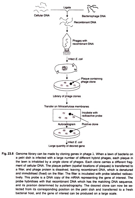
One can expect that the small-sized tetra-nucleotide sequence would occur by chance at very high frequency, so that every portion of the DNA could yield fragments. After partial digestion of DNA, the fragments are separated by gel electrophoresis. Fragments about 20 kb in length are incorporated into phage λ heads from which about half a million plaques are generated, to ensure that every portion of the genome is represented (Fig. 23.8).
The important point in this method is that, due to low concentration of the restriction enzyme, the DNA is randomly fragmented, and each and every target site is not cleaved. The phage recombinants produced constitute a permanent collection of all the DNA sequences in the genome.
Whenever a particular sequence is required for isolation from the library, phage can be grown in bacteria. Each of the plaques produced would have originated from infection of a single recombinant phage, and can be screened for the presence of a particular sequence.
Positional Cloning or Chromosome Walking:
When there is no biological information about a gene, but its position can be mapped relative to other genes or markers, it is called positional cloning. The approach involves cloning a gene from its known closest markers. It requires only the mapped position of the gene. On the basis of this information, researchers can locate the nearest physical markers.
The closest linked marker is used to probe the genomic library. DNA cleaved randomly, as described above, generates overlapping fragments that can be used in the analysis of regions of the chromosome extending out in both directions from a particular sequence, the gene of interest. These extended regions or linked markers serve as starting points for the process of chromosome walking (Fig. 23.11).
Using a small sequence at the end of the linked marker, let us call it M1, as a labelled probe, find a clone in the library. The positive clone that is found will lead to identification of an adjacent gene segment which is now a second marker. Isolate the end sequence of second marker (M2) and use it to probe library and find another adjacent genomic segment which becomes the third marker.
Isolate end of third marker (M3) and use it to again probe for the next adjacent segment, and so on. Using the new fragments as labelled probes the process is repeated in successive screening steps, leading to isolation of more and more of the original DNA molecule. Because this process consists of steps, hence the name chromosome walking. This approach allows study of the organisation of linked sequences over a considerable length of the chromosome.
Chromosome jumping is a variation of chromosome walking using larger high capacity vectors to bridge un-clonable gaps. Whereas in chromosome walking each step is an overlapping DNA clone, in chromosome jumping each jump is from one chromosome location to another without “touching down” on the intervening DNA.
The gene for cystic fibrosis (CF), a severe autosomal recessive disorder in humans, was identified by chromosome walking and jumping. Cloning of the CF gene was a breakthrough for studying the biochemistry of the disorder (abnormal chloride channel function), for designing probes for prenatal diagnosis, and for potential treatment by somatic gene therapy or other means.
cDNA Libraries:
The method described above for making genomic DNA libraries can also be used for generating cDNA libraries. A cDNA library would contain hundreds of thousands of independent cDNA clones, representating collections of cDNA inserts. It may be recalled that cDNA is produced from the mRNA by using reverse transcriptase.
If a specific gene that is being actively transcribed in a particular tissue is desired for study, then it is considered useful to convert its mRNA into cDNA and make a cDNA library from that sample. A cDNA library represents a subset of the transcribed regions of the genome, hence the cDNA library would be smaller than a complete genomic library.
Screening DNA Library for a Specific Clone:
Any sequence for which a probe is available can be isolated from a recombinant library. Two types of probe can be used, those that recognise a specific DNA sequence and those that recognise part of a specific protein.
Probes for DNA Sequences:
A probe that consists of a single strand of DNA would be able to find and bind to other complementary denatured (single-stranded) DNAs in the library and specifically hybridise with it. The procedure for identification of a specific clone in a library is carried out in two steps. First, the recombinant phages are plated on E.coli, and each phage replicates to produce a plaque on the lawn of bacteria.
The pattern of plaques of the library on the petri dish are transferred to an absorbent nitrocellulose membrane by laying the membrane directly on the surface of the medium. When the membrane is peeled off, the plaques remain clinging to its surface, are lysed in situ and DNA is denatured. The membrane is incubated in a solution containing radiolabeled probe that is specific for the sequence being searched (Fig. 23.9).
Generally, the probe is a cloned piece of DNA that has a sequence homologous to that of the desired sequence. The single-stranded probe will bind to the DNA of the clone being searched. To determine the position of the positive clone, the position of the radioactive label can be found out by placing the membrane on the X-ray film.
Emissions from the decay of radioactive label will reduce the grains in X-ray film, seen as a dark spot after developing the film. The procedure is called autoradiography, the exposed film an autoradiogram. The probe used for finding sequence of interest in a library can also be labelled with a fluorescent dye.
In that case the membrane is exposed to a particular wavelength of light that would excite fluorescence and a photograph of the membrane is taken. The position of the spot of label (radioactive or fluorescent) indicates the location of the DNA segments containing sequences complementary to the probe.
Probes for Protein Products of Genes:
Expression Cloning:
The protein product of a gene can be used to find the clone of its corresponding gene in a library. This can be accomplished if the amino acid sequence of the protein product is known, and the protein can be isolated in a purified form. Antibodies that bind specifically with unique protein molecules are used as probes to screen an expression library. These libraries are special cDNA libraries generated by using expression vectors.
An expression vector is a cloning vector containing the regulatory sequences necessary to allow transcription and translation of a cloned gene. Expression vector produces the protein encoded by a cloned gene in the transformed host. Expression vectors are essentially derivatives of a phage or plasmid cloning vector that has been modified by addition of a promoter specific to the host.
The cloned gene in such an expression vector is placed under the control of the promoter that ensures transcription of the cloned gene in the appropriate host cell. Expression vectors designed in this manner can yield high levels of recombinant protein in host cells that can be purified for structural and functional characterisation.
To make the cDNA library, the cDNA to be cloned is inserted into the special phage vector downstream from the bacterial promoter, in the correct triplet reading frame which ensures that the foreign DNA is transcribed and translated during infection.
Phages that have incorporated the gene of interest form plaques that contain the protein encoded by that gene. A membrane is laid over the surface of the medium and removed with some cells of each colony attached to the membrane.
The locations of these cells are identical to their positions in the original petri dish (replica plating). The membrane is dried, and immersed in a solution containing antibody. The antibody will bind only to the protein product of the gene of interest.
For detection of positive clones, a second antibody is prepared that is specifically against the bound antibody and labeled radioactively or with a fluorochrome. The plaque having the gene of interest is thus located on the replica plate through detection of bound antibody.
It is frequently considered useful to express high levels of a cloned gene in eukaryotic cells rather than in bacteria. The reason is that post-translational modifications of the protein (such as addition of carbohydrates or lipids) that take place in eukaryotic cells would take place normally.
One system frequently used for protein expression in eukaryotic cells involves infection of insect cells by baculovirus vectors, which yield very high levels of protein product of a gene. High levels of protein expression can also be achieved by using appropriate vectors in mammalian cells.


