ADVERTISEMENTS:
Read this article to learn about the importance of DNA fingerprinting / DNA profiling in the modern medical forensic.
DNA Fingerprinting:
DNA fingerprinting is the present day genetic detective in the practice of modern medical forensics. The underlying principles of DNA fingerprinting are briefly described. The structure of each person’s genome is unique. The only exception being monozygotic identical twins (twins developed from a single fertilized ovum).
The unique nature of genome structure provides a good opportunity for the specific identification of an individual. It may be remembered here that in the traditional fingerprint technique, the individual is identified by preparing an ink impression of the skin folds at the tip of the person’s finger. This is based on the fact that the nature of these skin folds is genetically determined, and thus the fingerprint is unique for an individual. In contrast, the DNA fingerprint is an analysis of the nitrogenous base sequence in the DNA of an individual.
History and Terminology:
ADVERTISEMENTS:
The original DNA fingerprinting technique was developed by Alec Jaffrey’s in 1985. Although the DNA fingerprinting is commonly used, a more general term DNA profiling is preferred. This is due to the fact that a wide range of tests can be carried out by DNA sequencing with improved technology.
Applications of DNA Fingerprinting:
The amount of DNA required for DNA fingerprint is remarkably small. The minute quantities of DNA from blood strains, body fluids, and hair fiber or skin fragments are enough. Polymerase chain reaction is used to amplify this DNA for use in fingerprinting. DNA profiling has wide range of applications—most of them related to medical forensics.
Some important ones are listed below:
i. Identification of criminals, rapists, thieves etc.
ADVERTISEMENTS:
ii. Settlement of paternity disputes.
iii. Use in immigration test cases and disputes.
In general, the fingerprinting technique is carried out by collecting the DNA from a suspect (or a person in a paternity or immigration dispute) and matching it with that of a reference sample (from the victim of a crime, or a close relative in a civil case).
DNA Markers in Disease Diagnosis and Fingerprinting:
The DNA markers are highly useful for genetic mapping of genomes. There are four types of DNA sequences which can be used as markers.
1. Restriction fragment length polymorphisms (RFLFs, pronounced as rif-lips).
2. Minisatellites or variable number tandem repeats (VNTRs, pronounced as vinters).
3. Microsatellites or simple tandem repeats (STRs).
4. Single nucleotide polymorphisms (SNPs, pronounced as snips).
The general aspects of the above DNA markers are described along with their utility in disease diagnosis and DNA fingerprinting.
Restriction Fragment Length Polymorphisms (RFLPs):
ADVERTISEMENTS:
A RFLP represents a stretch of DNA that serves as a marker for mapping a specified gene. RFLPs are located randomly throughout a person’s chromosomes and have no apparent function. A DNA molecule can be cut into different fragments by a group of enzymes called restriction endonucleases .These fragments are called polymorphisms (literally means many forms).
An outline of RFLP is depicted in Fig. 14.5. The DNA molecule 1 has three restriction sites (R1, R2, R3), and when cleaved by restriction endonucleases forms 4 fragments. Let us now consider DNA 2 with an inherited mutation (or a genetic change) that has altered some base pairs. As a result, the site (R2) for the recognition by restriction endonuclease is lost. This DNA molecule 2 when cut by restriction endonuclease forms only 3 fragments (instead of 4 in DNA 1).
As is evident from the above description, a stretch of DNA exists in fragments of various lengths (polymorphisms), derived by the action of restriction enzymes, hence the name restriction fragment length polymorphisms.
ADVERTISEMENTS:
RFLPs in the Diagnosis of Diseases:
If the RFLP lies within or even close to the locus of a gene that causes a particular disease, it is possible to trace the defective gene by the analysis of RFLP in DNA. The person’s cellular DNA is isolated and treated with restriction enzymes. The DNA fragments so obtained are separated by electrophoresis.
The RFLP patterns of the disease suspected individuals can be compared with that of normal people (preferably with the relatives in the same family). By this approach, it is possible to determine whether the individual has the marker RFLP and the disease gene. With 95% certainty, RFLPs can detect single gene-based diseases.
Methods of RFLP scoring:
ADVERTISEMENTS:
Two methods are in common use for the detection of RFLPs (Fig. 14.5).
1. Southern hybridization:
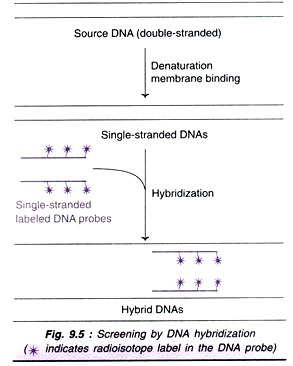
The DNA is digested with appropriate restriction enzyme, and separated by agarose gel electrophoresis. The so obtained DNA fragments are transferred to a nylon membrane. A DNA probe that spans the suspected restriction site is now added, and the hybridized bands are detected by autoradiograph. If the restriction site is absent, then only a single restriction fragment is detected. If the site is present, then two fragments are detected (Fig. 14.6A).
2. Polymerase chain reaction:
ADVERTISEMENTS:
RFLPs can also be scored by PCR. For this purpose, PCR primers that can anneal on either side of the suspected restriction site are used. After amplification by PCR, the DNA molecules are treated with restriction enzyme and then analysed by agarose gel electrophoresis. If the restriction site is absent only one band is seen while two bands are found if the site is found (Fig. 14.6B).
Applications of RFLPs:
The approach by RFLP is very powerful and has helped many genes to be mapped on the chromosomes, e.g. sickle- cell anemia (chromosome 11), cystic fibrosis (chromosome 7), Huntington’s disease (chromosome 4), retinoblastoma (chromosome 13), Alzheimer’s disease (chromosome 21).
Variable Number Tandem Repeats (VNTRs):
VNTRs, also known as mini-satellites, like RFLPs, are DNA fragments of different length. The main difference is that RFLPs develop from random mutations at the site of restriction enzyme activity while VNTRs are formed due to different number of base sequences between two points of a DNA molecule. In general, VNTRs are made up of tandem repeats of short base sequences (10-100 base pairs). The number of elements in a given region may vary, hence they are known as variable number tandem repeats.
An individual’s genome has many different VNTRs and RFLPs which are unique to the individual. The pattern of VNTRs and RFLPs forms the basis of DNA fingerprinting or DNA profiling. In the Fig. 14.7, two different DNA molecules with different number of copies (bands) of VNTRs are shown. When these molecules are subjected to restriction endonuclease action (at two sites R1 and R2), the VNTR sequences are released, and they can be detected due to variability in repeat sequence copies. These can be used in mapping of genomes, besides their utility in DNA fingerprinting.
VNTRs are useful for the detection of certain genetic diseases associated with alterations in the degree of repetition of microsatellites e.g. Huntington’s chorea is a disorder which is found when the VNTRs exceed 40 repeat units.
Limitations of VNTRs:
The major drawback of VNTRs is that they are not evenly distributed throughout the genome. VNTRs tend to be localized in the telomeric regions at the ends of the chromosomes.
Use of RFLPs and VNTRs in Genetic Fingerprinting:
RFLPs caused by variations in the number of VNTRs between two restriction sites can be detected (Fig. 14.8). The DNAs from three individuals with different VNTRs are cut by the specific restriction endonuclease. The DNA fragments are separated by electrophoresis, and identified after hybridization with a probe complementary to a specific sequence on the fragments.
Microsatellites (Simple Tandem Repeats):
Microsatellites are short repeat units (10-30 copies) usually composed of dinucleotide or tetra nucleotide units. These simple tandem repeats (STRs) are more popular than mini-satellites (VNTRs) as DNA markers for two reasons.
ADVERTISEMENTS:
1. Microsatellites are throughout the genome.
2. PCR can be effectively and conveniently used to identify the length of polymorphism.
Two variants (alleles) of DNA molecules with 5 and 10 repeating units of a dimer nucleotides (GA) are depicted in Fig. 14.9.
By use of PCR, the region surrounding the microsatellites is amplified, separated by agarose gel electrophoresis and identified.
Single Nucleotide Polymorphisms (SNPs):
SNPs represent the positions in the genome where some individuals have one nucleotide (e.g. G) while others have a different nucleotide (e.g. C). There are large numbers of SNPs in genomes. It is estimated that the human genome contains at least 3 million SNPs. Some of these SNPs may give rise to RFLPs. SNPs are highly useful as DNA markers since there is no need for gel electrophoresis and this saves a lot of time and labour. The detection of SNPs is based on the oligonucleotide hybridization analysis (Fig. 14.10).
An oligonucleotide is a short single-stranded DNA molecule synthesized in the laboratory with a length not usually exceeding 50 nucleotides. Under appropriate conditions, this nucleotide sequence will hybridize with a target DNA strand if both have completely base paired structure. Even a single mismatch in base pair will not allow the hybridization to occur. DNA chip technology is most commonly used to screen SNPs hybridization with oligonucleotide
Current Technology of DNA Fingerprinting:
In the forensic analysis of DNA, the original techniques based on RFLPs and VNTRs are now largely replaced by microsatellites (short tandem repeats). The basic principle involves the amplification of microsatellites by polymerase chain reaction followed by their detection. It is now possible to generate a DNA profile by automated DNA detection system (comparable to the DNA sequencing equipment).


