ADVERTISEMENTS:
Notes on Chromosomes. After reading this article you will learn about: 1. Introduction to Chromosomes 2. Shape of Chromosomes 3. Size 4. Number 5. Morphology 6. Characteristic Features 7. Classification 8. Special Types 9. Some Other Chromosomes 10. Role of Chromosomes in Heredity 11. Change in Chromosomes Structure.
Contents:
- Notes on Introduction to Chromosomes
- Notes on the Shape of Chromosomes
- Notes on the Size of Chromosomes
- Notes on the Number of Chromosomes
- Notes on the Morphology of Chromosomes
- Notes on the Characteristic Features of Chromosomes
- Notes on the Classification of Chromosomes
- Notes on the Special Types of Chromosomes
- Notes on Some Other Chromosomes
- Notes on the Role of Chromosomes in Heredity
- Notes on Change in Chromosome Structure
Note # 1. Introduction to Chromosomes:
ADVERTISEMENTS:
The darkly stained, rod shaped bodies visible under light microscope in a cell during metaphase stage of mitosis are referred to as chromosomes. Strasburger was the pioneer man who discovered chromosomes in 1875, and the term chromosome was coined by Waldeyer in 1888.
The main features of eukaryotic chromosomes are given below:
i. Chromosomes are not visible during interphase under light microscope. During other stages of cell division, they are visible, but are more clearly visible during mitotic metaphase. Hence, they are studied during metaphase.
ii. Chromosomes bear genes in a linear fashion and thus are concerned with transmission of characters from generation to generation.
ADVERTISEMENTS:
iii. Chromosomes of eukaryotes are enclosed by a nuclear membrane, while in prokaryotes, they remain without such envelope free in the cytoplasm.
iv. Chromosomes vary in shape, size and number in different species of plants and animals.
v. Chromosomes have property of self-duplication, segregation and mutation.
vi. Chromosomes are composed of DNA, RNA an histones. DNA is the major genetic constituent of chromosomes.
Note #
2. Shape of Chromosomes:
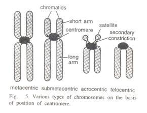
Chromosome shape is usually observed during anaphase. The shape of chromosomes is determined by the position of centromere, a part of chromosome on which spindle fibres are attached during metaphase.
Chromosomes have generally three different shapes, viz., rod shape, J shape and V shape. These shapes are observed when the centromere occupies terminal, sub-terminal and median (middle) position on the chromosomes, respectively.
Note #
3. Size of Chromosomes:
Chromosome size is measured with the help of micrometer at mitotic metaphase. It is measured in two ways, viz., in length and in diameter. Plants usually have longer chromosomes than animals. Moreover, species or individuals which have fewer chromosome numbers have larger chromosomes.
ADVERTISEMENTS:
The maximum length of chromosome is observed during interphase and minimum during anaphase. Thus chromosome size varies from species to species (Table 4.1). Giant chromosomes have length up to 300 µ.
Note #
4. Number of Chromosomes:
There are three types of chromosome number, viz., haploid, diploid and basic number as given below:
i. Haploid:
ADVERTISEMENTS:
It represents half of the somatic chromosome number of a species and is denoted by n. Since haploid chromosome number is usually found in the gametes, it is also known as gametic number.
ii. Diploid:
It refers to somatic chromosome number of a species and is represented by 2n. Since diploid chromosome number is found in zygotic or somatic cells it is also referred to as zygotic or somatic number.
iii. Basic Number:
The gametic chromosome number of a true diploid species is called basic number. It is the minimum haploid chromosome number of any species which is denoted by x. For example, in wheat, the basic number is 7, whereas the haploid number is 7, 14 and 21 for diploid, tetraploid and hexaploid species, respectively.
Thus haploid chromosome number differs from basic number. Both are same in case of true diploid species but differ in case of polyploid species. Thus, basic number can be a haploid number but all haploid numbers cannot be basic number. Chromosome number differs from species to species (Table 4.2). In plant kingdom, chromosome number usually is higher in dicots than in monocots.
Note #
5. Morphology of Chromosomes:
Chromosome morphology is studied in the cells of root tip during metaphase under light microscope.
Each chromosome consists of seven parts, viz:
ADVERTISEMENTS:
(i) Centromere,
(ii) Chromatids,
(iii) Secondary constriction and satellite,
(iv) Telomere,
(v) Chromomere,
(vi) Chromonema and
ADVERTISEMENTS:
(vii) Matrix (Fig. 4.1).
A brief description of these parts is given below:
i. Centromere:
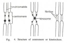
The region of chromosome with which spindle fibres are attached during metaphase is known as centromere or primary constriction or kinetochore.
Centromere has four important functions, viz:
(a) Orientation of chromosomes at metaphase,
ADVERTISEMENTS:
(b) Movement of chromosomes during anaphase,
(c) Formation of chromatids, and
(d) Chromosome shape.
Since centromere is associated with movement of chromosomes at anaphase, it is also called as kinetochore. Centromere may occupy various positions on the chromosome, viz., terminal, sub-terminal, median, etc.
Generally, each chromosome has one centromere, but in some cases, the number of centromere may vary from nil to many. Depending upon the position and number of centromere, chromosomes are given various names (Table 4.3).
A chromosome with a diffused centromere is called holokinetic or holocentric chromosome. Such chromosomes do not have localized centromere, but the entire body of the chromosome exhibits centromeric activity.
The spindle fibres get attached to the entire body of such chromosomes. Sister chromatids of such chromosomes are not associated at any point by centric connection and have autonomous entity. Such chromosomes can move towards either pole during anaphase.
ii. Chromatid:
One of the two distinct longitudinal subunits of a chromosome is called chromatid. These subunits of a chromosome get separated during anaphase. Chromatids are of two types viz., sister chromatids and non-sister chromatids.
Sister chromatids are derived from one and the same chromosome, while non-sister chromatids originate from homologous chromosomes. Chromatids are formed due to chromosome and DNA replications during interphase. Two chromatids of a chromosome are held together by centromere. After separation at anaphase each chromatid becomes a chromosome.
iii. Secondary Constriction:
The constricted or narrow region other than that of centromere is called secondary constriction. It has constant position and, therefore, can be used as useful marker. It is generally found on the short arm of a chromosome, away from the centromere. But in some cases, it is located on the long arm.
A chromosome segment separated from the main body of chromosome by one secondary constriction is known as satellite. A chromosome with secondary constriction is referred to as satellite chromosome or Sat-chromosome. The Sat-chromosomes are associated with nucleolar organizer.
iv. Telomere:
The terminal region of a chromosome on either side is known as telomere. These are not visible in the light or electron microscope, they are rather conceptual structures. Each chromosome has two telomeres. The telomere of one chromosome cannot unite with the telomere of another chromosome due to polarity effect. In other words, translocations can occur when the ends of two chromosomes are damaged.
v. Chromomeres:
The linearly arranged bead like structures found on the chromosomes is known as chromomeres. These are clearly visible in the polytene chromosomes. Available evidences indicate that chromomere represents a unit of DNA replication, chromosome coiling, RNA synthesis and RNA processing.
vi. Chromonema:
Under light microscope, thread like coiled structures are found in the chromosomes and chromatids which are called chromonema (Plural chromonemata). Chromonema is considered to be associated with three main functions. It controls size of chromosomes, results in duplication of chromosomes and is the gene bearing portion of chromosomes. Chromonema is a structure of sub-chromatid nature.
vii. Matrix:
A mass of acromatic material in which chromonemata are embedded is called matrix. Matrix is enclosed in a sheath which is known as pellicle. Both matrix and pellicle are non-genetic materials.
Note #
6. Characteristic Features of Chromosomes:
i. Karyotype:
Karyotype refers to the characteristic features of chromosomes of a species. In other words, karyotype is a phenotypic appearance of chromosomes of a particular species. It is represented by a diagram which is known as idiogram (Fig. 4.2).
The karyotype is generally identical for a species, but it differs from species to species.
In the study of karyotype, various features of chromosomes are taken into account, viz:
(i) Number,
(ii) Position of centromere,
(iii) Size,
(iv) Position of satellite,
(v) Degree and distribution of heterochromatin.
Karyotype is of two types, viz., symmetrical and asymmetrical. In the former case, all the chromosomes have median or sub-median position of centromere and less variation in the size of chromosomes. Plant species with this type of karyotype are considered as primitive ones.
In case of asymmetrical karyotype, the chromosomes have sub terminal centromere and show wide variation in the size of the smallest and the longest chromosome.
Plant species with this type of karyotype are considered advanced from evolution point of view. The karyotype is represented by gametic chromosome number. The idiogram is generally depicted in descending order of chromosome length. Thus, study of karyotype helps in understanding the evolutionary process.
ii. Heterochromatin and Euchromatin:
Marked or clear-cut differences are observed in the staining behaviour of different regions of chromosomes during interphase. Some regions of chromosomes get dark stain while other regions get light stain.
The darkly stained regions are known as heterochromatic regions and lightly stained portions are referred to as euchromatic regions. Besides staining, there are several differences between heterochromatin and euchromatin (Table 4.4).
The genes in heterochromatin region perhaps become active for a short period. Brown (1966) has described two distinct types of heterochromatin. viz., constitutive and facultative. The heterochromatin which is found in the same area of both homologous chromosomes in a cell is called constitutive.
It is often present in specific regions of chromosomes such as centromere, telomere, nucleolus organizing region and other secondary constrictions. Biochemical experiments suggest that coding sequences in constitutive heterochromatin are inactive. When only one chromosome of a homologous pair is heterochromatic (inactive), it is known as facultative heterochromatin.
Note #
7. Classification of Chromosomes:
Chromosomes can be classified in different ways.
The various criteria which are usually used for the classification of chromosomes include:
Classification and brief description of chromosomes
i. Position of Centromere:
a. Metacentric Chromosome:
A chromosome in which centromere is located in the middle portion, such chromosomes assume V shape at anaphase.
b. Sub-Metacentic:
A chromosome in which centromere is located slightly away from the centre point or has sub-median position. Such chromosomes assume J shape at anaphase.
c. Acrocentric Chromosome:
A chromosome in which centromere is located very near to one end or has subterminal position. Also called as sub-terminal chromosome. Such chromosome assumes J shape or rod shape during anaphase.
d. Holokinetic Chromosom:
A chromosome with diffused centromere. Centromere does not occupy a specific position, but is diffuses throughout the body of chromosome. Whole body of such chromosome exhibits centromenc activity. Also called holocentric chromosome.
ii. Number of Centromere:
a. Acentric Chromosome:
A chromosome without centromere. Such chromosome remains as laggard during cell division and is eventually lost.
b. Monocentric Chromosome:
A chromosome with one centromere. It represents normal type of chromosomes.
c. Dicentric Chromosome:
A chromosome having two centromeres. Such chromosome makes dicentric bridge at anaphase and are produced due to inversion and translocations.
iii. Shape at Anaphase:
a. V Shaped Chromosome:
A chromosome which assumes V shape at anaphase. It includes metacentric chromosome.
b. J Shaped Chromosome:
A chromosome which assumes J shape at anaphase. It includes sub-metacentric and sub-terminal chromosomes.
c. Rod Shaped Chromosome:
A chromosome which assumes rod like shape during anaphase. It includes telocentric chromosome.
iv. Structure and Appearance:
a. Linear Chromosome:
A chromosome with linear structure or having both the ends free. Such chromosomes are found in eukaryotes.
b. Circular Chromosome:
A chromosome with circular shape and structure. They are found in bacteria and viruses.
v. Essentiality:
a. A-Chromosome:
Normal members of chromosome complements of a species which are essential for normal growth and development.
b. B-chromosome:
Chromosomes which are found in addition to normal chromosome complements of a species. They are also called as accessory, supernumerary or extra chromosomes and are not essential for normal growth and development.
vi. Role in Sex Determination:
a. Allosomes:
Chromosomes which differ in morphology and number in male and female sex and contain sex determining genes. They are generally of two types, viz., X and Y or Z and W types.
b. Autosomes:
Chromosomes which do not differ in morphology and number in male and female sex and rarely contain sex determining genes.
vii. Structure and Function:
a. Normal Chromosome:
Chromosomes with normal structure (shape and size) and function.
b. Special Chromosome:
Chromosomes which significantly differ in structure and function from normal chromosomes. Such chromosomes include lampbrush chromosomes, polytene chromosomes and B-chromosomes.
Note #
8. Special Types of Chromosomes:
Chromosomes which significantly differ in structure and function from normal chromosomes are known as special chromosomes. Special chromosomes include lampbrush chromosome, polytene chromosome and B chromosome.
These are described below:
i. Lampbrush Chromosome:
These are special types of chromosomes in which large number of loops are projected out from the chromatin axis giving a lampbrush appearance. Such chromosomes are called lampbrush chromosomes. They are found in oocyte nuclei of both vertebrates and invertebrates and spermatocyte nuclei of Drosophila during diplotene stage. These chromosomes have three main features.
a. Extra Ordinary Length:
Lampbrush chromosomes have remarkable length. They are sometimes larger than polytene chromosomes. The length has been recorded up to 1 mm in urodele amphibian.
b. Large Number of Loops:
Lampbrush chromosomes have large number of loops. Loops are projected in pair from the chromomere (Fig. 4.3). One to nine loops may arise from a single chromomere. The chromomere are connected by inter- chromomere fibres.
c. Lamp-Brush Appearance:
Projection of large number of pairs of loops from chromomeres leads to lampbrush appearance. The loops increase gradually in numbers, reach maximum in diplotene and gradually decline after diplotene and ultimately disappear.
In diplotene stage, lampbrush chromosomes consist of two homologous chromosomes which are in contact only at certain points, called chiasmata. Each chromosome of the pair consists of two chromatids which lie together and form the chromosome axis or main axis.
The axis is differentiated into chromomeres (dark colour) and loops (light colour). Loops are formed on both sides of chromosomal axis. Each chromatid has one chromomere. The chromosomal axis, the chromomere and the loop axis all are made up of DNA and have hereditary function or are considered regions of genetic activity.
ii. Polytene or Giant Chromosomes:
The multiple replicates of the same chromosome holding together in a parallel fashion resulting in very thick chromosome are known as polytene chromosomes and such condition is referred to as polyteny. They were first reported by Balbiani (1881) in salivary glands of dipteran insects.
Later on they were reported in salivary glands of Drosophila and several other insects. Since these chromosomes are generally found in salivary gland, they are also known as salivary gland chromosomes.
These chromosomes have three main features as given below:
a. Bands:
The strips which are found in these chromosomes are known as bands. Some of the bands are visible in a swollen or expanded form which is known as puffs (Fig 4.4). When a puff becomes very much enlarged it is called a Balbiani ring.
b. Puffs:
The swollen regions are known as chromosome puffs or Balbiani rings. The puffs are reversible and considered as regions of genetic activity. Recently it has been found in Drosophila that each band contains the genetic material of a single gene.
Origin of Puffs:
Puffs originate from single band and are involved in RNA synthesis. The process of puff formation at different sites of polytene chromosomes is referred to as puffing. The first sign of puffing is the accumulation of an acidic protein at the pre-puff site followed by an increase in the rate of RNA synthesis at that site.
There is a characteristic puffing in different tissues and at different time during larval development. The presence of a specific puff is related with the appearance of a specific protein, for example, salivary proteins were found to be associated with a particular puff.
Significance of Puffs:
The puffs represent sites of DNA synthesis, i.e., gene transcription. Transcription also occurs in the bands, but to a very small extent. The accumulation of ribonucleo protein has been demonstrated in the region of puff. Inhibitors of transcription such as actinomycin D and alpha aminitin prevent puff formation and lead to some amount of reduction of existing puffs.
There is an increase in puffing during those stages of larval development when moulting hormone ecdysone is released from the prothorasic gland. This has also been shown experimentally by injection of ecdysone into fourth instar larvae which responds by increased formation of puffs.
c. Giant Size:
Polytene chromosomes have giant size. The size may be observed up to 200 times or more than the normal chromosomes. Because of their giant size, they are also referred to as giant chromosomes. These chromosomes are somatically paired and their number in the salivary gland cells always appears to be half of the normal somatic cells.
Now these chromosomes have also been reported in malpighian tubes, larval fat bodies, debte gut epithelia, etc. These chromosomes can be easily studied in the salivary gland of Drosophila. For this purpose, the salivary glands are dissected out from third instar larvae and squashed in aceto-carmine. The slides can be viewed under light microscope.
iii. B-Chromosomes:
The normal members of chromosome complements are known as A chromosomes. Some species possess extra chromosomes which are not members of normal chromosome complements. These are called as B chromosomes or supernumerary chromosomes or accessory or extra chromosomes.
The B chromosomes were first reported in maize and later on in some other species. Now B chromosomes have been reported in 42 families of angiosperms including 163 genera and 475 spp.
B chromosomes have been classified in two different ways, viz:
(1) On the basis of their stability, and
(2) On the basis of their size.
On the basis of stability, they are of two types, viz., stable and unstable as given below:
a. Stable:
The B chromosomes of this group are mitotically stable and all cells of an individual have the same number of B chromosome.
b. Unstable:
The B chromosomes of this group are mitotically unstable and give rise to cells with different numbers of B chromosomes within the same individual.
On the basis of size, B chromosomes have been classified in Festuca in four group, viz:
(I) Standard type,
(II) Small type,
(III) Very small type and
(IV) Large type.
These are described below:
a. Standard Type:
It is a predominant type. Its length is usually one fourth of the length of normal chromosome. These B chromosomes possess median centromere and are of uniform thickness.
b. Small Type:
This group consists of all accessory chromosomes with smaller size but not smaller than half of the standard B chromosomes described above.
c. Very Small Type:
They appear as small dot like structures and are smaller than standard B chromosomes described above.
d. Large Type:
In some plants, chromosomes of double the size are found which are known as large accessory chromosomes.
B chromosomes are usually shorter than shortest of the normal chromosomes. Mostly, B chromosomes are heterochromatic as reported by various workers in Festuca, maize and Sorghum.
Behaviour of B-Chromosomes at Mitosis and Meiosis:
The behaviour of B chromosomes during mitosis differs from species to species. For example, in rye B chromosomes occupy central position at metaphase while in maize, they occupy peripheral position at metaphase. The meiotic behaviour of B chromosomes is studied during pachytene stage.
They have been observed to behave in four different ways as given below:
i. They do not pair with ‘A’ chromosome.
ii. Lower degree of pairing is observed among ‘B’ chromosomes. Whenever they pair long B will pair with long B and short B with short B.
iii. When single B chromosome is present, it remains univalent during pachytene. When two B chromosomes are present, they pair at pachytene but at metaphase remain as univalent. When more number of B chromosomes are present, they result in clumping of chromosomes at early stages of meiosis, but these chromosomes act as univalent at diakinesis and metaphase I.
iv. Sometimes they show preferential non-disjunction during meiosis and post meiotic divisions as in case of Lilium and Trillium.
Effects of B-Chromosomes:
B chromosomes are not essential for the normal growth and reproduction of plants and thus have no beneficial effects. However, effects of B chromosomes differ from species to species.
In maize, B chromosomes lead to:
(i) Reduction in vigour and fertility,
(ii) Production of defective seeds with partially developed endosperm, and
(iii) Increase in the percentage of abortive pollens.
In Festuca, 1 or 2 B chromosomes have beneficial effect on vegetative growth. B chromosomes were found to have deleterious effects in rye and positive effects in wheat.
B chromosomes are believed to originate from A chromosomes. Breakage of A chromosome and subsequent hetero chromatization leads to formation of B chromosomes.
Note #
9. Some Other Chromosomes:
i. Isochromosomes:
A chromosome with two identical arms is known as isochromosome. In such chromosomes, both the arms are similar in respect of morphology and gene contents. In other words, both the arms are mirror images of one another. Isochromosomes originate by misdivision of centromere (Fig. 4.5A).
Normally, the centromere divides longitudinally at the time of separation of two chromatids. Sometimes, the centromere divides vertically or in a transverse manner resulting in formation of two chromosomes each with terminal centromere. The two chromatids of each part are joined together. If both the sister chromatids remain together and do not separate during anaphase, two isochromosomes originate (Fig. 4.5A).
Isochromosomes may pair in three ways, viz:
(i) Internal,
(ii) Fraternal and,
(iii) Internal fraternal (Fig. 4.5B).
In case of internal pairing, the two arms of an isochromosome pair with each other. This leads to formation of a univalent ring. In case of fraternal pairing, one arm of an isochromosome pairs with a homologous arm of another chromosome. In internal-fraternal pairing, an isochromosome pairs partly as internal and partly as fraternal.
In some cases, two ends of a chromosome are genetically identical due to reciprocal translocations between end segments of opposite arms of homologous chromosomes. Such chromosomes are known as pseudo isochromosomes.
ii. Ring Chromosome:
A physically circular chromosome is known as ring chromosome. Ring chromosome is a normal feature in some prokaryotes like E. coli and some viruses. Since prokaryotes divide mitotically, a ring chromosome gives rise to two daughter rings of equal size which are passed on to the daughter nuclei.
Thus ring chromosomes are staple in prokaryotes. In eukaryotes, a ring chromosome may be formed when both the ends of chromosome are broken and the damaged ends of the segment bearing centromere unite together. Such chromosomes exhibit ring appearance at metaphase and lead to bridge formation at anaphase.
iii. Chromosome Models:
Chromatin fibres are the basic units of chromosome structure. Chromosome model refers to organization of chromatin fibres in a chromosome.
Two models, viz.,
(1) Folded fibre model arid
(2) Nucleosome solenoid model are widely accepted to explain chromosome structure and organisation of chromatin fibre in a chromosome.
These models are briefly described below:
a. Folded Fibre Model:
Chromatin fibres are basic units of chromosome which are about 230 Å in diameter. A single chromatin fibre is found in each chromatid which consists of a single coiled double DNA helix. The folding of chromatin fibre in different ways results in the development of chromatin structure which is observed at metaphase.
Thus according to this model folding of a very long chromatin fibre leads to formation of chromatin structure visible at metaphase. Two copies of chromatin fibre are formed from a single chromatin as result of DNA replication during interphase. The replication of chromatin is initially restricted to the chromosome arms so that both the sister chromatins are held together.
The replication of chromatin in the centromere region takes place where two chromatins have to separate out. Two sister chromatids originate from extensive folding of these sister chromatins. Extensive folding of chromatin fibres leads to significant reduction in their length and increase in thickness and stainability.
This folded structure further undergoes super coiling, which leads to further reduction in the length and increase in the thickness of chromosomes. This model was originally proposed by DuPraw in 1965. Electron micrographs of metaphase chromosome support this model. Hence this is widely accepted.
b. Nucleosome-Solenoid Model:
Chromatin is composed of DNA, RNA, histones and other proteins. Chromatin fibres are 300 Å in diameter. The nucleosomes are sub units of chromatin and have bead like appearance. Each nucleosome is composed of a histone octamer and 146 base pairs (bp) of DNA.
Each nucleosome consists of (1) a core particle and (2) linker or spacer DNA (Fig. 4.6). The core particle has two copies each of H2B, H3 and H4 histone molecules. Thus it has a histone octamer. The core particle is about 100 Å in diameter and 60 Å in height.
A duplex DNA strand is tightly wound around this core particle making two circles. Spacer or linker DNA has four base pairs. One molecule of histone H1 is connected with linker DNA. The super coiled nucleosome fibre is known as solenoid.
According to this theory, a very long molecule of DNA (146 bp) is packed into a single unit of nucleosome and several units of nucleosome constitute chromatin fibre. The chromatin fibre of 300 Å which is visible under electron microscope at metaphase develops from the nucleosome fibres as a consequence of super coiling of latter.
This model of chromatin organisation was put forth Kornberg and Thomas in 1974. This model is universally accepted as a model of chromatin fibre organisation.
iv. Chromosome Banding:
The differentially stained regions or sections of chromosomes which are visible under light or fluorescence microscope as a result of chromosome treatment with various dyes are known as chromosome bands.
The artificial production of such bands by treatment with specific dyes is referred to as chromosome banding. The pattern of chromosome banding is highly specific in each chromosome of a species. Thus it is a useful tool for identification of chromosomes.
This technique was first developed by Caspersson and his colleagues in 1971 in Sweden for identification of human chromosomes. Later on this technique was used in some other animals as well as plant species. First quinacrine dye was used for study of banding pattern. Subsequently, a number of dyes were identified which are able to produce chromosome bands.
There are four types of banding techniques, viz:
(a) Q bands,
(b) G bands,
(c) R bands and,
(d) C bands.
These are briefly described below:
a. Q Bands:
In this technique, the chromosomes are treated with quinacrine mustard, quinacrine or Hoechst 33258 or some other dyes. Then the chromosomes are examined for banding pattern under fluorescence microscope. Some regions show intensely fluorescent bands. These regions are considered to be rich in adenine and thymine. The less fluorescent bands indicate regions which are rich in G + C.
b. G Bands:
In this technique, first the chromosomes are treated with saline. Detergents and urea may also be used for this purpose. The chromosomes are then treated with Giemsa stain. The slides of such chromosomes are examined for banding pattern under light microscope. G bands appear to reflect variation of protein sulphur in different regions of a chromosome.
c. R Bands:
In this technique, first the chromosomes are treated in a buffer at a high temperature. These chromosomes are then treated with Giemsa stain. This gives altogether a different pattern known as R banding. The R bands are reverse of the G bands.
d. C Bands:
The chromosomes are first treated with a moderately strong alkali and then by warm saline solution. The chromosomes are then stained with Giemsa dye. This technique is called C band technique. In this technique, the region around the centromere which contains satellite DNA exhibits dark stain under microscope.
The banding pattern for a given chromosome is constant but different with different banding procedures.
The banding pattern studies are useful in the identification of chromosomes in three different ways:
i. Such studies help in identification of individual chromosome of a species with certainty because banding pattern is highly specific for a chromosome.
ii. It helps in identification of structural chromosomal changes, viz., deletion, duplication, translocation and inversion.
iii. It is also useful in assigning various linkage groups to specific chromosome and in accurate gene mapping.
Note #
10. Role of Chromosomes in Heredity:
Chromosomes are considered as physical basis of inheritance. The first conclusive evidence that chromosomes carry the units of inheritance was put forward by Sutton in 1903.
Working with grasshopper, he gave a hypothesis that chromosomes contain genes and their behaviour during meiosis is the physical basis of Mendelian laws of heredity. Thus his work formed the basis of chromosomal theory of heredity. Now this theory is universally accepted.
Various evidences which support that genes are located on the chromosomes which form the physical basis of heredity are briefly presented below:
i. Similarities between Chromosomes and Genes:
There are some similarities between chromosomes and genes, viz:
(i) Two copies of each in somatic cells one copy in gametic cell,
(ii) Self-duplication or replication capacity,
(iii) Segregation during meiosis and
(iv) Mutability.
All these parallel features between gene and chromosomes suggest that chromosomes carry genes and represent the physical basis of heredity.
ii. Studies on Sex Chromosomes:
Chromosomes play an important role in sex determination. In unisexual diploid organisms, there are two sex chromosomes and rest are autosomes. In case of Drosophila and man the sex chromosomes are of X and Y type. The XX sex is female and XY is male. This has also proved beyond doubt that genes are located on the chromosomes.
iii. Linkage Studies:
The linkage studies of Morgan in Drosophila clearly demonstrated that genes are located on the chromosomes in a linear fashion and genes of one chromosome are linked together, if crossing over does not occur.
iv. Studies on Structural Chromosomal Changes:
Studies on structural chromosomal changes especially deficiency and duplication suggest that genes are located in chromosomes. Because loss of some part of a chromosome leads to alteration in a specific character. Similarly, duplication of chromosome segment affects the phenotypic expression of a particular trait.
This provides strong evidence in favour of chromosomal theory of inheritance. Translocations and inversions lead to change in the normal sequence of genes. Such changes in gene sequence are reflected through the pairing of trans-located or inverted chromosomes during meiosis which is observable cytologically. This provides strong support in favour of chromosomal theory of inheritance.
v. Monosomic and Nullisomic Analysis:
Studies of monosomic and nullisomic analysis are useful in locating genes on different chromosomes. Loss of one chromosome from one set leads to alteration of some characters in an individual indicating the presence of genes on the missing chromosome.
Nullisomics, if viable are also useful in assigning the genes to different chromosomes of an individual. Monosomic series can be developed for all the chromosomes of an individual and genes for different characters can be easily located.
vi. Studies on Crossing Over:
Cytological studies of Stern on Drosophila and McClintock on maize proved that crossing over occurs due to exchange of segments between homologous chromosomes. This clearly demonstrated that genes are located in chromosomes. The concept that crossing over depends on the distance between two genes further supports the chromosomal theory of inheritance.
vii. Studies on Bar Locus:
The studies on bar locus of Drosophila provide strong evidence that genes are present in the chromosome. Because single dose of bar segment produces normal eye, double dose leads to bar eye and triple dose results in ultra-bar eye.
viii. Studies on Polyploids:
The polyploidy individuals have more than two copies of each chromosome and thus exhibit complex inheritance. For example, allotetraploids have four copies of each type of chromosome. Cytological studies of such individuals show quadrivalent formation during meiosis which indicates that each gene also’ has four copies in such individuals.
ix. Biochemical Studies:
Biochemical studies reveal that hereditary units (genes) are composed of DNA in eukaryotes and RNA in some prokaryotes. The major part of DNA is found in chromosomes which prove beyond doubt that chromosomes are the carriers of hereditary units what we call genes.
x. Non-Disjunction Studies:
Studies on non-disjunction of chromosomes also reveal that genes are located on the chromosomes.
xi. Transformation and Transduction Studies:
Transformation and transduction studies have clearly brought out that genes are located on the chromosomes and are composed of DNA in eukaryotes and RNA in some prokaryotes.
ADVERTISEMENTS:
The above evidences clearly show that genes are located on the chromosomes and thus chromosomes are the physical carriers of hereditary units which are responsible for transmission of characters from one generation to other generation.
Note #
11. Change in Chromosomes Structure:
Any change which alters the normal structure of a chromosome is known as structural chromosomal change. Such changes are also referred to as chromosomal mutations or structural chromosomal aberrations.
Important points about structural chromosomal changes are given below:
i. Changes in chromosome structure may take place both in somatic as well as germ cells.
ii. Structural changes usually take place either during interphase or early prophase.
iii. Structural changes occur due to breakage and reunion of the chromosome. The breaks may be caused by radiations and various chemicals. Sometimes breaks also occur under natural conditions due to cosmic irradiation, temperature treatment etc.
iv. The chromosomal breaks are of two types, viz., restituted and non-restituted. In case of restituted breaks, reunion restores the original sequence of genes in a chromosome, while non-restituted breaks lead to various changes in chromosome structure.
v. Two non-restituted breaks in one chromosome can lead to deficiency, duplication and inversion. A non-restituted break in each of two non-homologous chromosomes may lead to reciprocal translocations.
vi. Structural chromosomal changes are detected either by pairing of chromosomes at pachytene stage or by pollen sterility.
vii. Structural changes lead to alteration in phenotype, fertility, viability and karyotype of an individual. Thus structural changes are of great evolutionary significance.
Type of Structural Changes in Chromosome:
Changes which occur in chromosome structure are of two types, viz:
(i) Those which alter gene number in the chromosome, and
(ii) Those which alter the sequence of genes in the chromosome.
The first group includes deletion and duplications and the second group consists of translocations and inversions.


