ADVERTISEMENTS:
Free-living cells and the cells of multi-cellular organisms are subdivided into two major classes— eukaryotes (i.e., “true nucleus”) and prokaryotes (i.e., “before nucleus”).
In eukaryotes, the constituents of the cell nucleus are separated from the rest of the cell by a membranous envelope, whereas in prokaryotes these materials are not separated.
Although the presence or absence of a true nucleus is the most obvious distinction between eukaryotic and prokaryotic cells, it will soon become clear that these two groups of cells also differ in many other important respects.
ADVERTISEMENTS:
Essentially all animal and plant cells are eukaryotic, whereas prokaryotic cells include bacteria, blue-green algae (or cyanobacteria), and the so-called pleuro-pneumonia-like organisms (PPLO) or mycoplasmas.
Eukaryotic Cells: The Composite Animal Cell:
Animal cells vary considerably in size, shape, organelle composition, and physiological roles. Consequently, there is no “typical” cell that can serve as an example of all animal cells. There are, however, a number of cell structures common to the majority of animal cells that are similar or identical in organization. These structures are depicted in the composite animal cell diagrammed in Figure 1-20 and described briefly in the following sections.
They are dealt with in greater that are individually devoted to the structure and functions of cell organelles. Figure 1-21 is an electron photomicrograph of an animal cell containing many of the structures to be discussed.
The Plasma Membrane:
The contents of the cell (cytoplasm and cytoplasmic organelles) are separated from the external surroundings by a limiting membrane, the plasma membrane (also called cell membrane or plasma lemma), which is composed of protein, lipid, and carbohydrate. This structure regulates the passage of materials between the cell and its surroundings and in some tissues is involved in intercellular communication (e.g., nerve tissue).
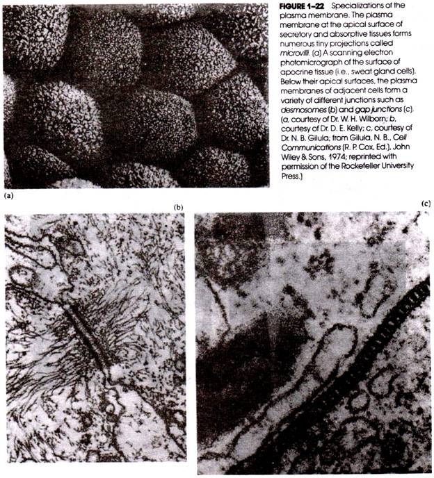
In some tissue cells, portions of the plasma membrane are modified to form a large number of fingerlike projections called microvilli (Fig. 1-22a) because of their resemblance to the much larger villi of the wall of the small intestine. The microvilli greatly increase the surface area of the cell and provide for the increased passage of materials across the plasma membrane.
When large numbers of cells are in close contact with one another (for example, in a tissue), it is not unusual to observe special junctions between neighboring plasma membranes. These take the form of tight junctions, desmosomes, and gap junctions.
The plasma membrane should not be thought of as a homogeneous structure that has the same chemical composition over its entire surface. Instead, the composition and organization vary in different regions of the membrane. Some areas of the plasma membrane of a liver cell, for example, face the plasma membranes of neighboring liver cells; other areas face the bile channels (bile canaliculi) into which substances are secreted by the liver cell.
Still other portions of the plasma membrane face the epithelial lining of capillaries from which substances are absorbed. Each of these regions of the plasma membrane is differently composed and differently organized and, in fact, is continually undergoing change and reorganization.
The Cytoskeleton and Micro trabecular Lattice Radiating through the cytosol of many cells are components of the cytoskeleton and micro trabecular lattice. The cytoskeleton consists of arrays of thin filaments, intermediate filaments, thick filaments and microtubules.
ADVERTISEMENTS:
These structures give shape and form to the cell and are also involved in cell movement. The cytoskeletal elements appear to be interconnected by a network of finer threadlike structures comprising what is called the micro trabecular lattice. This lattice also interconnects a number of membranous organelles and ribosomes.
The Endoplasmic Reticulum and Ribosomes:
ADVERTISEMENTS:
Within the cytoplasm of most animal cells is an extensive network of branching and anastomosing membrane- limited channels or cisternae collectively called the endoplasmic reticulum (abbreviated ER) (Fig. 1-23). The membranes of the endoplasmic reticulum divide the cytoplasm into two phases: the lumenal or intracisternal phase and the hyaloplasmic phase or cytosol.
The lumenal phase consists of the material enclosed within the cisternae of the endoplasmic reticulum, whereas the cytosol surrounds the ER membranes. In the typical cell, the surface area that is represented by ER membranes is more than ten times that of the plasma membrane.
In the cytosol are large numbers of small particles called ribosomes. These particles are distributed either along the hyaloplasmic surface of the endoplasmic reticulum (“attached” ribosomes) or free in the hyaloplasm (“free” ribosomes). There is some evidence that the free ribosomes are interconnected by fine filaments of the micro trabecular lattice.
Endoplasmic reticulum with associated ribosomes is called rough ER (RER), the membranes of the RER typically being sheet-like (as in Fig. 1-23e). Endoplasmic reticulum that is devoid of attached ribosomes is called smooth ER (SER), the membranes usually forming a network of branching and fusing tubes. Smooth ER appears to be the site for the synthesis of lipids destined to be included in lipoproteins. Certain portions of the endoplasmic reticulum may be continuous with the plasma membrane and the nuclear envelope.
ADVERTISEMENTS:
Mitochondria Within the cytoplasm are numerous vesicular organelles called mitochondria. Each mitochondrion is bordered by two membranes. The outer membrane is smooth, but the inner membrane displays numerous in-folding’s called cristae that greatly increase the surface area of the inner membrane. The space between neighboring cristae is called the mitochondrial matrix and often contains inclusions. Mitochondria are engaged in numerous metabolic functions in the cell, including the energy-producing phases of carbohydrate and fat metabolism (called respiration), ATP synthesis, and porphyrin synthesis.
The Golgi apparatus:
The Golgi apparatus (also called Golgi body or Golgi complex) consists of a set of smooth, flattened cisternae that are usually stacked together in parallel rows; in this state, the organelle is sometimes referred to as a dictyosome. The Golgi apparatus is frequently surrounded by vesicles of various sizes, some of which are discharged from the margins of the main body of the organelle (Fig. 1-25).
A variety of functions are ascribed to the Golgi apparatus, including the processing, packaging, and dispatchment of proteins destined for secretion, inclusion in the plasma membrane, and inclusion in lysosomes.
Lysosomes:
Many cells contain vesicular structures that are generally smaller than mitochondria and are called lysosomes (Fig. 1-26). Lysosomes are bounded at their surface by a single membrane and contain quantities of various hydrolytic enzymes capable of digesting protein, nucleic acid, polysaccharide, and other materials. Under normal conditions, the activity of these enzymes is confined to the interior of the organelles and is therefore isolated from the surrounding cytosol.
However, if the lysosomal membrane is ruptured, the released enzymes can quickly degrade the cell. Among their various roles, lysosomes take part in the intracellular digestion of particles that are ingested by the cell during endocytosis and the intracellular scavenging of worn and poorly functioning organelles.
Peroxisomes and Glyoxysomes:
ADVERTISEMENTS:
Many cells contain small numbers of peroxisomes and/or glyoxysomes. These small organelles, which are bounded by a single membrane, contain a number of enzymes whose functions are related to the metabolism of hydrogen peroxide and glyoxylic acid. The structure and functions of peroxisomes, glyoxysomes.
The Nucleus:
The nucleus is a relatively large structure frequently but not always located near the center of the cell. The contents of the nucleus are separated from the cytosol by two membranes that together form the nuclear envelope. At various positions, the outer membrane of the envelope fuses with the inner membrane to form pores (Fig. 1-27).
Nuclear pores provide continuity between the cytosol and the contents of the nucleus (sometimes referred to as the nucleoplasm). Often, the nuclear pores are plugged by a granular material (Fig. 1-28). The outer membrane of the nuclear envelope may have ribosomes attached to its hyaloplasmic side (Fig. 1-29) and may also form continuities with the membranes of the endoplasmic reticulum.
Because the latter may be continuous with the plasma membrane, the perinuclear space (i.e., the space between the inner and outer membranes of the nuclear envelope) corresponds to the lumenal phase of the cell and may be considered external to the cell (see Fig. 1-30).
The nuclear envelope and the pores that penetrate it are dramatically revealed in freeze-fracture preparations. Fig. 1-31 shows the cytosol-contacting half of the outer nuclear membrane fractured away, exposing the inner half of that membrane (i.e., face EF1 of Fig.’ 1-30). Also fractured away are pieces of the inner nuclear membrane (the half-membrane that faced the perinuclear space), leaving only the half-membrane that faced the nucleoplasm (i.e., PF2 of Fig. 1-30). The nuclear pores penetrate both membranes and in Figure 1-31 appear to be non-randomly distributed in the nuclear envelope.
The nucleus contains the genetic machinery of the cell (chromosomal DNA, chromosomal proteins, etc.), Using either the light microscope or the electron microscope, the nucleus often reveals one or more dense, granular structures called nucleoli (Fig. 1-32). Nucleoli are not bounded by a membrane and are formed in part from localized concentrations of ribosomal precursors.
Flagella and Cilia:
Many free-living cells (such as protozoa and other microorganisms) possess locomotor organelles that project from the cell surface; these are either flagella or cilia (Fig. 1-33). The tissue cells of multicellular animals may also contain cilia, but they are employed here to move a substrate across the cell surface (such as mucus in the respiratory tract or the egg cell during its passage through the oviduct) and not for cell locomotion.
The organelles are called cilia when they are short but present in large numbers and are called flagella when long but few in number. Each cilium or flagellum is covered by an extension of the plasma membrane. Internally, these organelles contain a specific array of microtubules that run from the basal body or kinetosome toward the tip of the structure. This array consists of two central microtubules and nine pairs of peripheral (outer) microtubules (Fig. 1-33b). Just below the basal body are rows of membrane particles referred to as the ciliary necklace.
Eukaryotic Cells: The Composite Plant Cell:
All the organelles described in the preceding section as regular constituents of animal cells are also found in similar form in many plant cells. Several other organelles are unique to plant tissues and include the carbohydrate-rich cell wall, plasmodesmata, chloroplasts, and large vacuoles. A composite plant cell is depicted in Figure 1-34a.
The Cell Wall:
The cell wall is a thick, polysaccha- ride-containing structure immediately surrounding the plasma membrane (Fig. 1-34). In multicellular plants, the plasma membranes of neighboring cells are separated by these walls, and adjacent plant cells have their walls fused together by a layer called the middle lamella. The cell wall serves both a protective and supportive function for the plant.
The degree to which the cell wall may be involved in the regulation of the exchange of materials between the plant cell and its surroundings is difficult to assess but is most likely restricted to macromolecules of considerable size. As in animal cells, most of the regulation of exchanges between the cytoplasm and the extracellular surroundings of plant cells is a function of the plasma membrane.
Plasmodesmata:
At intervals the plant cell wall may be interrupted by cytoplasmic bridges between one cell and its neighbor (Fig. 1-35). These bridges are called plasmodesmata and represent regions in which channel like extensions of the plasma membranes of neighboring cells merge. The channels serve in intercellular circulation of materials.
Chloroplasts:
The ability to use light as a source of energy for sugar synthesis from water and carbon dioxide is a special feature of certain plant cells. This process, termed photosynthesis, is carried out in organelles called chloroplasts (Fig. 1-36). These organelles are commonly ellipsoidal structures bounded by two membranes. The outer membrane is smooth and continuous, whereas the inner membrane gives rise to extensive parallel infoldings called lamellae supported in a homogeneous matrix called the stroma.
Most of the lamellae are arranged to form disk-shaped sacs (called thylakoids) that contain chlorophyll and may be stacked on top of one another, forming structures called grana. Lamellar membranes connecting the grana are called stroma lamellae (Fig. 1-36).
Vacuoles:
Although vacuoles are present in both animal and plant cells, they are particularly large and abundant in plant cells (Fig. 1-34), often occupying a major portion of the cell volume and forcing the remaining intracellular structures into a thin peripheral layer. These vacuoles are bounded by a single membrane and are formed by the coalescence of smaller vacuoles during the plant’s growth and development. Vacuoles serve to expand the plant cell without diluting its cytoplasm and also function as sites for the storage of water and cell products or metabolic intermediates.
Prokaryotic Cells: Bacteria:
The bacteria are structurally distinct from eukaryotic microorganisms such as protozoa and contain a number of unique cellular organelles. The typical bacterium is about the size of a mitochondrion of an animal or plant cell, and in view of this small size, it is to be expected that the organelles of bacteria would be correspondingly smaller. The composite structure of a bacterium is depicted in Figure 1-37.
The Bacterial Cell Wall and Capsule:
The bacterial cell is enclosed within a wall that differs chemically from the cell wall of plants in that it contains protein and lipid as well as polysaccharide. The presence of a particular peptidoglycan is the basis of the histo- chemical classification of bacteria, being high in the so-called “gram-positive” bacteria (such as Bacillus sub-tiles) and low in the “gram-negative” bacteria (such as Escherichia coli and the Simonsiella).
In some bacteria, the cell wall is surrounded by an additional structure called a capsule. The cell wall and capsule confer shape and form to the bacterium and also act as a physical barrier between the cell and its environment. This is important because osmotic forces usually result in a positive hydrostatic pressure inside the bacterium; in the absence of a cell wall and capsule, mechanically fragile bacteria would readily rupture.
Plasma Membrane Intrusions:
Infoldings of the plasma membrane of gram-positive bacteria give rise to structures called mesosomes (or chondrioids) (Fig. 1-38). Mesosomes are believed by many investigators to play a role in the division of the cell. Intrusions of the plasma membrane also form the photosynthetic organelles (chromatophores) of the photosynthetic bacteria.
Cytoplasmic Lamellae:
In some bacteria, there is a lamellar arrangement of membranes within the cytoplasm (Fig. 1-38b). However, there are no structures comparable to the endoplasmic reticulum of animal and plant cells. Bacteria contain large numbers of ribosomes, but most of these organelles are free in the bacterial cytosol; some ribosomes may be attached to the interior surface of the plasma membrane.
Although bacterial ribosomes, like the ribosomes of eukaryotic cells, are the sites of protein synthesis, considerable differences exist between the organelles of these two groups. Cytoplasmic lamellae are particularly abundant in the autotrophic bacteria, which support their growth through photosynthesis or similar processes.
Nucleoids:
In bacteria the nuclear material is not separated from the cytosol by membranes as it is in eukaryotic cells. However, the nuclear material is usually concentrated in a specific region of the cell, referred to as a nucleoid. During bacterial cell division, nucleoidal DNA becomes anchored to the plasma membrane and is distributed to the daughter cells without formation of observable chromosomes. Nucleoli are not present in the nucleoid.
Bacterial Flagella:
Many bacteria contain one or more flagella employed for cellular locomotion. These organelles arise from the cytoplasm and penetrate the plasma membrane and cell wall (and capsule, if present). Bacterial flagella are smaller than the flagella of animal and plant cells and are simpler in organization, containing a single filament of globular proteins (called flagellin) surrounded by a sheath. Some bacteria are multi-flagellated.
Flagella like axial filaments are characteristic of some spirally shaped bacteria; these structures do not project away from the cell but are wrapped around the cell surface. Some bacteria contain non-flagellar appendages called fimbriae that arise from the cytosol and project a short distance above the cell surface (Fig. 1-37a). These structures are believed to play a role in the attachment of the bacterium to a surface or to other bacteria.
Some bacteria, such as E. coli, Proteus mirabilis, and B. subtilis, occur as separate, individual cells. However, in a number of groups, the daughter cells remain attached to each other following division, so that chains (e.g., streptococci) or filaments are formed. An example of a filamentous genus is Simonsiella, which colonizes the mucosal epithelial surface of the mouth
The individual cells of some filamentous bacteria reveal a dorsal-ventral differentiation, that is, the ventral surface (which in Simonsiella attaches to and glides along the epithelium) is structured differently than the dorsal surface (which faces away from the epithelium).
This is apparent not only in the scanning electron micrographs of whole filaments but also in transmission electron photomicrographs of thin sections through the filaments (Fig. 1-39). Individual cells of a filament exhibit features common to single-cell (i.e., non-filamentous) forms like E. coli and B. subtilis.
Prokaryotic Cells: Cyanobacteria:
The cyanobacteria (also called blue-green algae) are photosynthetic prokaryotes and occur as individual cells, as small clusters or colonies of cells, or as long, filamentous chains (Figs. 1-40 and 1-41). Cyanobacteria lack flagella but are able to glide over a gelatinous layer secreted through the cell surface.
The photosynthetic apparatus consists of lamellae called thylakoids; the thylakoids are lined with pigment granules referred to as phycobilosomes. Cyanobacteria also contain several kinds of membrane- bound inclusions such as gas vacuoles (which provide buoyancy in aquatic cyanobacteria) and carboxysomes (which contain enzymes involved in carbon dioxide fixation).
Prokaryotic Cells: Mycoplasmas:
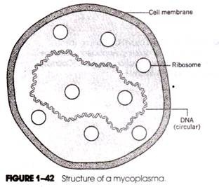
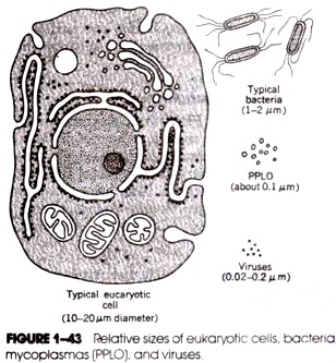
The mycoplasmas or PPLO (i.e., pleuropneumonia- like organisms), which cause a number of diseases in humans and other animals, are the smallest (i.e., about 0.1 µm in diameter) and simplest of all cells capable of autonomous growth. They are smaller even than some of the larger viruses.
A mycoplasma is bounded at its surface by a membrane composed of proteins and lipid, but there is no cell wall. Internally the cell’s composition is more or less diffuse. The only microscopically discernible features within the cell are its genetic complement, which consists of a double-helical strand of circular DNA, and a number of ribosomes (Fig. 1-42).
Mycoplasmas appear to contain the bare minimum of structural organization required for a viable, free-living cell and may represent a form intermediate between viruses and bacteria. The relative sizes of typical eukaryotic cells, bacteria, PPLOs, and viruses are compared in Figure 1-43, which dramatizes the differences that exist.


