ADVERTISEMENTS:
Lecture notes on Cells. After reading these notes you will learn about: 1. Introduction to Cell 2. Origin and Evolution of Cell 3. Shape 4. Size 5. Number 6. Structure.
Contents:
- Notes on Introduction to Cell
- Notes on the Origin and Evolution of Cell
- Notes on the Shape of Cell
- Notes on the Size of Cells
- Notes on the Number of Cells
- Notes on the Structure of Cells
Note # 1. Introduction to Cells:
ADVERTISEMENTS:
Cells were first seen and described by Robert Hooke, the English scientist in 1665. The cells he saw were the boxlike cavities he found in cork. In the next hundred years or so, many other scientists made observations on the cell and some of its components. Among these were Lamarck (1809), Detrochet (1824) and Turpin (1826). Brown (1831) noticed that the nucleus was a regular constituent in all plant cells.
In 1838, Schleiden, a German botanist studied the plant cells and emphasised that “cells are organisms and entire animals and plants are aggregations of these organisms arranged according to definite laws”. In 1839, Schwann, a German zoologist stated, “we have seen that all organisms are composed of essentially like parts namely of cells”.
The deductions of two microscopists (Schleiden and Schwann) formed the basis of what came to be known as the cell theory.
The cell theory holds that “all living matter, from the simplest of unicellular organisms to very complex higher plants and animals, is composed of cells and that each cell can act independently but functions as an integral part of the complete organism.”
ADVERTISEMENTS:
In 1841, A. Kollikar suggested that sperms and ova are histological elements originating in the organisms. In 1858, Virchow reported, “where a cell exists there must have been a pre-existing cell, just as the animal arises only from an animal and the plant only from a plant.”
According to cell theory and organismal theory, the cell may be considered as a smallest mass of protoplasm having permeable plasma membrane and nucleus which is capable of energy transformation, biosynthesis and self-reproduction.
But certain primitive units of life such as viruses do not fulfil the fundamental requirements of the cell as suggested by the cell theory, therefore, in the present state of knowledge the cell has been defined as the smallest but complete expression of the fundamental organisation and functions of all living organisms, delimited by a permeable plasma membrane and capable of reproducing in a medium free of other living systems unlike the viruses.
The body of all living organisms except the viruses are composed of one or many cells.
The organisms may have two types of cells, viz., prokaryotic cells and eukaryotic cells:
1. Prokaryotic Cells:
The prokaryotic (Gr., pro = before, primitive; karyon=nucleus) cells are the most primitive cells from morphological point of view. These cells have primitive nuclei without nuclear membrane and the nuclear contents as proteins, nucleic acids (DNA and RNA), etc., and have direct contact with the cytoplasm.
The prokaryotic cells also lack in well defined cytoplasmic organelles. The prokaryotic cells occur in viruses, bacteria and blue green algae.
ADVERTISEMENTS:
2. Eukaryotic Cells:
The eukaryotic (Gr., eu = good or well; karyotic = nucleus) cells are the true cells which occur in the plants (from algae to angiosperms) and the animals (from Protozoa to mammals).
Though the eukaryotic cells have different shape, size, and physiology but all the cells typically composed of plasma membrane, cytoplasm and its organelles, viz., mitochondria, endoplasmic reticulum, ribosomes, Golgi apparatus, etc., and a true nucleus. Here the nuclear contents such as DNA, RNA and nucleoproteins remain concentrated and separated from the cytoplasm by the thin, perforated nuclear membranes.
Before going into the details of cell and its various components, it will be advisable to consider the general features of different types of cells which are as follows:
ADVERTISEMENTS:
Note # 2. Origin and Evolution of Cells:
ADVERTISEMENTS:
Chemical Evolution of Cell (Evolution of Organic Molecules/Prebiotic synthesis of Bio- Molecules):
The conditions on the prebiotic earth were favourable to a sort of continuing chemical evolution that gave rise to a special class of organic molecules. Organic molecules must have evolved before the origin of life from primordial atmosphere which was anoxic and chemically reducing.
The atmospheric composition would include CH4, NH3, H2S, H2, CO, H2O and N2. Miller and Urey (1953) devised an experiment simulating pre-biological conditions to synthesize important biomolecules like amino acids (constituents of proteins) and carbohydrates from a mixture of gases (methane, ammonia, hydrogen and water) (Fig. 2.1).
Later experiments with varying chemical compositions resulted in the synthesis of many molecules that are central to life. Amino acids (e.g., alanine, glycine, leucine, etc.) produced are important constituents of protein (Fig. 2.2).
ADVERTISEMENTS:
Volatile fatty acids like formate, acetate, propionate, butyrate are produced. Porphyrins, basic molecular structure of respiratory enzymes, chlorophylls, hemoglobin’s, have also been synthesized. Subsequent to abiotic synthesis of organic matter, seas were converted into what Haldane (1954) called as hot dilute prebiotic soup or primordial soup.
This was followed by the development of a simplest physicochemical system which has the characteristics of self-organisation, self-regulation, self- perpetuation, energy transfer mechanism and isothermal open system.
Protobiont:
ADVERTISEMENTS:
The first primitive living cell must have been far from the modern cell in structural organisation. A number of models have been suggested, but an acceptable one should fit in the framework of the above set of characteristics.
Oparin’s Coacervate Model (1938):
Coacervates are submicroscopic structures of colloidal solution of proteins and carbohydrates which show osmotic activity and could give rise to larger aggregates. They sustain more rapid reaction bringing about oxidation, reduction and polymerization. These molecular aggregates could have been easily surrounded by a boundary layer, akin to membrane (Fig. 2.3A). These coacervates have interesting properties.
In one experiment, enzyme phosphorylase, which was trapped inside coacervates, releases the phosphate form glucose-1-phosphate and polymerizes the glucose units into starch deriving energy from phosphate bond (Fig. 2.3B).
The coacervates, therefore, grow and eventually divide into two coacervates. In another experiment, the red-ox enzyme NADH dehydrogenase was trapped inside the coacervates, catalyses the reduction of methyl red coupled to oxidation of NADH, analogous to electron transfer system.
ADVERTISEMENTS:
Thus coacervates have the interesting properties:
(a) Spatial organisation of simple bio-energetic process;
(b) Maintain a certain size and, therefore, divide if their volume increases.
(c) A spatial structure in which replicators could reside.
ADVERTISEMENTS:
Fox’s Proteinoid Microspheres Model (1969):
Proteinoid microspheres are spherical structures of varying size made up of protein like polymers which were able to carry out a variety of enzymatic reactions, especially oxidation, decarboxylation, amination and hydrolysis. These could grow in size by accumulating more organic materials and give rise to small protuberances like yeast.
The most important structure is the double membranous semipermeable envelope around the proteinoids (Fig. 2.4).
Nucleic Acid Hypothesis:
Though coacervates and proteinoids had unique characteristics that brought them close to living cell, but they do not have the potential capacity to code for proteins, to undergo self-replication and to evolve through natural selection.
As the nucleic acid can undergo mutations, only the cell possessing them can have the tendency to evolve. Five organic bases of nucleic acid, e.g., adenine, guanine, cytosine and thymine (in DNA) or uracil (in RNA) which constitute the letters of genetic code, have been synthesized in Miller type experiments.
Simulation experiments using appropriate bases, pentose sugars and phosphoric esters have demonstrated that nucleotides can be synthesized (Fig. 2.5). Orgel et al. (1980) were able to demonstrate polymerization of nucleotides on nucleic acid templates in enzyme-free system.
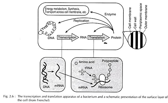
Nucleic acid originated abiotically on the primitive earth was the prime requirement for evolving a genetic apparatus and suitable metabolic machinery (Fig. 2.6).
Note # 3. Shape of Cells:
The plant and animal cells exhibit various forms and shapes. Typically the animal cell is spherical in shape but the shape of the cell may be irregular, triangular, tubular, cuboidal, polygonal, cylindrical, oval, rounded or elongated in different animals and plants. The shape of the cells may vary from animal to animal and from organ to organ. Even the cells of the same organ may display variations in the shape.
Generally the shape of the cell remains correlated with its functions. For example, the epithelial cells have flat shape and the muscles are elongated. Moreover, external or internal environment may also cause shape variations in the cell due to internal or mechanical stress or pressure and surface tension, etc.
Note # 4. Size of Cells:
Mostly the eukaryotic cells are microscopic in size but definitely they are larger in size than the bacterial cells. The size of cells varies from 1 µ. to 1,75,000 µ (175 mm). The ostrich egg cell is usually considered as largest cell (175 mm in diameter) but certain longest nerve cells have been found to have the length of 92 cm to 1.06 metre.
Note # 5. Number of Cells:
The unicellular or acellular animals (protozoans) consist of single cell. Most of the animals and plants have many cells in the body and are known as multicellular animals or plants. The number of cells in the multicellular organisms usually remain correlated with size of the organism and, therefore, small-sized organisms have less number of cells in comparison to large-sized organisms.
Note # 6. Structure of Cells:
Recently with the introduction of electron microscope and the use of new biological techniques for an analysis of the parts of a cell, the finer detailed structures, properties and functions of the cell have come to light. Although the animal cells differ greatly in shape, size, structure and function, yet all types of cells have some common features.
A generalized animal cell is a translucent speck of protoplasm containing an inner nucleus and an outer cytoplasm enclosed within an external plasma membrane.
The cell consists of the following parts:
i. Plasma membrane
ii. Cytoplasm
iii. Nucleus
i. Plasma Membrane:
The plasma membrane is a living, ultra-thin, elastic, porous and semi-permeable membranous covering of the cell. The plasma membrane is a trilaminar (three-layered) membrane of lipoprotein. The trilaminar nature of plasma membrane was proposed by Danielli and Davson in 1935.
In 1938, Harvey and Danielli produced a hypothetical model of plasma membrane which has shown a bimolecular lipid structure sandwiched by two (outer and inner) layers of protein molecules. The electron microscopic studies have confirmed this protein-lipid-protein arrangement in plasma membrane.
The plasma membrane of most cells varies from 100 to 215A0 in thickness. The plasma membrane of most cells composed of mainly carbohydrates, lipids and proteins.
Molecular Structure of the Plasma Membrane:
The plasma membrane is composed of two layers of protein molecules and two layers of lipid molecules. The lipid molecules occur in chains. In plasma membrane, two molecular chains of lipids remain parallel to each other and form a bimolecular or double-layered structure.
Both lipid layers remain linked with each other by the inner ends of lipid molecules which are non-polar and hydrophobic (Gr., hydr= water; phobe=hate) in nature. Both the layers of lipids are able to be held together due to Vanderwaal’s forces at these non-polar ends.
The lipid layers are enclosed by an outer and inner layer of proteins. The lipid molecules remain linked with the molecules of protein layers by their outer, polar and hydrophilic (Gr., hydr – water; phil-loving) ends. In the hydrogen bonds, ionic linkages or electrostatic forces bind the molecules of lipids and proteins together.
The carbohydrate molecules occur in the association of protein molecules and provide stability to lipoprotein complex. The protein layers provide elasticity and mechanical resistance to the plasma membrane.
Intercellular space:
In the tissues of multicellular animals, the plasma membrane of two adjacent cells usually remains separated by a space of 110 to 150 A0 wide. This intercellular space is uniform and contains a material of low electron density which can be considered as a cementing substance. The exact chemical nature of this substance is still unknown.
ii. Cytoplasm:
The plasma membrane is followed by the cytoplasm which is distinguished into following structures:
A. Cytoplasmic Matrix:
The space between plasma membrane and the nucleus is filled by an amorphous, translucent, homogeneous liquid known as cytoplasmic matrix or hyaloplasm. The cytoplasmic matrix consists of various inorganic molecules such as water, salts of Na, K and other metals and various organic compounds, viz., carbohydrates, lipids, proteins, nucleoproteins, nucleic acids (RNA and DNA) and a variety of enzymes.
The peripheral layer of cytoplasmic matrix is relatively non-granular, viscous, clear and rigid and is known as the plasmagel, or cortex or cortical layer or ectoplasm (Gr., ecto = outside; plasm = form). The inner portion of the cytoplasmic matrix is granular, less viscous and is known as endoplasm (Gr., endo-inner; plasm=form).
B. Cytoplasmic Structures:
In the cytoplasmic matrix, certain non-living and living structures are suspended. The non-living structures are called paraplasm, deutoplasm or inclusions, while the living structures are membrane bounded and are called organoids or organelles.
Both kinds of cytoplasmic structures can be studied under the following headings:
(a) Cytoplasmic Inclusions:
The cytoplasmic matrix contains many refractile granules of various sizes and shapes known as trophoplasm. These granules in animal cells are known as cytoplasmic inclusions or deutoplasm (Gr., deuteros = second) or paraplasm. The cytoplasmic inclusions include oil drops, yolk granules, pigments, secretory granules and glycogen granules.
(b) Cytoplasmic Organoids or Organelles:
Besides the cellular inclusions and plastids, the cytoplasmic matrix contains many large-sized living structures known as cytoplasmic organelles or organoids which perform various important biosynthetic and metabolic activities such as respiration, storage, synthesis, transportation, support and reproduction.
These cytoplasmic organelles are the endoplasmic reticulum, ribosomes, Golgi apparatus, lysosomes, mitochondria, plastids, centrioles, cilia, etc.
iii. Nucleus:
The nucleus (Fig. 4.7) is a centrally located and spherical cellular component. It is most significant component of the cell which controls various metabolic activities of the cell and contains the genetic material DNA. The nucleus is found in all the eukaryotic cells of the animals and plants. The location of the nucleus in a cell usually is the characteristic of the cell type and it is often variable.
Usually the nucleus is located in the centre of the cell. Usually the cells contain single nucleus but the number of nucleus may vary from cell to cell. The shape of the nucleus usually remains related with the shape of the cell but certain nuclei are almost irregular in shape.
The size of the nucleus of various cells is also variable. The nucleus is composed of following structures, viz., nuclear membrane, nucleoplasm, chromatin fibres and nucleolus.
(i) Nuclear Membrane:
The nuclear membrane or karyotheca forms an envelope-like structure around the nuclear contents and is commonly known as the nuclear envelope. The nuclear envelope separates the nucleoplasm from the cytoplasm. The nuclear envelope also contains many pore-like perforations through which to and fro movement of chemical substances takes place.
The outer nuclear membrane of nuclear envelope is continuous with the membranes of endoplasmic reticulum, Golgi apparatus and mitochondria, etc.
The electron microscopic studies of the nuclear envelope have shown that it is composed of two unit membranes, viz., an outer membrane and an inner membrane. Each membrane is about 75 to 90A° thick and lipoproteinous in nature. The outer and inner nuclear membranes are separated by a space of 100 to 150A° (Robertis et al., 1970) 100 to 300A0 (Cohn, 1970), or 400 to 700A° (Burke, 1970).
(ii) Nucleoplasm and Chromatin Fibres:
The space between the nuclear envelope and nucleolus is filled by a transparent, semi-solid, granular and slightly acidophilic ground substance known as nucleoplasm or karyolymph. The nucleoplasm contains dissolved phosphorus, ribose sugars, proteins, nucleotides and nucleic acids. The nucleoplasm also contains many thread-like, coiled, much elongated structures which readily take basic stains such as basic fuchsin.
These thread-like structures are known as chromatin fibres. Such chromatin fibres are observed only in the interphase nucleus. During cell division (mitosis and meiosis), chromatin fibres become thick ribbon-like structures which are known as the chromosomes. The chromatin granules or chromosomes fundamentally consist of large molecules of deoxyribonucleic acid (DNA) and many proteins.
(iii) Nucleolus:
The nucleoplasm contains a conspicuous darkly stained spherical body known as the nucleolus. The position of nucleolus in the nucleus is accentric. Chemically nucleolus is composed of large amount of ribosomal proteins and ribosomal RNA. The Nucleus, nucleolus plays a very important role in the process of protein synthesis and cell division.
The main function of nucleolus is the biogenesis of ribosomes. It stores all the proteins of ribosomes and rRNA which is transcribed by rDNA of nucleolar organizer.


