ADVERTISEMENTS:
The following points highlight the nine components of a typical plant cell.
The nine components of a typical plant cell are: (1) Protoplasm (2) Protoplast (3) Cytoplasm (4) Nucleus (5) Plastids (6) Chondriosomes or Mitochondria (7) Centrosomes (8) Golgi Bodies and (9) Lysosomes.
Component # 1. Protoplasm:
Protoplasm is the essence of life. It has been very fitly described by Huxley in the nineteenth century as ‘the physical basis of life’. All the characteristics of the living things, such as metabolism, irritability, growth and reproduction are nothing but the external manifestations of the internal changes of this mysterious fluid protoplasm.
ADVERTISEMENTS:
Every plant or animal has its own type of protoplasm, but fundamentally it is same in all organisms. Protoplasm was first recognised by Hugo von Mohl in the middle of nineteenth century. Even today in spite of all the advances and attainments in science our knowledge about this mysterious fluid is rather vague and superficial. In recent years special techniques have been employed for unraveling the mystery.
The investigators have taken the help of dark-field illumination, ultra-violet photography, polarised light, high-speed centrifuge, micromanipulator and the electron microscope for the purpose. Protoplasm is composed of water and various substances like proteins, fats, carbohydrates and inorganic salts, but it is, by no means, a mere mixture of all these materials.
It is continually undergoing constructive and destructive changes, a dual process of waste and repair. Thus it is a complex dynamic system of substances. Sharp has described it as ‘a system in dynamic equilibrium’. Thomson has called it ‘a marvellous form of matter in motion’.
Physical Properties:
It appears under the microscope as a clear, optically homogeneous fluid saturated in a high percentage of water. The specific gravity of protoplasm is slightly above that of water. A large number of minute granules and globules of varying size and shape remain-distributed in water. Thus it looks granular under the microscope. Due to occurrence of many vacuoles the protoplasm of plant cells has often foamy or alveolar appearance.
ADVERTISEMENTS:
It is a viscous and elastic fluid. The viscosity has been found to be like that of glycerine or light machine oil. The elastic nature of protoplasm has been determined with the help of a micromanipulator. It has been found that protoplasm can stand stretching and can be drawn out like a thread, which would return to its normal position when released.
Protoplasm has the power of responding to external stimuli like heat, electric shocks, applications of chemicals, what is referred to as irritability, an inherent character of protoplasm.
Protoplasm exhibits streaming movements of different types which are commonly noticed in amoebas, slime moulds, and particularly in the plant cells with large vacuoles.
Rotatory movements in the cells of leaves of aquatic plants like Vallisneria and Elodea of family Hydrocharitaceae and circulatory movements in the staminal hairs of Rhoeo disiblor of family Commelinaceae are the glaring examples.
In rotation protoplasm moves in a definite direction along the cell wall (Fig. 486); whereas in circulation the course of movement is irregular (Fig. 487). Besides the movements stated above, commonly called cyclosis, the streaming movements of slime moulds have been observed with the help of a motion picture camera and have been found to be due to rhythmic contraction and relaxation of protoplasm. This type is called amoeboid movement. Rather rapid movement caused by highly sensitive whip-like protoplasmic extensions called cilia, is common in many lower organisms.
Attempts to ascertain the nature of protoplasm had been going on since a long time and early workers proposed a number of theories, viz. (i) reticular theory, stating that it is something like a fine network, (ii) fibrillar theory, claiming that it consists of fine fibrils, (iii) granular theory, demanding that it is composed of innumerable minute granules, and (iv) alveolar or foam theory, stating that it is a collection of minute droplets of a liquid distributed in another liquid as an emulsion.
The most important physical character of protoplasm, as known today, is that it is a complex colloidal system. The properties like viscosity, surface tension, alterations of physical state due to variations in external conditions and internal reactions are clear indications of a colloidal system.
Though the type of colloidal structure is not clearly known, it is probably an emulsion with proteins, lipides and other substances suspended in the water medium.
Chemical Composition:
The chemical composition of protoplasm, itself a dynamic system, cannot be determined without destroying it during the process of analysis. A general idea about its composition may be had after subjecting it to normal procedure of analysis.
ADVERTISEMENTS:
It is found that protoplasm is a very complex substance, which in normal active state consists of more than 75% of water and less than 25% of other materials. Of the dry matters nearly 90% are organic and the rest inorganic. The reactions are either slightly alkaline or neutral but seldom acidic.
As already stated water forms the most important constituent of protoplasm. In fact, life is inconceivable without water. In aquatic plants the proportion of water may go up to 95%. Low water content is noticed in the inactive and dormant organs like spores and seeds when it is something like 10 to 15%.
The organic matters present in protoplasm are proteins, fats and carbohydrates, of which the proteins are the most important ones. They are very complex matters, composed of carbon, hydrogen, oxygen, nitrogen, often sulphur and sometimes phosphorus.
ADVERTISEMENTS:
The nucleoproteins occurring in the nucleus in particular are of special significance. The fatty substances entering into the composition of protoplasm are the true fats, composed of carbon, hydrogen and oxygen, and more commonly the phospholipids which are complex derivatives of fats containing nitrogen and phosphorus.
The carbohydrates which directly enter into actual constitution of protoplasm are the pentose sugars. But other sugars, the hexoses, and their condensation products, the insoluble polysaccharides, have also importance in the activities of protoplasm.
They usually combine with proteins and lipides. The inorganic salts are of different varieties; in plants they occur mainly as phosphates, sulphates, chlorides and carbonates of magnesium, potassium, sodium, calcium and iron. Many other elements may be present in minute traces.
It hardly needs mentioning that the account of the chemical composition given above does not reveal any information as regards the organization of protoplasm. It simply gives a .general idea about the kinds and proportions of compounds present.
Component # 2. Protoplast:
Protoplast is the organized mass of protoplasm of each cell. It is the smallest part of die essential living substance. It possesses a highly organized more or less rounded body known as nucleus, what figuratively means the centre of activities.
ADVERTISEMENTS:
The remaining portion goes by ‘he name cytoplasm, which may be looked upon as the ground substance of protoplasm (Fig. 488). The term cytoplasm was used by Kolliker in 1862 to designate the living material surrounding the nucleus.
Component # 3. Cytoplasm:
Cytoplasm is hyaline and jelly-like in nature and looks granular under the light microscope, due to suspension of many droplets and granules in the watery matrix. It is very complex, both physically and chemically.
ADVERTISEMENTS:
Various organic and inorganic substances remain distributed in the watery background either in true solution or in colloidal condition. The hyaline ground substance is also called hyaloplasm.
The outermost layer of cytoplasm is, however, non-granular and firmer in consistency. It goes, by the name plasma membrane or ectoplasm. The term ectoplasm is not very happy, as it has been used in a different sense in case of Amoeba.
Plasma membrane is an extremely delicate layer of ultra-microscopic thinness, comparable to the surface film of a drop of water. It is semipermeable in nature.
Though invisible under light microscope it can be seen as a single layered smooth and continuous membrane under electron microscope. This layer plays a great role in the metabolic activities. It regulates the entry and exit of fluid to and from the cells.
The plasma membrane practically covers the protoplast, even including the plasmodesmata connections. It goes without saying that absorption of dissolved mineral matters from the soil would have been impossible, if the plasma membrane were perfectly semipermeable. But it allows certain substances to pass through it under one set of circumstances while under another set their entry is restricted. It appears that plasma membrane is formed by proteins and fatty substances particularly lipides.
Electron microscopy revealed that plasma membrane is three-layered, made of two dense layers of protein, each roughly 20Å (2 nm) wide, with a lighter layer of lipid, 35Å (35 nm) thick, sandwiched between them (Robertson—1959; unit membrane theory). The remaining granular portion of cytoplasm is called endoplasm.
ADVERTISEMENTS:
The cytoplasmic portion of the protoplast is referred to as cytosome. A good number of technical expressions like poplioplasm, have been suggested from time to time to designate the different parts of cytoplasm.
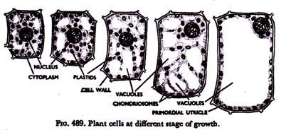
Liquid-filled cavities, called vacuoles, are of universal occurrence in the cytoplasm of plant cells. In the meristematic cells the vacuoles are small, they may be even at the limits of microscopic visibility. With gradual maturation of the cells the vacuoles become increasingly larger and prominent as cell enlargement is associated with a massive uptake of water. Frequently the major portion of an adult cell may be occupied by a large central vacuole (Fig. 489).
The protoplast in that case is pushed towards the cell wall forming a lining layer, what is known as primordial utricle. The origin of vacuoles has been a subject of diverse opinions. It is, however, believed that vacuoles arise by hydration and collection of some cytoplasmic colloids. It is, at any rate, true that vacuoles are already present in all cells, even meristematic cells.
So the old idea that young cells have no vacuoles and they become vacuolated’ with the growth of the cell has been found to be incorrect. Some vacuoles, called contractile vacuoles, commonly present in lower animals and also plants exhibit a peculiar phenomenon of rhythmic filling and discharge of contents.
They appear to be excretory passages. The layer of cytoplasm forming the vacuole membrane is known as tonoplasm. It is also a surface film of extreme fineness. Some people consider that tonoplasm is similar to plasma membrane in nature and composition both being relatively rich in lipoidal matter; whereas others are of view that the two differ both in structure and function. But it is admitted at any rate that tonoplasm is a distinct entity and that it is different from inner cytoplasm.
ADVERTISEMENTS:
Finer details of the cell structure are revealed by the electron microscope. It appears that optically clear hyaloplasm is made of two components—viz., the matrix or ground substance and a membrane-enclosed vesicular system known as endoplasmic reticulum (Porter—1945).
They usually occur in three forms—cistemae, vesicles and tubules. The cisternae are elongated, flattened and arranged in parallel lines, the vesicles, are circular and the tubules are of various shapes. This system appearing as slender vesicles is continuous from the nuclear envelop throughout the cytoplasm to the surface membrane, even through the wall in meristematic cells, whereas in adult cells it appears as discontinuous vesicles.
Numerous small particles, ribosomes, occur on the outer surfaces of endoplasmic reticulum or freely in the matrix. They are often 150Å (15 nm) in diameter and rich in ribonucleoprotein. Ribonucleic acid—RNA and basic proteins play significant role in protein synthesis. The ribosomes with fragments of endoplasmic reticulum form which are called microsomes. Fig. 489A represents the diagram of a meristematic cell under electron microscope.
The watery content of the vacuole is called cell-sap. It is mainly water with various non-protoplasmic substances either in solution or in a colloidal condition. Inorganic salts, plant food matters like carbohydrates, amides; waste products like tannins, organic acids and mineral crystals are of common occurrence in the cell-sap of the vacuoles.
Vacuoles are quite helpful in maintaining the turgor of the cells, which is necessary for the support of the herbaceous plants. Further, they serve as storehouse of water, food matters and various by-products of metabolic changes of protoplasm. The cell-sap is normally slightly alkaline or neutral (pH 7), but with gradual enlargement during differentiation of the cell it becomes acidic (pH 5 or even lower). Water-soluble pigments, called anthocyanins, are often present in the cell-sap of the vacuoles.
Anthocyanin usually imparts fed to blue colorations to the plant parts particularly to flowers and fruits. These are glycosidic compounds formed by chemical reaction between a sugar—generally a glucose and one or more non-sugars.
With changes in the acidity and alkalinity the anthocyanins change their colour. Cyanin, the pigment of dahlia, rose, etc., is red when sap is acidic (pH 3), violet in slightly alkaline sap (pH 8, 5) and blue in highly alkaline sap (pH 11). Anthocyanins always occur in nature in form of chlorides. Besides attraction of insects for pollination, it is also supposed to serve as a screen against intense sunlight, particularly ultra-violet rays.
Component # 4. Nucleus:
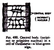
The nucleus is much denser than cytoplasm and is usually round or elliptical in shape. It is regarded as the ‘brain’ of the cell, as it is the controller or regulator of all the activities of the protoplast. The nucleus is of universal occurrence in all plant cells. In lower plants like blue- green algae (Fig. 490) and bacteria (procaryotes) a well-formed nucleus may be absent; even in those cases corresponding nuclear materials must be present.
In the sieve-tubes of the complex tissue phloem the nucleus disintegrates with maturity. Nucleus never arises de novo or afresh, but always originates from a pre-existing one. There are different methods of formation of a nucleus from a pre-existing one. Some methods are quite complicated, which will be discussed in succeeding chapters.
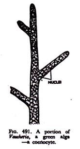
Usually one nucleus occurs in a cell. In that case the cell is called uninucleate. Presence of more than one nucleus in a cell, what is called multinucleate condition, cannot be altogether ruled out. Non-septate multinucleate bodies, technically called coenocytes, occur in the order Siphonales of grass-green algae Chlorophyceae (Fig. 491) and some fungi of the group Phycomycetes.
Even in some higher plants more than one nucleus may arise due to disease and other reasons. Formation of many nuclei in a cell may be due to (i) repeated division of the nucleus without corresponding cytoplasmic division; and (ii) absorption of walls between the cells.
The nucleus is usually round or elliptical in shape (Fig. 488). In highly elongated cells the nuclei are also correspondingly elongate. Peculiar shapes like crescent, dumb-bell, are found on rare occasions.
It occupies comparatively a small space of the protoplast. In a meristematic cell it is quite prominent (Fig. 489). As the gradual enlargement of the cell is not accompanied by corresponding growth of the nucleus, the latter does not look so conspicuous in an adult cell.
As regards size, it shows wide variations. In lower plants like Mucor the diameter of a nucleus is hardly more than 1 micron (micron, written as µ is equivalent to one thousandth part of an mm.). The egg cell of Dioon, a gymnosperm, may have a nucleus as large as 600µm in diameter. But consideration of a large number of cells reveals that average diameter of a nucleus varies from 10 to 15µm.
The variations in the size and shape of the nucleus are not confined to different types of plants. Curiously enough considerable variations may be noticed in the different tissues of the same plant.
In meristematic cells with small vacuoles the nucleus is located more or less at the centre of the cell (Fig. 484B). In adult cells with a number of vacuoles the nucleus occupies more or less the same position; but when the vacuoles coalesce to form a large central vacuole, the nucleus with cytoplasm, etc. is pushed towards the cell wall (Fig. 489).
Very peculiar position of nucleus is found in some algae like Spirogyra, where it is kept suspended in the midst of a large central vacuole by fine cytoplasmic threads (Fig. 493A).
Whatever may be its position, the nucleus always remains associated with cytoplasm, never touching either the vacuole or the cell wall. The nucleo-cytoplasmic ratio usually ranges between 1: 7 to 1: 10. Hartwig (1960) suggested nucleo-cyto- plasmic index—
NP = Vn / Vc – Vn
where NP is nucleo-cytoplasmic index, Vn—nucleus volume, Vc—cytoplasm volume.
The nucleus is much more viscous than cytoplasm. In fact, with the help of micromanipulator it may be pushed from one side to other in some cells without any visible injury. It has been observed that the nucleus may be stretched many times its normal size, which would come back to its original size on removal of the micromanipulator needles. In living condition it appears homogeneous.
But it may be fixed and suitably stained for observation of detailed structure.
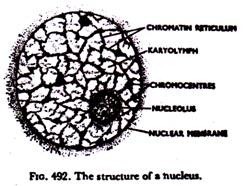
A metabolic nucleus after suitable staining presents the following structure (Fig. 492):
The main body of the nucleus is surrounded by a fine delicate membrane called nuclear membrane. It demarcates the main body of the nucleus from surrounding cytoplasm and has been found by electron microscopy to be double-layered with pores and projections into the cytoplasm continuous with the endoplasmic reticulum (Fig. 489A).
Each layer is usually 80-100A (8-10 nm) wide and the space between two layers —perinuclear space, is 100-300A (10-30 nm). Chemically it is made of proteins and lipids. The membrane disappears at certain stage of nuclear division only to reappear at the end of the process, it is regarded as a structure formed by the karyolymph or jelly-like fluid filling up the main body of the nucleus. This fluid is also known as nuclear sap or nucleoplasm.
A definite number of highly crooked threads, called chromonemata, remain suspended in the karyolymph. These threads anastomose and form a network which is called chromatin reticulum. The threads have strong affinity for taking up stains. The chromonemata are the most important bodies, because they represent the chromosomes at this stage.
The most important constituent of the chromosome seems to be the nucleic acid—DNA, deoxyribose nucleic acid, which is the actual chromatic material causing staining of the chromosomes. Differential staining at different regions of chromosome means difference in its chemical nature. The nucleus contains the basic genetic information of the cell in form of long strands of this chemical material DNA, the molecules of which are believed to be in the form of double strands of intertwined helices in the chromosomes.
At a certain stage of nuclear division another stainable substance, matrix, is associated with the chromonemata giving the chromosomes compact forms (Fig. 521). At that stage chromosomes lose water and acquire a fibrous coat of nucleic acid (stainable matrix).
Densely stainable lumps are often found in the midst of chromonemata in some nuclei. These are called chromocentres, the regions where chromonemata are more closely packed and have more matrix.
Besides these, there may be one or two highly stainable bodies inside the nucleus. This internuclear body is called nucleolus or plasmosome. Like nuclear membrane it also disappears at a certain stage of nuclear division and reappears at the close of the process. The number of nucleoli is variable. It may so happen that a number of them fuse at the end of cell division to form one nucleolus. Its function is not definitely known.
According to one school, the nucleolus is nothing but accumulated materials, somehow related to metabolic activities of the nucleus. It is formed at a particular locus on one of the chromosomes and is mainly composed of ribonucleic acid—RNA.
Chemically, the nucleus practically resembles protoplasm, excepting that it contains nucleoproteins made of nucleic acid and certain basic proteins. Though proportion of proteins is higher in cytoplasm, the nucleus is more rich in nucleic acids, DNA and RNA which are combinations of phosphoric acids with some carbohydrates and bases.
DNA, as already reported, is the carrier of genetic information and RNA is concerned with protein synthesis. RNA is the prevailing type of nucleic acid in cytoplasm. It has been found that in an electric field the nucleus carries negative charge, whereas the cytoplasm is positive.
Functionally, the nucleus is of highest importance. It plays very prominent part in cell division, growth and reproduction. In fact, the physiological activities depend on the interaction of the nucleus and cytoplasm, the nucleus being the controlling centre.
It is manifest in the formation of epidermal hairs in the roots, where the nucleus always moves, on to the tip. It may be shown by microdissection that the dissected part of the cell without the nucleus is destined to die, whereas the other part continues quite well secreting a new cell wall. The chromosomes of the nucleus are the carriers of hereditary characters which occur in form of genes in the chromosomes.
Component # 5. Plastids:
Plastids are distinct living bodies or organs embedded in the cytoplasm of the plant cells. They are much smaller than the nuclei and usually a good number of them occur in a cell. Plastids are of universal occurrence in the plant cells with the exception of lower plants like bacteria, fungi, slime moulds and blue-green algae. Like nucleus plastids do not arise de novo, but originate from pre-existing ones.
They have viscous bodies which may change shape in amoeboid fashion. Plastids are perfectly distinct from cytoplasm in which they remain embedded, possibly due to presence of a membrane resembling the plasma membrane on their outer margins.
They have distinct protoplasmic basis and so are intimately connected with the metabolic activities of protoplasm. The body or ground substance of the plastid appears like colourless cytoplasm and is known as stroma (Fig. 493).
Different pigments remain distributed on the stroma, and accordingly the plastids attain different colours. Broadly speaking, plastids are of two types, viz., the colourless plastids, called leucoplasts, and the coloured ones called chromoplasts or chromatophores.
The term chromatophore, meaning colour-bearer, though suggestive is now used to refer to the plastids of algae; so the other term is preferred. The coloured plastids or chromoplasts again may be green due to the pigment .chlorophyll, or coloured other than green, the colour usually ranging between red to yellow.
The green ones are called chloroplasts and others are referred to as chromoplasts.
For the sake of convenience it is said that plastids are of three types, viz., chloroplasts, chromoplasts and leucoplasts.
Chloroplasts:
Chloroplasts are the most important plastids, because they are responsible for the all important function, photosynthesis. For that wonderful and unique property chloroplasts occupy die most strategic position in the living world.
They are green due to the presence of the pigment chlorophyll on the stroma. Chlorophyll is really a collection of four pigments, chlorophyll a, chlorophyll b and the yellow pigments, carotin and xanthophyll.
Under light microscope chloroplasts look granular or homogeneous. Recent studies with electron microscope have revealed that chlorophyll remains distributed in the form of delicate granules or platelets called grana (Fig. 493) in the colourless proteinaceous ground substance stroma.
Chloroplast is bounded by a two-layered membrane. An elaborate system of membranes called lamellae run parallel to one another throughout the stroma along the length of the chloroplast. They are differentiated into thin stroma lamellae and more dense and thick disc-like grana lamellae piled one above the other like a stack of coins. The latter form the grana. Minute granules, osmiophilic droplets and starch grains are present in the stroma.
Fig. 493A shows a chloroplast under electron microscope. Alter extraction of chlorophyll grana have been found to consist chemically of protein and lipide. Chlorophyll has resemblance with haemoglobin, the efficient oxygen carrier of animal bodies—the place of magnesium in chlorophyll being occupied by iron in haemoglobin.
Chloroplasts occur only in those organs which are exposed to sunlight. They are abundant in the mesophyll cells of the leaves and young stems, and are normally absent in the underground organs and deep-seated regions of aerial organs where sun’s rays do not penetrate.
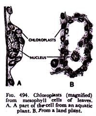
But they may be found in the parenchymatous cells of vascular tissues and in the embryo and endosperm of seeds. Chloroplasts have regularity of shape, usually being round, elliptical and discoidal (Fig. 494).
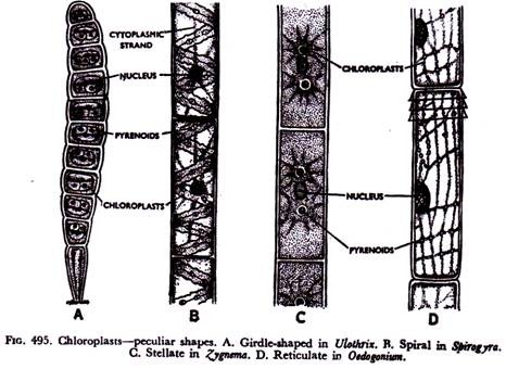
Very peculiar chloroplasts are characteristics of lower plants, particularly grass green algae (Chlorophyceae). In Spirogyra chloroplasts occur in form of spiral bands (Fig. 495B); Zygnema (Fig. 495C) and desmids have stellate or star-shaped ones; those of Ulothrix are girdle-shaped (Fig. 495A); Oedogonium (Fig. 495D) has peculiarly reticulate green plastids.
Chloroplasts of the liverwort Anthoceros (Fig. 496) are spindle shaped. In higher plants, as already stated, they have regular shapes and the average diameter usually ranges between 4 to 6 micra.
The number of chloroplasts is also variable—it may be one, as in Anthoceros, two in Zygnema, and more than that in many plants. In most of the cells they usually aggregate near the nucleus, so much so that in many cases they form a lump almost covering the nucleus.
The most important function performed by chloroplasts is photosynthesis involving elaboration of complex carbohydrate food matters out of water and carbon-dioxide gas in presence of sunlight.
To start with, simple soluble sugar, glucose, is synthesized. They also convert the simple glucose into complex insoluble starch grains. These starch grains, called assimilatory starch, are re-converted into sugar with nightfall.
Chloroplasts of some plants bear special refractive bodies called pyrenoids (Figs. 495 & 496). These have protein centres usually surrounded by starchy layers. They are found in lower plants like algae and liverworts.
In many members of grass-green algae (Chlorophyceae) one or more pyrenoids are present on the chloroplasts. In red algae (Rhodophyceae), pyrenoids are naked, where surrounding starchy layers are absent. These bodies may arise de novo or by division of pre-existing pyrenoids.
Chromoplasts:
Chromoplasts are coloured other than green, the colours usually ranging between red and yellow. These colours are due to yellow pigments, carotin and xanthophyll. Chromoplasts have no definite shape. They may be angular, forked, rod like, acicular (Fig. 497), etc.
The carotenoids may remain diffused in the stroma, may- occur in form of granules or may even appear to be crystalline. Chromoplasts are common in brightly coloured parts of the plants like the petals of the flowers and fruits. Attractive hues of all flowers are not due to chromoplasts alone.
Water-soluble pigments, anthocyanin, present in the cell- sap of the vacuoles also impart colours to the plants. Tomato fruits, when ripe becomes red due to appearance of a pigment, lycopen, in the chloroplasts.
The main function of chromoplasts is colour manifestation for attracting insects and other visitors which bring about pollination and dispersal of fruits and seeds. They are often present in the underground organs like carrots. Here the function is rather obscure.
Leucoplasts:
These are colourless plastids. They do not bear any pigment. Leucoplasts occur only in those organs which are not exposed to sunlight. Thus they are found in the underground organs and deep-seated parts of the aerial organs.
They may also occur in the epidermal cells. Leucoplasts are irregular in shape (Fig. 498)—may be small, granular and often larger. Small leucoplasts are converted into chloroplasts when exposed to sunlight.
The larger ones, called amyloplasts, are responsible for conversion of simple sugars into insoluble starch grains for the purpose of storage. These starch grains are called reserve starch or storage starch, and function performed by the amyloplasts is referred to as amyloplastic function.
Leucoplasts connected with the formation and storage of fatty substances arc known as elaioplasts (Fig. 499) which occur mainly in the liverworts and monocotyledons. Aleuronoplasts concerned with formation of proteinous matters have been reported in Ricinus. Leucoplasts -are abundantly present in immature cells and usually aggregate near the nucleus.
The origin of plastids has been a subject of keen investigation since a long time. In meristematic cells they are usually very small and non-pigmented. These are proplastids or plastid primordia which may be called the precursors of all the three different types of plastids. With gradual maturity and elaboration of the organs the plastids multiply and grow into different types of mature plastids. Possibly they multiply by constriction. Division of the proplastid is not usually associated with the division of other parts of the cell.
But in some plants like the green alga, Zygnema and the liverwort, Anthoceros the plastid divides regularly b fore or during the division of other parts of the cell. The entire plastid- complex of an organism has been referred to as plastidome.
Component # 6. Chondriosomes or Mitochondria:
These are very small living bodies, much smaller than plastids usually ranging in size from 0’2 to 3’0 µm universally present in the cytoplasm of plant, and animal, cells. They are more dense than cytoplasm and appear as small granules, rod-lets or nets. Mitochondria occur more abundantly in the cells which are metabolically more active.
Electron microscopy reveals that they have a double membrane, the outer limiting one and the inner one being convoluted into many plate-like folds called cristae projected into the lumen of the mitochondrion, which remains filled with a proteinous ground substance—matrix (Fig 500).
The outer and inner membranes are 40-75A (4-7 5 nm) wide and the space between them being 20-60A. (2-6 nm). Very small granules—rough and smooth are present on the outer and inner membranes.
The nature and function of chondriosomes has been a subject of long-drawn controversy amongst cytologists. Some workers regard them as permanent ‘organelles’ of cytoplasm which originate from pre-existing ones and multiply in number.
As they are often constantly appearing and disappearing, the suggestion that they may be formed anew has also been put forward. Chemically they are mainly composed of proteins and lipids, mainly phospholipids, and small amount of RNA and often DNA.
The function of chondriosomes has also been a vexing problem. The idea that they are proplastids or at least some of them develop into plastids has not received universal support. Due to occurrence of a large number of enzymes and co-enzymes they are regarded as the ‘power-house’ of the cell capable of breaking down complex molecules by oxidation and phosphorylation.
Through oxidation carbohydrates, proteins and fats are broken down with consequent release of energy. The stored energy remains bound in Adenosine triphosphate or ATP. Thus mitochondria are connected with a variety of activities like respiration, secretion and enzyme action.
Component # 7. Centrosomes:
These are minute cytoplasmic inclusions invariably present in animal cells. In some lower plants, algae and fungi, they occur near the nucleus.
A centrosome (Fig. 500A) has a deeply-stained central granule, called centriole, which usually remains embedded in a mass of hyaline substance known as centrosphere; but both of them may occur alone. During cell division a number of radiating rays, called astral rays, collect round the centrosome. They usually divide before cell division and move to the two opposite poles of the bipolar spindle.
Centrosomes may also be associated with the formation of cilia, the organs for locomotion, in some reproductive cells. In view of the fact that in many cases centrosomes are found to appear during cell division and disappear after the process, they are not regarded as permanent part of the cell.
Component # 8. Golgi Bodies:
These cytoplasmic inclusions, named after the discoverer Golgi, have been definitely found in animal cells. Special techniques with osmic acid and silver salts have to be employed for demonstrating them.
Using similar device certain cytoplasmic bodies with double membrane and vesicles, often dilated at the ends, have been reported in plants, particularly in the meristematic cells, which are supposed to be homologous to the Golgi bodies of animal cells (Fig. 501).
They have been noticed with the aid of electron microscope in the root tip of Allium spand root cells of Pisum sativum. Doubts exist in certain quarters that these are mere artifacts—non-living materials collected and rendered visible during staining.
The role of Golgi bodies in secretion in animal cells has been accepted. It has also been established that the Golgi complex, also called dictyosomes, has positive role in enzyme synthesis and regeneration of membrane systems.
Component # 9. Lysosomes:
These are small mitochondrion-like bodies having densely-packed granules with membraneous covering. Apart from their abundance in different animal cells they have been- reported in yeasts. Due to pressure of hydrolytic enzymes lysosomes take active role in breakdown of different cell substances. The materials in the lysosome often form crystals. The function of lysosome membrane is that of separating the hydrolytic enzymes from other parts of the cell, thus avoiding self-digestion.
On death of the cells enzymes are released which rapidly digest the remainder of the cell. They may be considered analogous to the digestive vacuoles in the unicellular organisms.


