ADVERTISEMENTS:
The importance of cell division can be appreciated by realizing the following facts:
1. Cell division is a pre-requisite for the continuity of life and forms the basis of evolution to various life forms.
2. In unicellular organisms, cell division is the means of asexual reproduction, which produces two or more new individuals from the mother cell. The group of such identical individuals is known as clone.
ADVERTISEMENTS:
3. In multi-cellular organisms, life starts from a single cell called zygote (fertilized egg). The zygote transforms into an adult that is composed of millions of cells formed by successive divisions.
4. Cell division is the basis of repair and regeneration of old and worn out tissues.
Cell Division:
Cell division, cell reproduction or cell multiplication is the process of formation of new or daughter cells from the pre-existing or parent cells.
ADVERTISEMENTS:
It occurs in three ways:
1. Amitosis or Direct cell division.
2. Mitosis or Indirect cell division.
3. Meiosis or Reductional cell division
Amitosis (Direct Cell Division):
(GK. a = no, mitosis = thread, osis = state)
Amitosis is a mode of division in which nucleus elongates, constricts in the middle and divides directly into two daughter nuclei. This is followed by centripetal constriction of cytoplasm to form two daughter cells.
Amitosis is characterized by:
i. Intact nuclear envelope is found through nut the division.
ADVERTISEMENTS:
ii. Chromatin does not condense into definite chromosomes.
iii. A spindle is not formed.
iv. Chromatin distribution occurs unequally which causes abnormalities in metabolism and reproduction.
v. Cytokinesis may or may not follow karyokinesis.
Amitosis occurs in mega-nucleus of paramecium, nuclei of internodal cells of Cham, endosperm cells of seeds, cartilage cells and diseased cells. Remak (1955) discovered amitosis in RBCs of chick embryo, but the term was coined by Flemming (1882). 
Mitosis (Indirect Cell Division)/ (Equational Cell Division):
(GK. Mitos = thread, osis = state)
Synonymes:
Indirect cell division, somatic or vegetative cell division, equational division or duplication division.
ADVERTISEMENTS:
Definition:
Mitosis is a type of cell division in which chromosomes are equally distributed resulting in two genetically identical daughter cells.
History:
Mitosis was first discovered in plant cells by Strasburger (1875). Later on, W. Flemming (1879) discovered it in animal cells. The term mitosis was coined by Flemming (1882).
ADVERTISEMENTS:
Occurrence:
The cells undergoing mitosis are called mitocytes. In plants, the mitocytes are mostly meristematic cells. In animals, the mitocytes are stem cells, germinal epithelium & embryonic cells. It also occurs during regeneration. Root tip is the best material to study mitosis.
Duration:
It varies from 30 minutes to 3 hours.
Steps of Mitosis:
Mitosis is a continuous process and for better understanding the whole process is divided into following six stages:
1. Prophase:
(i) Nucleus becomes spherical and cytoplasm becomes more viscous.
(ii) The chromatin slowly condenses into well-defined chromosomes.
(iii) Each chromosome appears as two sister chromatids joined at the centromere.
(iv) The spindle (microtubules) begins to form outside nucleus. In plants the spindle apparatus or mitotic spindle is anastral. In animals and brown algae the mitotic spindle is amphiastral which include two asters in opposite poles of the spindle. Each aster consists of two centrioles surrounded by astral rays.
2. Prometaphase:
ADVERTISEMENTS:
(i) Nuclear envelop breaks down into membrane vesicles and the chromosomes set free into the cytoplasm.
(ii) Chromosomes are attached to spindle microtubules through kinetochores. Specialized protein complexes that mature on each centromere are called Kinetochores.
(iii) Nucleolus disappears.
3. Metaphase:
(i) Kinetochore microtubules align the chromosomes in one plane to form metaphasic plate or equatorial plate. The process of formation of metaphasic plate is called congression.
(ii) Centromeres lie on the equatorial plane while the chromosome arms are directed away from t he equator called auto orientation.
(iii) Smaller chromosomes remain towards the centre while larger ones occupy the periphery.
4. Anaphase:
(i) Chromosomes split simultaneously at the centromeres so that the sister chromatids separate. They are now called daughter chromosomes. Where each one consists of single chromatid.
(ii) The separated sister chromatids move towards opposite poles at the speed of 1µm per minute.
(iii) Pole-ward movement of daughter chromosomes occurs due to shortening of kinetochore microtubules; appearance and elongation of inter-zonal fibers.
(iv)Daughter chromosomes appear V-shaped (metacentric), L-shaped (sub-metacentric), I-shaped (acrocentric) and I-shaped (telocentric).
(v) It is the shortest of all stages of mitosis.
5. Telophase:
(i) Daughter chromosomes arrive at the poles.
(ii) Kinetochore microtubules disappear.
(iii) Nuclear envelope reforms around each chromosome cluster of each pole.
(iv) Chromosomes uncoil into chromatin.
(v) Nucleolus re-appears.
(vi) It is considered as the reverse of prophase.
6. C-phase or Cytokinesis:
(i) It is the cytoplasmic division that starts during anaphase and completed by the end of telophase.
(ii) It takes place by two bare different methods i.e. cell plate method and cleavage or cell furrowing method.
i. Cell plate cytokinesis.
It occurs in plant cells. The spindle fibres persists at equatorial plane. The Golgi vesicles fuse at the centre to form barrel shaped phragmoplast. Further addition of vesicles causes the phragmoplast to grow centrifugally till it meets with plasma membrane of the mother cell. The contents of phragmoplast solidify to become cell plate or future middle lamella which separates the two daughter cells. The daughter protoplast secretes primary wall materials on both sides of the cell plate or middle lamella.
ii. Cleavage cytokinesis.
It occurs in animal cells and pollen mother cells of some angiosperms. In this process, a cleavage furrow appears at the middle, which gradually deepens and breaks the parent cell into two daughter cells. A special structure called mid body is formed in the centre, and it is a centripetal process.
Significance of-Mitosis:
1. Genetic Stability:
Mitosis maintains constant chromosome number and genetic stability in all somatic or vegetative cells of the body.
2. Growth:
Mitosis increases cell number so that a zygote transforms into a multicellular adult.
3. Surface-Volume ratio:
As the size (volume) of a cell increases, the surface area decreases accordingly. By mitosis, the cell becomes smaller in size and the surface volume ratio is restored.
4. Nucleo-plasmic ratio:
When a cell grows in size, nucleocytoplasmic ratio decreases. 11 are restored by mitosis.
5. Mitosis is a method of asexual reproduction and vegetative propagation.
6. Mitosis provides new cells for repair, regeneration and wound healing.
7. DNA content is reduced to half from parent cell to daughter cell.
Abnormal Mitosis:
(a) Intra nuclear mitosis (= Promitosis):
In this type of division nuclear envelope does not disappear and spindle developed within the nucleus, e.g., many protozoans, yeast, some fungi etc.
(b) Endomitosis (=Endopolyploidy):
Reduplication of chromatids within intact nucleus forming polytene chromosome, e.g. some liver cells in human.
(c) Dinomitosis:
In this mitosis, nuclear envelope remains intact and intra nuclear spindle is not formed. Condensed chromosomes are present even in interphase, e.g., Dinoflagellates.
(d) Free nuclear division:
In this case, repeated karyokinesis occurs without subsequent cytokinesis. This causes multinucleate condition called coenocyte (e.g… Rhizopus, Mucor etc.), Syncytium (e.g. Opalina) orplasmodium (e.g., slime moulds).
(e) C-Mitosis:
Doubling of chromosome number without cytokinesis by the application of alkaloid colchicine is known as C-Mitosis.
Meiosis (Reductional Cell Division):
(Gr. meioum = to reduce, osis = state)
Synonymes:
Reduction or dis-junctional division.
Definition:
Meiosis is a double division in which a diploid cell divides twice to form four haploid daughter cells.
History:
Vanzeneden (1883)-First reported meiosis Farmer & Moore (1905) Coined the term meiosis
Occurrence:
The cells undergoing meiosis are called meiocytes. In plants, the meiocytes are microsporocytes (Pollen mother cell) of anthers and megasporocytes (megaspore mother cell) of ovules. In animals, the meiocytes are primary spermatocytes in testes and primary oocytes in ovaries.
lnterphase I:
(i) Physiologically most active stage
(ii) Nuclear envelop remains intact
(iii) Nucleoli is prominent
(iv) Chromosomes appear in form of chromatin reticulum
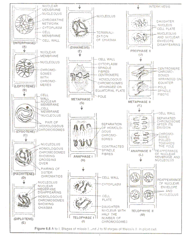
Prophase I:
It is typically longer and more complex phases… On the basis of chromosomal behaviour, it is divided into 5 sub-stages: Ieptotene, zygotene, pachytene, diplotene and diakinesis.
(a) Leptotene (= Leptonema-thin thread)
(i) Chromatin gradually condenses to form chromosomes.
(ii) Chromosomes appear as thin thread like structures with series of beads called Chromomeres also called Bouquet formation or synezesis.
(iii) Although chromosomes are replicated chromatids are not distinguished.
(iv) Both ends of each chromosome attached to nuclear envelope via specialized structures called attachment plaque.
(b) Zygotene (=Zygonema-paired\pairing)
(i) Pairing or synapsis of homologues chromosomes takes place in a zipper-like manner.
(ii) Each synapsed chromosome pair is called a bivalent.
(iii) During synapsis, a ladder like proteinous structure appears called synaptonemal complex (SC) between the homologues of a bivalent. Synaptonemal complex = Bivalent + U-Protein
(iv) Shortening of chromosomes continues.
(c) Pachytene (= Pachynema)
(i) Bivalents now appear as terrads.
(ii) Recombination nodules appear at intervals on the SC (synaptonemal complex)
(iii) The recombination nodules are thought to contain enzymes for crossing over or genetic recombination
(iv) Between non-sister chromatids.
(v) Nucleolus dissapears.
(d) Diplotene (= Diplonema)
(i) The SC dissolves so that the homologues in a bivalent separate from each other except at the cross-over points or chiasmata.
(ii) Homologues condense and detach from the nuclear envelope.
(iii) This stage lasts for months or years.
(e) Diakinesis
(i) The chromosomes further contract.
(ii) The chiasmata start moving towards the ends of tetrad. This process is called tenninalization.
(iii) Number of chiasmata reduce.
(iv) RNA synthesis stops
(v) Spindle formation occurs.
(vi) By the end, the homologues are held together only at their ends, nucleolus disappears and the nuclear envelope breaks down.
Metaphase I:
(i) Bivalents arrange on two equatorial plates.
(ii) The centromeres are directed towards poles and the arms of chromosomes face the equatorial plate called co-orientation.
(iii) The microtubules (chromosomal fibers) from opposite poles of the spindle attach to the bivalents.
Anaphase I:
(i) Half of the homologues chromosome separate and move to opposite pole. This process is known as disjunction.
(ii) Chromosomal fibres contract causing attraction while interzonal spindle fibres elongate causing repulsion.
Telophase I:
(i) Each pole possesses a group of dyad chromosomes.
(ii) Spindle fibers disappear
(iii) Nucleoli reappear and nuclear envelope reformed.
(iv) In Trillium telophase I is absent. The chromosomes pass from anaphase I lo prophase
Interkinesis:
(i) It is a very brief interphase between meiosis I and meiosis II.
(ii) There is no DNA replication i.e. S-phase absent.
Meiosis II (Equational or Homotypic D. vision):
The meiosis II is similar to mitosis in which chromosomes number remains constant.
Prophase II:
(i) Chromosomes shorten and thicken
(ii) Nucleoli disappear.
(iii) Nuclear envelope breaks down
(iv) The spindle fibres appear at right angles to the spindle of meiosis-I.
(v) In animal cells, centrosome divides and moves to opposite poles.
Metaphase-II:
i. Chromosomes aligned in one equatorial plate.
ii. Spindle fibres attached to kinetochores of sister chromatids.
iii. Centromeres remain on the metaphasic plate while the chromatids are extended towards the poles.
Anaphase-II:
(i) The centromere divides and the two chromatids of each chromosome separate and pulled towards opposite poles.
(ii) The separated chromatids are now called as daughter chromosomes.
Telophase II:
(i) The daughter chromosomes reach at the opposite poles.
(ii) The chromosomes uncoiled to form chromatin.
(iii) Nucleoli and nuclear envelope reappear.
(iv) Spindle fibers dissapear.
Cytokinesis:
In meiosis, 2 types of cytokinesis can be seen.
(a) Successive type:
In this case, cytokinesis occurs after both meiosis I and meiosis 11. As a result four haploid cells are formed. In plants the cells are arranged in form of isobilateral tetrad or in a linear manner.
(b) Simultaneous type:
In this case, cytokinesis occurs twice only after meiosis II. The four haploid cells arranged in form of a tetrahedral tetrad.
In plant cells, cytokinesis takes place by cell plate method, while in animal cells cleavage or furrowing method generally occurs.
Significance of Meiosis:
1. Meiosis essentially maintains constancy in chromosomes from generation to generation.
2. Crossing over and disjunction bring genetic variation within the species. The variations are important raw materials for evolution and also help in improvement of races.
3. Meiosis causes segregation and random assortment of genes.
4. Meiosis causes conversion from sporophytic generation to gametophytic generation in plants.
5. It leads to the formation of haploid gametes (n) which is an essential process in sexually reproducing organisms. Fertilization restores the normal somatic (2n) chromosome number.
Types of Meiosis:
There are three types of meiosis based on the variations in time and place of the division in the life- cycle of the plant.
1. Zygotic or Initial Meiosis (Haplontoic Pattern):
During the process of fertilization, the two gametes fuse to form zygote which represents the only diploid stage in the life-cycle. The zygote undergoes meiosis and forms four haploid cells which later on develop into haploid individuals, e.g., Thallophyta.
2. Gametic or Terminal Meiosis (Diplontoic Pattern):
This type of meiosis can be seen in animals and some lower plants. Here the meiotic division takes place immediately before gamete formation and the haploid cells thus formed are transformed into sperm (male gamete) and egg (female gamete).
3. Sporic or Intermediate Meiosis (Diplo- haplontoic Pattern):
We come across this type of meiosis in higher plants and in some thallophyta but not in animals. The life-cycles of these organisms are characterised by alternation of haploid and diploid generations (i.e., gametophytic and sporophytic generations).
Meiosis occurs in the sporogenous cells (micro-and megaspore mother cells) of the sporophyte producing haploid spores. The spores on germination form gametophytes (male and female). Cells of the gametophyte form gametes. Fusion of these gametes again leads to diploid or sporophytic generation, and in this way alternation between gametophytic and sporophytic generations keeps on going.

