ADVERTISEMENTS:
In this article we will discuss about:- 1. Introduction to Dynamics of Chromosome 2. Events of Chromosome Movement 3. Mechanism.
Introduction to Chromosome Movement:
Chromosomes are involved in a series of directed movements during both mitosis and meiosis. With the separation of the sister chromatids/homologues at anaphase, the equilibrium is broken, the chromosomes move towards the poles at the rate of about 1 pm/min.
By hydro-dynamic analysis it has been calculated that a force of about 10-11 dynes is needed and the entire displacement from the equator to the pole of a chromosome requires the use of about 30 ATP molecules.
Events of Chromosome Movement:
ADVERTISEMENTS:
During mitosis, where chromosome movements have been studied in some detail, following events have been observed. In prometaphase, the chromatid pair is first drawn to the spindle pole, then migrates towards the centre of the spindle.
At the onset of anaphase, the chromatids separate and migrate slowly towards the spindle poles (Fig. 5.18). Anaphase movement of chromosomes comprises of two distinct processes — Anaphase A and Anaphase B.
Anaphase A (early anaphase) involves the shortening of microtubules associated with kinetochore (discontinuous microtubules);
ADVERTISEMENTS:
Anaphase B (late anaphase) involves elongation of entire spindle fibres nearly to double length to reach to the poles (continuous microtubules).
Role of Kinetochore:
Kinetochore is a trilaminar plate like structure situated at the primary constriction of chromosome. The chromosomes get attached with microtubules by kinetochore before proceeding towards poles. Contact between microtubule and kinetochore takes place laterally and then converts to polar attachment (Fig. 5.19).
Kinetochore is supposed to be a passive structure remains attached to microtubule ends while microtubules are shortened during chromosomal movement in anaphase. This shortening may be due to disassembly or de-polymerization.
Kinetochore-spindle pole attractions result in chromosome movement so that when two chromatids separate, they move automatically towards the poles due to de-polymerization. The discontinuous spindle fibres carrying the kinetochores contract and pull the chromosomes towards the poles while the continuous fibres elongate and hold the poles apart.
Removal, of spindle polar region affects the movement of chromosomes whereas removal of spindle fibres does not affect the chromosomal movement towards the pole. These findings suggest that kinetochore region is mostly responsible for anaphasic movement providing the motive force for this.
Mechanism of Chromosome Movement:
ADVERTISEMENTS:
Several hypotheses have been attempted to interpret the molecular mechanism of movement of chromosome (Fig. 5.20).
Ostergreen (1949) proposed the dynamic equilibrium hypothesis where the events of polymerization and de-polymerization of microtubules is directly responsible for the movement of chromosomes. According to this hypothesis the energy released (from GTP hydrolysis) is sufficient to drive the chromosome movement during anaphase.
ADVERTISEMENTS:
This hypothesis postulates that the microtubules are firmly anchored at kinetochore, while the polar ends are free from disassembly. In this anchorage-translocation system the motive force would be provided by the polarized assembly-disassembly of the microtubules.
According to sliding hypothesis, the spindle microtubules assemble and disassemble but the driving force is not directly generated to move the chromosomes. This hypothesis postulates that involvement of lateral interactions of microtubules can play a role analogous to dynein in flagella.
Many of the observations, after using the protein like actin and myosin, suggested that the interaction of spindle actin with myosin or dynein with microtubules could provide the motive force while the direction is provided by the microtubules.
ADVERTISEMENTS:
Role of Motor Protein:
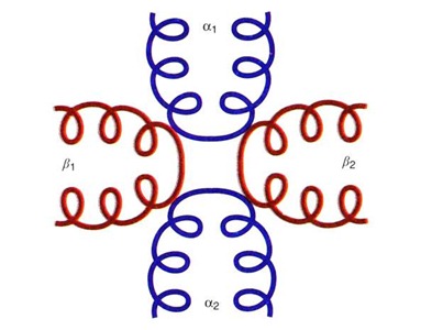
Recently two proteins have been identified, namely kinesin and cytoplasmic dynein (Fig. 5.21), which are associated with kinetochore, have been shown to be dynamic structures. The forces generated by these proteins are opposite in direction — one is directed towards the pole and the other is away from it.
The cytoplasmic dynein may be involved in pole ward movements, whereas kinesin may be involved in reverse movement towards centre during congression. Both these proteins are members of larger families and that microtubule based movements require a variety of motor proteins, which need to be targeted and activated for initiation of their function.
Kinesin has two headed motor domains which have an ATPase activity and thus catalyzes movement by hydrolysis of ATP. Dynein consists of force producing heavy chains and a variety of accessory subunits.
Role of Phosphorylation and Dephosphorylation:
Recent investigations on the forces involved for chromosome movements have been- studied using in vitro techniques involving association of isolated chromosomes and microtubules. It has been shown that microtubules become associated with kinetochores and in presence of ATP travel towards the minus end (pole ward direction).
Using the antibody (anti- thiophosphate) technique, the thiophosphate group could be located in the kinetochore, suggesting that phosphorylation of a protein component of kinetochore actually leads to reversal of the direction of chromosome movement.
It is suggested that both kinase and phosphatase may be associated with kinetochore and two motor proteins (dynein and kinesin) are involved in the development of forces in opposite directions (Fig. 5.22).
It has been shown that differential phosphorylation and dephosphorylation of two ATPases (phosphorylation facilitated by kinase and dephosphorylation facilitated by phosphatase) make an unique balance to support the movement of chromosome. Though the minus end motor activity is given by dynein located at kinetochore but the plus end motor activity may be due to some other protein like kinesin.
ADVERTISEMENTS:
Role of De-polymerization and Polymerization of Microtubules:
Besides phosphorylation and de-phosphorylation, microtubules grow and shrink due to polymerization and de-polymerization at the kinetochore. This phenomenon suggests that motor activity and microtubule dynamics are coordinated away from and towards the spindle pole during cell division.
Two models have been proposed to explain the de-polymerization of microtubules:
(i) Pac-man model (microtubules are chewed up at the kinetochore end);
(ii) Pole ward flux model (microtubules are depolymerized at the poles) (Fig. 5.23).


