ADVERTISEMENTS:
In this article we will discuss about:- 1. External Features of Riccia 2. Anatomy of Gametophyte in Riccia 3. Sex Organs.
External Features of Riccia:
Study dorsal and ventral surface of the thallus; separate scales and mount in glycerine; also separate rhizoids from the ventral surface, stain in safranin, mount in glycerine and study:
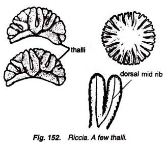
1. Gametophytic plant body is thalloid, prostrate, linear or ribbon-shaped and flattened structure.
ADVERTISEMENTS:
2. It is dichotomously branched (Fig. 152) and green to light green in colour.
3. Thallus is thick in the middle and becomes thin towards the margins.
4. An apical notch is present on the terminal portion of thallus. Gametophytic plant body has two surfaces, dorsal and ventral.
ADVERTISEMENTS:
(a) Dorsal Surface:
5. A median longitudinal furrow or depression is present on the dorsal surface (Fig. 153).
6. Sex organs are also present on this surface.
(b) Ventral Surface:
Scales and rhizoids (Fig. 154) are present.
Scales:
1. These are generally present (Fig. 154) on the margins in one row, or near the growing points.
ADVERTISEMENTS:
2. Scales are multicellular but one-celled thick structures (Fig. 155).
3. The colour of the scales is violet.
4. Function of scales is to protect the young growing points.
ADVERTISEMENTS:
Rhizoids:
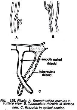
1. They are of two types, i.e., smooth-walled and tuberculate rhizoids (Fig. 156).
2. Smooth-walled rhizoids are unicellular, hyaline and fine hair-like structures.
ADVERTISEMENTS:
3. Tuberculate rhizoids are also unicellular, but some peg-like ingrowths rise in the lumen of the cell of these rhizoids.
4. Functions of the rhizoids are absorption and fixation.
Anatomy of Gametophyte in Riccia:
Cut thin T.S. of thallus, stain in safranin, mount in glycerine and study.
Following two distinct regions are visible (Figs. 157,158):
(a) Photosynthetic region, and
(b) Storage region.
Photosynthetic Region:
1. It is situated on the upper or dorsal surface of the thallus and consists of loose green tissue. It is also known as assimilatory region.
2. It consists of a layer of epidermis, many air pores, air spaces or air chambers and many one-celled thick vertical rows of chlorophyll-containing cells.
3. In the cells of vertical rows are present many discoid chloroplasts.
ADVERTISEMENTS:
4. In between these vertical rows are present air spaces or air chambers.
5. Uppermost cells of these vertical rows of chlorophyll-containing cells remain devoid of chloroplast and thus form a hyaline discontinuous layer of upper epidermis.
6. Continuity of the upper epidermis is broken by many air pores.
Storage Region:
7. It is situated on the lower or ventral surface of the thallus and consists of colourless cells.
8. Cells are closely packed and parenchymatous.
ADVERTISEMENTS:
9. Cells contain starch and are without intercellular spaces.
10. The lowermost cells of this region form a regular lower epidermis, from which arise scales and rhizoids (Fig. 158).
11. Scales are present on the margins of the thallus.
12. Function of storage region is to store the food material and water.
Riccia fluitans:
External Features:
1. It is the only aquatic species of Riccia. It remains floating on or under the water surface.
2. Thallus is long, ribbon-like (Fig. 159A), and shows a clear dichotomous type of branching.
3. Scales and rhizoids are absent.
4. Sporogonia develop only when it comes in contact with mud of the pond.
5. Formation of adventitious branching is the means of vegetative reproduction.
Anatomy:
It differs from the terrestrial species in the following ways:
1. The upper epidermis is a continuous layer.
2. Air spaces are large and formed by irregularly arranged lamellae (Fig. 159B).
3. Lamella is single-layered.
4. Cells of upper epidermis and lamella contain chloroplasts.
5. Rhizoids and scales are absent.
Sex Organs of Riccia:
Cut L.S. of mature thallus through mid-dorsal groove and observe the sex organs under microscope.
If antheridia, archegonia or sporogonium are visible, stain in safranin, mount in glycerine and study:
1. Sexual reproduction is oogamous.
2. Sex organs are antheridia and oogonia. They are present in the region of mid-dorsal groove on the dorsal surface of the thallus.
3. Sex organs develop in acropetal succession.
4. Most of the species are homothallic but a few species are heterothallic, e.g., R. himalayensis.
5. Monoecious species are generally protandrous, i.e., antheridia develop first and oogonia later on.
Antheridium:
V.S. of gametophyte through antheridium (Fig. 160) reveals following structures:
1. Antheridium is present in a cavity on the dorsal surface called antheridial chamber.
2. Antheridial chamber has a narrow opening or pore on the apical side.
3. A mature antheridium is a stalked, club-shaped or pear-shaped body.
4. Antheridial stalk is multicellular.
5. It remains surrounded by an outermost layer of one- celled thick sterile jacket.
6. Inside the jacket layer are present many small, cubical androcyte mother cells.
7. Each androcyte mother cell contains dense cytoplasm and large nucleus. It divides diagonally into two androcytes.
8. Each androcyte metamorphoses into a single structure, variously called antherozoid, spermatozoid or sperm.
9. Each antherozoid is a minute, uninucleate body containing two long flagella at its anterior end.
10. Lower flagellum is slightly larger than upper one.
11. Dehiscence of antheridium takes place in the presence of water.
Archegonium:
A vertical section passing through archegonium reveals the following details (Fig. 161):
1. The archegonium remains embedded in the archegonial cavity on the dorsal surface of the gametophyte.
2. Upper part of the neck of archegonium generally protrudes out of the cavity.
3. An archegonium is a flask-shaped structure made up of a long, elongated neck and a globular venter.
4. Venter is sessile and surrounded by a one-celled thick layer, made up of 12 to 20 cells.
5. Neck consists of 4 to 6 neck canal cells, and remains surrounded by six vertical rows of cells.
6. Each longitudinal row consists of 6 to 9 cells.
7. At the tip of the neck are present four cover cells or lid cells.
8. Venter contains an upper, small ventral canal cell and a lower, large egg cell.
9. At the time of fertilization (Fig. 162) all the cells, except the egg, disintegrate and form a mucilaginous liquid, which gives entry to the spermatozoids. The ultimate product of the fertilization is zygote.
10. The ultimate product of the fertilization is zygote.
Sporogonium:
1. It is simple and made up of only capsule or spore-sac (Fig. 163).
2. Foot and seta are absent.
3. It remains embedded in the gametophyte, and it is a non-green structure, thus depending entirely on the gametophyte for food.
4. Inside sporogonium are present many spore mother cells which remain surrounded by a capsule wall and two-layered calyptra.
5. Spore mother cells divide reductionally, and each of them thus forms four haploid spores, arranged tetrahedrally.
6. Prior to the formation of spores, the wall of the sporogonium as well as the inner layer of calyptra dissolve, and thus only a single-layered calyptra is present outside the spores.
7. Elaters are absent (Fig. 163).
8. Neck of the long archegonium may remain outside for some time but it ultimately withers.
Spore:
1. It is the first cell of the gametophytic generation.
2. Shape of the spore is rounded or pyramidal.
3. Each spore is surrounded by a thick, black or sculptured wall.
4. Wall of the spore consists of three layers: outermost exine or exosporium, which is thick and sculptured; middle thin mesosporium and: innermost, thin intineorendosporium(Fig. 164).
5. Spores are unicellular and uninucleate structures.
6. In the cytoplasm are present many oil globules.
7. Spores germinate into gametophyte.


