ADVERTISEMENTS:
In this article we will discuss about the anatomy of marsilea.
Cut the transverse sections through different parts of the plant (rhizome, petiole, leaflet, and root), stain them separately in safranin-fast green combination, mount in glycerine and observe the anatomical details under microscope:
Rhizome:
ADVERTISEMENTS:
1. The outermost layer is epidermis which is continuous and without any stomata.
2. The cortex consists of four regions.
3. The outermost region of cortex is made up of one to many-celled thick parenchymatous tissue. Some tannin cells are also present in this region.
4. Below the parenchymatous region is the air storage region of cortex consisting of many air cavities separated by one-celled thick parenchymatous septa (Figs. 260,261).
5. Inner to the parenchyma is the region of sclerenchynia in the cortex.
6. The innermost region of cortex consists of many- celled thick compact parenchymatous tissue. In this region some tannin cells are also present.
7. The stele is amphipholic siphonostele, i.e., xylem is surrounded on both the sides by the phloem, and in the centre there is present the pith (Figs. 260, 261).
8. Just outside the central parenchymatous (in submerged species) or sclerotic (in species of dry conditions) pith there is a layer of inner- endodermis surrounded by a layer of inner pericycle.
9. Outside the inner pericycle is the region of inner phloem enveloped externally by the xylem ring.
10. Protoxylem elements in the xylem ring varies in their position. In some species the protoxylem is absent, e.g., Marsilea quadrifolia.
11. Outside the xylem ring is present the outer phloem surrounded by outer pericycle and outer endodermis.
12. In the section of rhizome passing through the node there is present a leaf gap.
ADVERTISEMENTS:
Petiole:
1. The outermost layer is epidermis which consists of rectangular cells.
2. Below the epidermis is the hypodermis followed by the cortex.
3. Cortex is differentiated into outer and inner cortex.
ADVERTISEMENTS:
4. Outer cortex consists of aerenchyma having many- air cavities or air chambers separated from each other with the help of one-celled thick trabeculae or septa (Fig. 262).
5. Inner cortex is parenchymatous and contains starch and tannin-filled cells.
6. The stele is triangular in shape, present in the centre and shows the protostelic structure.
ADVERTISEMENTS:
7. The stele is bounded by a layer of endodermis and unilayered pericycle. Xylem is ‘V’ shaped, and the arms of ‘V’ contain a metaxylem in the centre and protoxylem towards the ends. The xylem remains surrounded by the phloem.
Leaflet:
1. A layer of upper epidermis (Fig. 263) on the upper surface and lower epidermis on the lower surface is present.
2. Stomata are generally present on the upper epidermis but they may be present on both the layers.
ADVERTISEMENTS:
3. Mesophyll (Fig. 263) is differentiated into palisade tissue and many rounded cells of spongy parenchyma.
4. The vascular bundles are concentric. The xylem is surrounded by phloem and a layer of endodermis.
Root:
1. The outermost layer is the epidermis, which is a continuous layer.
2. The cortex consists of two zones, i.e., outer cortex and inner cortex (Fig. 264).
3. Outer cortex is aerenchymatous and consists of many air chambers separated by septa or trabeculae.
4. Inner cortex is parenchymatous but if the roots are present in dry situation, it is sclerenchymatous.
5. The stele is bounded by a layer of endodermis and unilayered pericycle.
6. The xylem is diarch and exarch. Two large strand of metaxylem elements are present in the centre having two groups of protoxylem towards the periphery.
7. The phloem is present in the form of bands.
Sporocarp:
ADVERTISEMENTS:
Spores are of two types (micro- and megaspores) and present in a special body called sporocarp (Figs. 258, 265).
1. Each sporocarp is a bean-shaped, ovoid or capsule-like structure.
2. Sporocarps (Fig. 265) are present at the base of or slightly higher on the petiole. They are attached to the petiole with the help of a long stalk called peduncle.
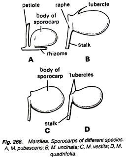
3. The place of attachment of peduncle with the body of the sporocarp is known as raphe (Fig.266).
4. Slightly above the raphe, on the body of the sporocarp, are present two protuberances or teeth- like structures called tubercles. In M. polycarpa, there is no tubercle on the sporocarp.
5. Marsilea is heterosporous.
6. The number of sporocarps may be one, two or more in different species.
General Anatomy of Sporocarp:
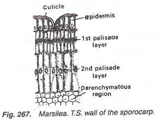
1. The sporocarp is surrounded by a thick wall, which consists of an outer layer of epidermis having many stomata, and two-layered hypodermis, the cells of which are radially elongated. The nuclei in the cells of hypodermal layers are arranged in a line (Fig. 267).
2. Inside the sporocarp wall is present a ring of gelatinous tissue (Fig. 268).
3. Sporocarp is a bivalved structure and each valve contains a row of sori.
4. Each sorus is surrounded by a thin delicate indusium contains microsporangia and megasporangia having microspores and megaspores respectively.
5. Each sporocarp gets vascular supply by a stalk bundle, dorsal bundles, many lateral bundles, placental branches and placental bundles (Fig. 269).
Anatomy of Sporocarp:
Cut the sections of the sporocarp in different planes (Y.T.S..H.L.S. and V.L.S.). Stain them separately in safranin mount in glycerine and study:
V.T.S. Sporocarp:
Vertical transverse section (V.T.S) is cut at right angle to the body of the sporocarp (Fig.270A):
1. The sporocarp is surrounded by sporocarp wall consisting of layer of epidermis with stomata and two layers of hypodermis.
2. The cells of the inner layer of hypodermis are more elongated and thin than that of the outer layer.
3. The gelatinous ring is observed in the form of two gelatinous masses on two sides. The gelatinous mass present on the dorsal side is more prominent and clear than that of ventral side.
4. In the centre of the sporocarp are present only two sori covered by their individual indusium.
5. In each sorus are present megasporangia having megaspores, and on each corner is present a microsporangium having microspores (Fig. 270 B, C).
6. Dorsal bundle, lateral bundles and placental bundles constitute the vascular supply of the sporocarp in V.T.S.
H.L.S Sporocarp:
Horizontal longitudinal section (H.L.S.) is cut perpendicularly to the flat surface of the sporocarp (Fig. 271 A).
1. The structure of the wall of the sporocarp and the gelatinous ring, present in the form of two gelatinous masses on dorsal and ventral sides, is the same as observed in vertical transverse section (V.T.S.).
2. A stalk bundle is present in stalk, which is cut transversely.
3. All the sori are observed in H.L.S.
4. Each sorus is covered by its indusium and the sori of two sides alternate with each other.
5. Two microsporangia on sides and one mega-sporangium at the apex are present in each sorus (Fig. 271 B, C).
6. Dorsal bundle, lateral bundles, placental branches and stalk bundle constitute the vascular supply of the sporocarp in H.L.S.
V.L.S. Sporocarp:
Vertical longitudinal section (V.L.S.) is cut dorsiventrally(Fig. 272 A).
1. The structure of sporocarp wall is same as discussed earlier (Fig. 267).
2. In the centre are present a sori surrounded by a gelatinous ring.
3. All the sori are visible in this section, and each sorus contains its individual indusium.
4. If the section is passing through the median line, only megasporangia are visible, but if it is slightly away from the median line only microsporangia are visible.
5. Many lateral bundles and a single stalk bundle constitute the vascular supply in V.L.S.
Microspores:
1. Each microspore is globose in shape, and is surrounded by a thick wall consisting of an inner endospore and an outer exospore (Fig. 273).
2. In the centre is present large conspicuous nucleus surrounded by cytoplasm having many starch grains?
3. A microspore ranges from 45 µ to 60-75µ in diameter in different species.
Megaspore:
1. Each megaspore is large (0.3 to 0.8 mm in diameter), ellipsoidal structure having a small projection at its anterior end (Fig. 274).
2. Megaspores are surrounded by four layers.
3. The nucleus is situated in apical papilla.
4. In the large basal part, the cytoplasmic material consists of starch grains, oil and many albuminous granules while in the upper papillate region it is finely granular.


